UIUC MCB 245 Practical 1
 Call Kai
 Learn
Learn Practice Test
 Spaced Repetition
 Match
Match Flashcards
 Knowt Play
1/231

There's no tags or description
Looks like no tags are added yet.
Name | Mastery | Learn | Test | Matching | Spaced | Call with Kai | Chat |
|---|
No analytics yet
Send a link to your students to track their progress
232 Terms
Front
Anterior
Rear
Posterior
Towards the midline
medial
away from the midline
lateral
closer to the attached trunk
proximal
farther from the attached trunk
distal
belly side
ventral
back side
dorsal
outside, closer to the surface
superficial
inside, farther from the surface
deep
toward the nose or mouth
rostral
toward tail
caudal
toward the head
cranial/cephalic
divides the body into left and right
sagittal
special type of sagittal plane that divides the body equally
midsagittal
divides body into anterior and posterior
coronal
divides the body into superior and inferior portions
transverse
ocular lens/ eyepiece

contains lens for viewing, also increases magnification by 10x
ocular lens
nosepiece of microscope
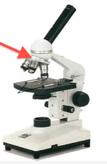
holds the objective lenses and can be rotated to change magnification
nosepiece of microscope
arm of microscope

provides support for the body and adjustment tubes
arm of microscope
objective lens

contains lenses of different magnifications
objective lenses
lamp light

provides light source which is projected up through the tissue to allow for viewing
lamp light
base

provides structural support for the microscope and houses the light source
base
control knob
#2
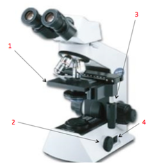
allows for adjustment of light being transmitted through tissue
control knob
power switch
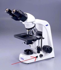
mechanical stage

supports microscope slide which can be adjusted to allow viewing of different sections of a slid
mechanical stage
mechanical stage controls

stage clips

condenser

focuses incoming light onto slides
condenser
course adjustment knob

moves the mechanical stage up and down in large increments to aid in focusing
course adjustment knob
condenser adjustment knob

regulates the amount of light being passed through the tissue
condenser adjustment knob/ iris diaphram
layers of cells covering internal or external surfaces
Epithelial Tissues
provide physical protection, control permeability, provide sensation, produce specialized secretions
Functions of epithelial tissues
cellularity- very little extracellular matrix, polarity, attachment, avascular, regeneration, layers (for stratified)
characteristics of epithelia
lining of blood and lymph vessels, lining of central cavities, portions of kidney tubules, inner lining of cornea, alveoli of lungs
simple squamous epithelium location
rapid diffusion (permeability), reduce friction
function of simple squamous epithelium
outer surface of the skin, lining of the mouth, throat, esophagus, rectum, anus, and vagina
location of stratified squamous epithelium
protection against physical abrasions, pathogens, chemical injury from exposure to outside environment
function of stratified squamous epithelium
glands (endocrine and exocrine), parts of kidney tubules, thyroid gland
location of simple cuboidal epithelium
secretion of hormones and other solutions
functions of simple cuboidal epithelium
lining of some ducts of sweat glands and mammary glands
location of stratified cuboidal epithelium
secretion of exocrine solutions like sweat and breast milk
functions of stratified cuboidal epithelium
stomach lining, intestinal lining, uterine tubes
location of simple columnar epithelium
absorption (primarily digestive) and secretion of mucus
function of simple columnar epithelium
salivary gland ducts and urethra
location of stratified columnar epithelium
protection and secretion of saliva
functions of stratified columnar epithelium
lining of nasal passages, trachea, and bronchi
location of pseudostratified ciliated columnar epithelium
protection- cilia traps particles
function of pseudostratified ciliated columnar epithelium
urinary bladder, ureters
location of transitional epithelium
allows expansion and recoil upon stretch
function of transitional epithelium
Diffusion of water through a selectively permeable membrane which occurs whenever there is a difference concentration on the two sides of a membrane
osmosis
water and solutes pass through a membrane from an area of higher hydrostatic (fluid) pressure into an area of lower hydrostatic pressure
filtration
passive transport of solutes from high to low concentration
diffusion
when a cell expends cellular energy (ATP) to move across its membrane
active transport
connective tissue proper, fluid connective tissue, supporting connective tissue
classification of connective tissues
structural framework
Transporting fluids and dissolved materials
Protecting
Supporting, surrounding, and interconnecting
Storing energy reserves
Defending against pathogens
functions of connective tissues
connect and protect
connective tissue proper function
loose and dense
two classes of connective tissue proper
more ground tissue and fewer fibers
loose connective tissue
more fibers, less ground substance (tendons)
dense connective tissue
highly vascularized, contains both collagen and elastic fibers, cushions organs, provides support but allows movement, defense
areolar connective tissue properties and functions
deep dermis of skin, beneath epithelial lining of GI tract, respiratory tract, urinary tract, between muscles, covers blood vessels, nerves and joints
location of areolar connective tissue
provides padding, cushions shock, insulator, storage of energy
functions of adipose connective tissue
deep to the skin, breast, buttocks, padding around kidney, abdominal region
location of adipose connective tissues
provides supporting framework
functions of reticular connective tissue
liver, spleen, lymph nodes, bone marrow
location of reticular loose connective tissue
poorly vascularized, mostly fibers with little ground substance, tightly packed with high numbers of collagen fibers, site of attachment for bones and muscles, reduces friction between muscles, stabilize relative position of bone
functions/properties of dense regular connective tissue
tendons and ligaments
locations of dense regular tissue
provides strength and reduces forces from multiple directions; helps prevent overexpansion of organs, contains collagen fibers
properties/functions of dense irregular tissue
periosteum, perichondrium, dermis of the skin, urinary bladder
locations of dense irregular tissue
stabilization permits contraction/expansion of organs, contain elastic fibers
properties of elastic connective tissue
in ligaments between vertebrae, ligaments supporting penis, trachea, vocal cords, and aorta
location of elastic connective tissue
strong framework, few cells, fibrous matrix, mature cells in lacunae
properties of supportive connective tissues
support and shape
functions of supportive connective tissue
bone and cartilage
types of supportive connective tissue
no innervation, avascular, surrounded by perichondrium
properties of cartilage
hyaline cartilage, fibrocartilage, elastic cartilage
types of cartilage
provides stiff but flexible support, reduces friction between bony surfaces
properties and function of hyaline cartilage
resists compression, prevents bone to bone contact and limits movement of vertebral column, contains rows of collagen fibers
functions and properties of fibrocartilage
pads in knee joint, pubic symphysis, cartilage of intervertebral discs
location of fibrocartilage
tolerates flexion and returns to normal state, contains web of elastic fibers
properties and functions of elastic cartilage
-Tip of ear
-Epiglottis
-Auditory canal
locations of elastic cartilage
lymph and blood
two types of Fluid Connective tissue
carries fluid and lymphocytes to lymph glands and then back to the venous system to be recirculated into blood
functions of lymph
long, short, flat, irregular
shapes of bone
long and thin bones found in arms, legs, hands, feet, fingers, and toes
long bones
Thin with parallel surfaces
Found in the roof of skull, sternum, ribs, and scapulae
flat bones
small and thick bones such as the ankle or wrist
short bones
have complex shapes such as vertebrae or pelvic bone
irregular bone