Acoustic artifacts
 Call Kai
 Learn
Learn Practice Test
 Spaced Repetition
 Match
Match Flashcards
 Knowt Play
1/24

There's no tags or description
Looks like no tags are added yet.
Name | Mastery | Learn | Test | Matching | Spaced | Call with Kai | Chat |
|---|
No analytics yet
Send a link to your students to track their progress
25 Terms
Noise/speckle
Caused by non-specular reflections from rough surface
More speckle seen with lower frequency transducers
Scattering causes wave interference, resulting in speckled appearance
Typically associated with liver, spleen, thyroid tissue
Side lobes
Weak off axis portions of the beam; seen with single PE element
Grating lobes
Weak off axis portions of the beam; seen with an element array
When wea, off axis portions of the beam encounter a string reflector, the reflected energy may become part of the main beam
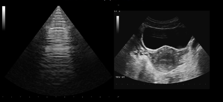
Reverberation
Caused by reflection of US back and forth between two closely spaced interfaces
Multiple echogenic lines equally spaced apart
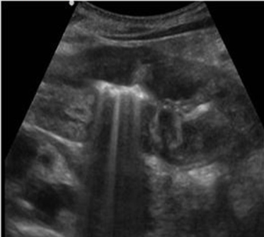
Ring down artifact
Type of resonance artifact
Occurs with gas/tissue interface
Vertical streak of parallel lines project posterior to interface
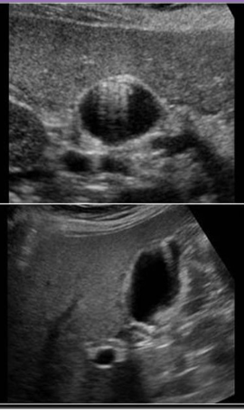
Comet tail artifact
Series of echoes caused by small multiple reflections from a highly reflective object
Forms a single long hyperechoic artifact
Posterior enhancement
Anechoic structures allow sound to be transmitted through and there is increased reflection from structures posteriorly
Posterior shadowing
Echogenic structures reflects majority of sound, no sound is transmitted posteriorly
Bayonet artifact
Propagation speed artifact that causes the appearance of a bent needle
Caused by variations of densities
Mirror image
Created as sound reflects off a strong reflector
Appears as a second copy of structure deeper to image
Range ambiguity
Shallow depth = High PRF
Shallow reflectors reflect sound energy that arrives at the transducer before the rest of the pulse finishes transmitting
Causes confusion on echo placement in near field
Refraction artifact
Refraction of the beam between two dissimilar media can cause a structure to appear more lateral than the actual structure
Volume averaging
AKA slice thickness or beam width artifact
Affect eval of superficial lesions
Reduced with standoff pad
Can cause solid lesions to appear anechoic
Echo texture of adjacent structures is then averaged to that of lesion
Aniostrphy artifact
Falsely hypoechoic areas of tendon
Occurs when transducer is not completely parallel to tendon
Reduced by rocking the transducer to make the face of the transducer more parallel to tendon
Element damage
If a PE element is not functioning, data will be missing from the image
If the probe is a single element, the entire image will not be displayed
A linear, sector or vector will demonstrate the area of no reflected info as a black vertical line
An annular array with demontsrate an anechoic horizontal arch of missing data
A curved array will demonstrate a vertical line of missing data
Nyquist limit
Maximum frequency that can be displayed is ½ PRF
Blooming
Color obscures gray scale display of soft plaque becuase of resolution of color Doppler is lower than the gray scale
The adjacent flow causes the pixel to be displayed as color instead of gray scale info
Crosstalk
Mirror image of Doppler signal
Which of the following is an artifact caused by gas-tissue interface
Ring down
Mirror
Speckle
Shadowing
Ring down
What acoustic artifact is associated with adenomyomatosis
Comet tail
Which of the following results in a copy of a structure displayed laterally on the image?
Ring down
Mirror
Enhancement
Refraction
Refraction
Anisotropy artifacts are seen on the evaluation of:
Tendons
How can you differentiate element damage from posterior shadowing when using a linear transducer?
Identify where the shadowing beings
What causes clutter artifact?
Low velocity scale settings
Blossoming of the spectral Doppler waveform can cause:
Overestimation of PSV