Special Senses
 Call Kai
 Learn
Learn Practice Test
 Spaced Repetition
 Match
Match Flashcards
 Knowt Play
1/166

There's no tags or description
Looks like no tags are added yet.
Name | Mastery | Learn | Test | Matching | Spaced | Call with Kai | Chat |
|---|
No analytics yet
Send a link to your students to track their progress
167 Terms
Sensory system overview
Sensory receptors receive stimuli from the extneral or internal environment, stignals carreid to brain or spinal cord, different parts of the brain process information
Somatosensory system
Concered with the conscious perception of touch, pressure, pain, temperature, position, movement, vibration which arise from muscles, joints, skin, fascia
Somatic sensation
Sensation from the skin, muscles, bones, tendons and joints initiated by somatic receptors
Stimulus motility
What we perceive after a stimulus ( a form of sensory perception)
- Ex. Light, sound, temperature
Somatosensation
Process that conveys information regarding body surface and interaction with environment
Propioception/Kinesthesia
Sense of posture and movement - position with body parts and muscle contraction in space
Individuals who lost propioception
Loss of sensory nerves (sensation of touch)
Modality
A form of sensory perception. Each modality has it's own sensory receptor
Receptor potentials
Graded potentials initiated by receptor cells for stimulus response
Photoreceptors
Respond to light
Mechanoreceptors
Respond to pressure
Thermoreceptors
Respond to temperatures
Auditory receptors
Respond to sound
5 types of somatosensory receptors
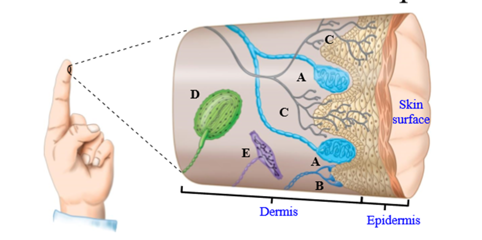
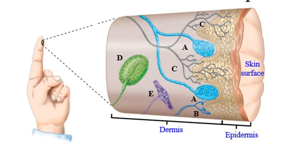
Meissner's Corpuscles, Merkel's corpuscles, Free neuron ending, Pacinian corpuscles, Ruffini corpuscle
Meissner's Corpuscles
mechanoreceptors that respond to touch and pressure (rapidly adapting)
Merkel's Corpuscles
mechanoreceptor that responds to touch and pressure (slowly adapting)
Free neuron ending
Close to skin surface (nociceptors, thermoreceptors, mechanoreceptors)
Pacinian corpuscle
Responds to vibration and deep pressure (rapidly adapting)
Ruffini corpuscle
Responds to skin stretch (slowly adapting mechanoreceptor)
How are afferents activated
Somsatosensory receptors are activated by touch or pressure (mechanoreceptors)
- Sensory receptor opens up cationic channels to move down concentration gradient. Sodium flows down concentration gradient, causing depolarization
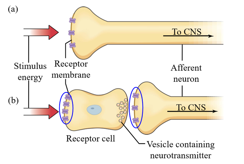
Types of sensory receptors
Sensory receptor located directly on afferent fiber, and sensory receptors located on specialized receptors
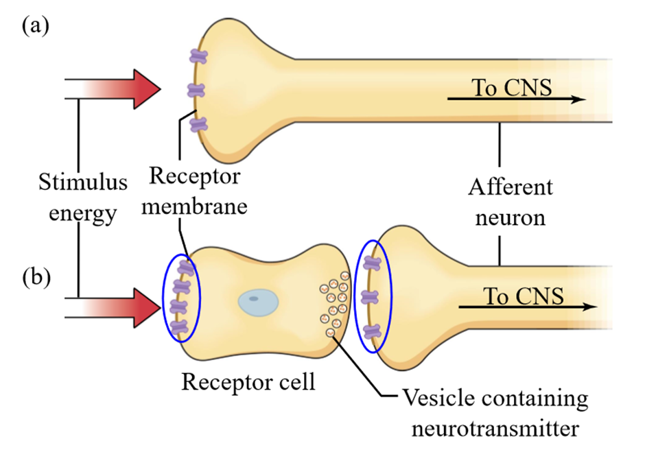
In both types of sensory receptors, where does the signal go?
To the spinal cord (CNS)
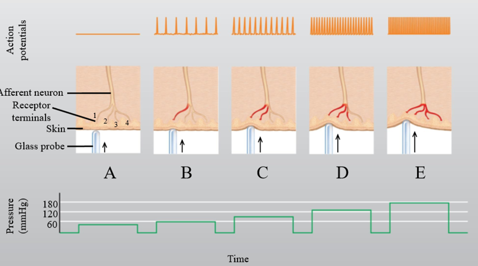
What is the receptor potential with a weak stimulus
Receptor potential is generated, but there is not enough stimulus to bring first node a Ranvier to threshold. No AP, no NT release
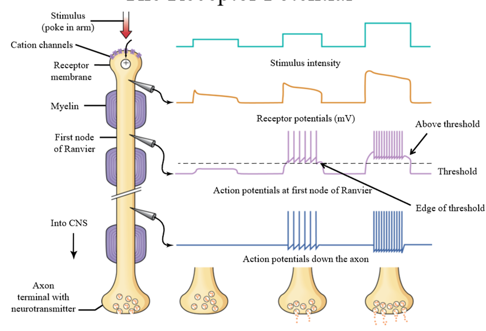
What is the receptor potential with a medium stimulus
Receptor potential is greater than in weak stimulus. There is enough graded polarization to bring neuron to threshold, and starting a AP. NT is released to projection neurons that stimulate the cortex, and we are aware of being poked
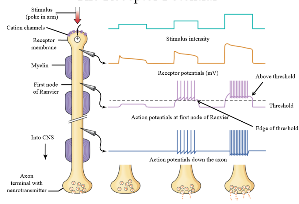
What is the receptor potential with a strong stimulus
Larger graded potential is generated, more action potentials are fired, causing more release of NT. Projection neurons send more signals to the cortex, and we are aware of a hard poke in the arm
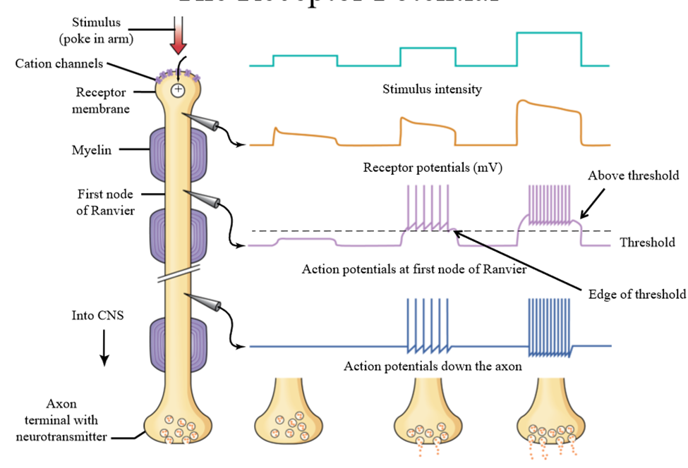
What determines stimulus intensirty
More receptor terminals are activated, causing activation of more action potentials.
- More graded depolarization of the afferent
Different types of somatic receptors in the same area
Receptors function in the same wall, but differ in the speed which they adapt to a stimulus (ex. Meissner's corpuscle rapid adaptation, Merkel's corpuscle slow adaption)
Merkel's corpuscle adaptation (slow)
Slow decay of receptor potential. Potential is activated during full time of stimulus
- Frequency of action potentials decrease over time, but they remain on during poke
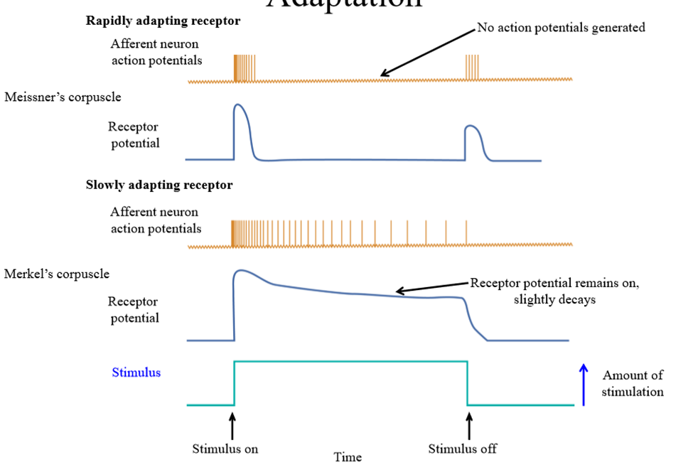
Meissner's corpuscle adaptation (fast)
Receptor potential immediately generated, but quickly decays. Action potentials are only generated at the start and end of an action potential
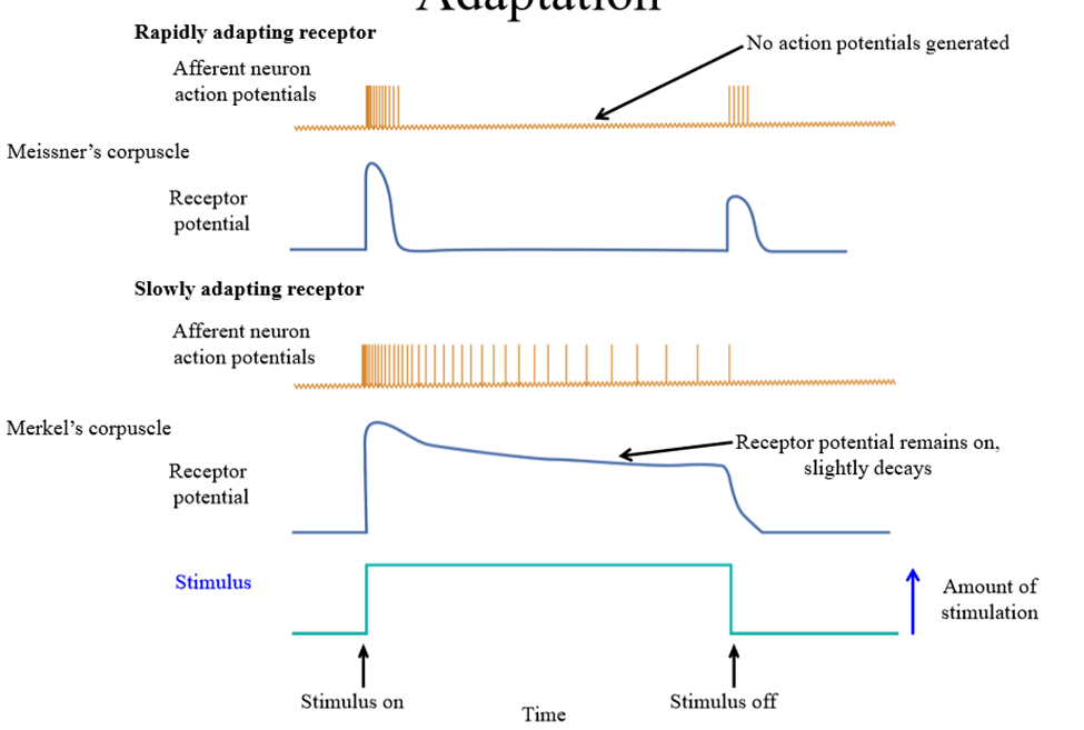
Use of rapidly adapting mechanoreceptors
Filter out unimportant information (time felt sitting in a chair). We know when we start sitting and when it ends
Example of a slowly adapting receptor
Proprioception - need to know where our body position is at all times (ex. arms sticking out straight in front of us)
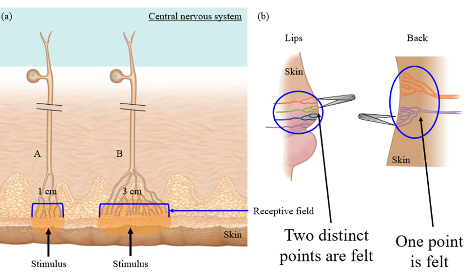
Factors that affect our ability to localize a stimulus
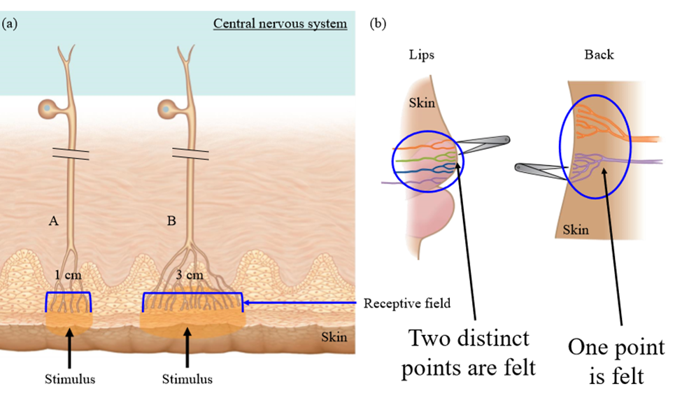
Receptive field size, density of innervations, overlapping receptive fields
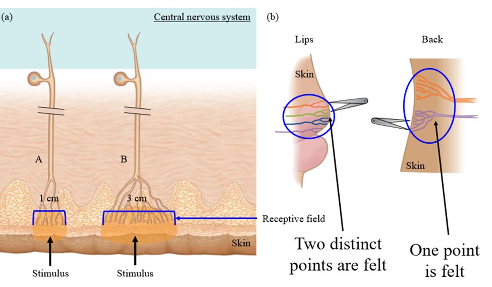
Receptive field size
Extent of the body that feels stimulus
Densitiy of innervation
Number of sensory receptors within a certain area that receive stimulus.
Overlapping receptive fields
if the branches of two adjacent units both sense the stimulus.
Density of inneration effect on stimulus localization
More dense receptors = greater ability to localize stimulus (lips poked with forceps, feel two touches. Back poked with forceps, only feel one touch)
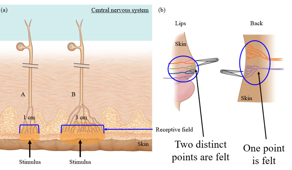
Receptive field size effect on stimulus localization
Smaller receptive fields allow for better localization (specific cortical neuron is activated)
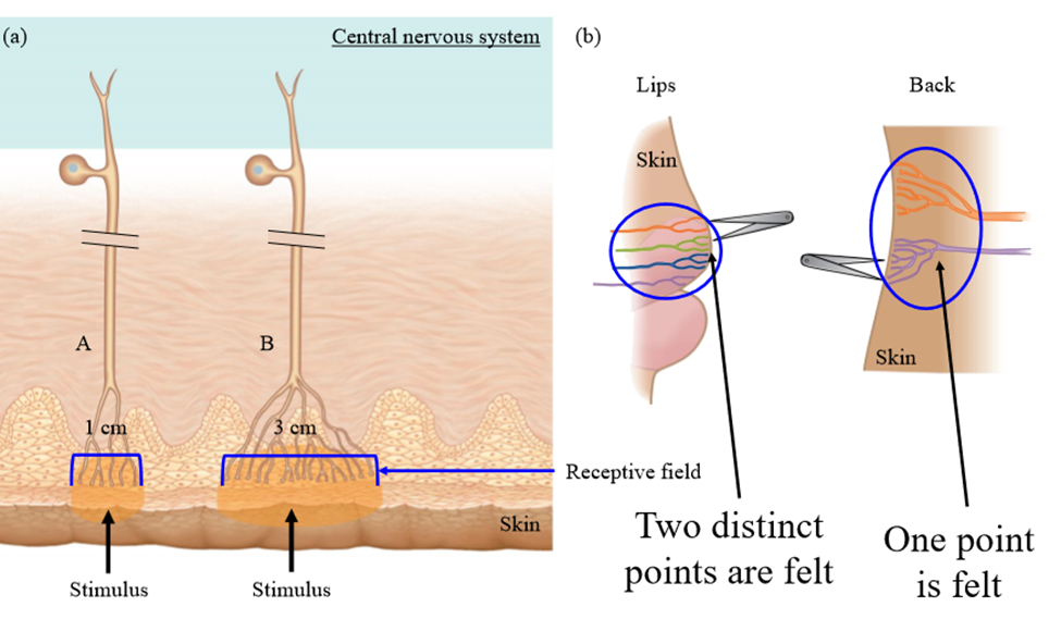
Overlapping receptive fields effect on stimulus localization
Poke in the middle of receptive field triggers many action potentials, and small amount in nearby fields. If between fields, medium amount of potentials generated in each field
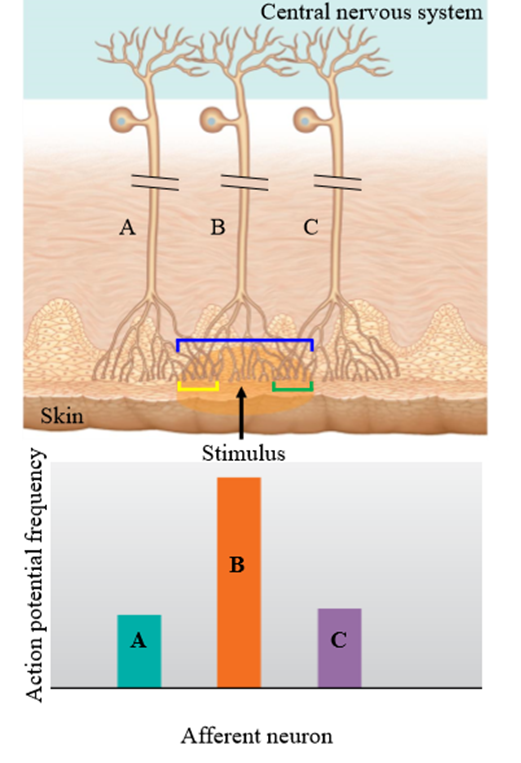
Lateral inhibition
Overlapping receptive fields help ID specific stimulus sites. Information from directly stimulated neurons is activated, while receptor neurons at edge of stimulus are inhibited
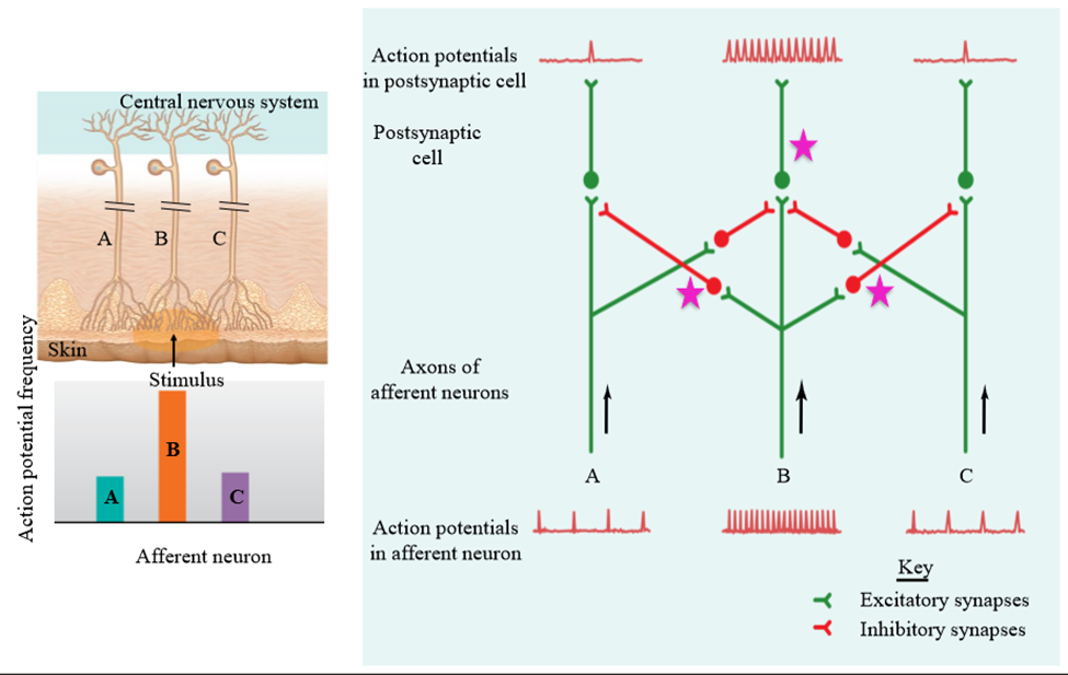
In which sensory systems is lateral inhibition present
Somatosensation and vision (not auditory)
How is localization possible
Lateral inhibition removes information from peripheral regions
What neurons are inhibited in lateral inhibtion
All of the neurons are inhibited, but the peripheral neurons are turned down a lot. The central neuron is only slightly
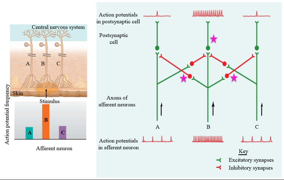
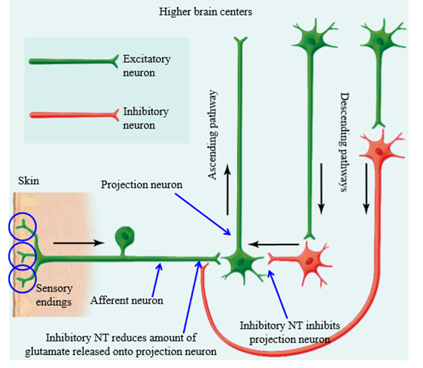
How are sensory signals modified before they reach higher levels of CNS
Lateral inhibition, pathways descending from higher centers of the brain, synapses on axon terminals of primary afferent neurons (presynaptic inhibition), indirectly by interneurons
Cortex role in signal modification
Inhibition to sensory fibers and projection neurons (turns down the volume of these neurons)
What happens when you remove inhibitory effect of cortex
Amplification of sensory input
Setup of neurons from skin to brain
Sensory endings --> afferent neuron --> projection neuron -->higher brain sensors
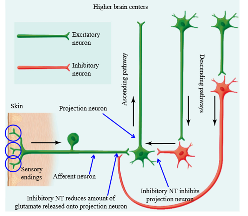
What does the afferent neuron synapse to
Travels to spinal cord and synapses with projection neuron
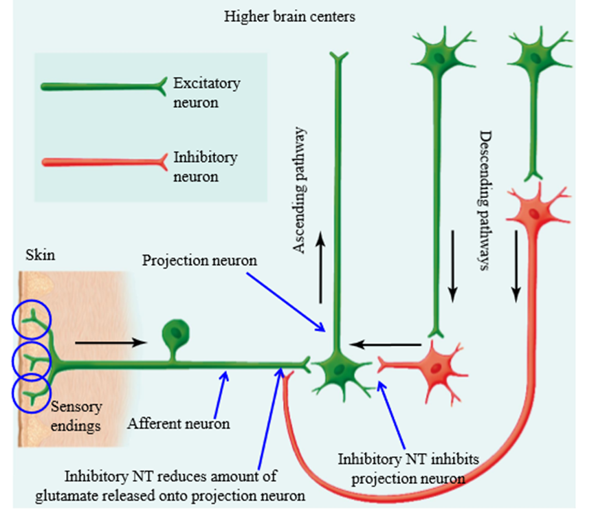
Where does the projection neuron synapse to
Brain centers, such as the cortex
How to inhibitory neurons work
Release inhibitory neurotransmitters onto projection neurons or sensory afferent neurons, reducing glutamate release. Turns down the response of projection neuron
What is the use of inhibition of pain signals
Based on the situation we are in, we will feel more or less stimulation from pain
- Stepping on our foot at school (hurts) vs at a track meet (not as much) vs in war (not at all)
What happens if we don't have pain tolerence
We have no signal to tell us to remove ourself from painful stimuli
What are the 2 ways information ascends to the cortex
Anterolateral system (spinothalamic system) and the Dorsal column system
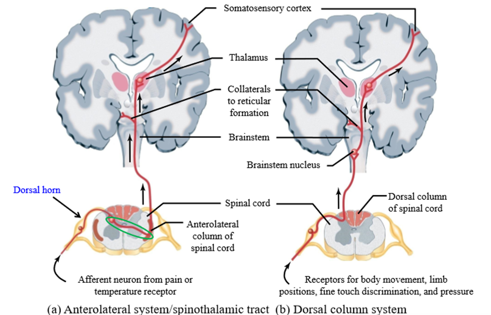
Anterolateral system (spinothalamic system)
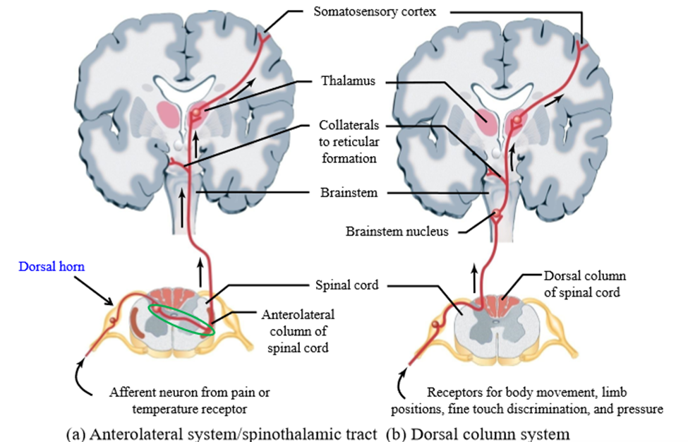
Pathway that carries pain and hot/cold information up to the somatosensory cortex
Dorsal column system
Pathway that carries information on fine touch mechanoreceptors to the somatosensory tract
Anterolateral system pathway
1. Painful stimulus from right side activates free neuron endings
2. Action potentials generated into mixed peripheral nerve
3. Synapse between sensory receptor neuron and second neuron on the dorsal horn of the right side of the spinal cord
4. Second neuron crosses spinal cord and travels up the left side of the spinal cord
5. Synapse in thalamus with a cortically projecting neuron
6. Cortically projecting neuron takes information to somatosensory cortex
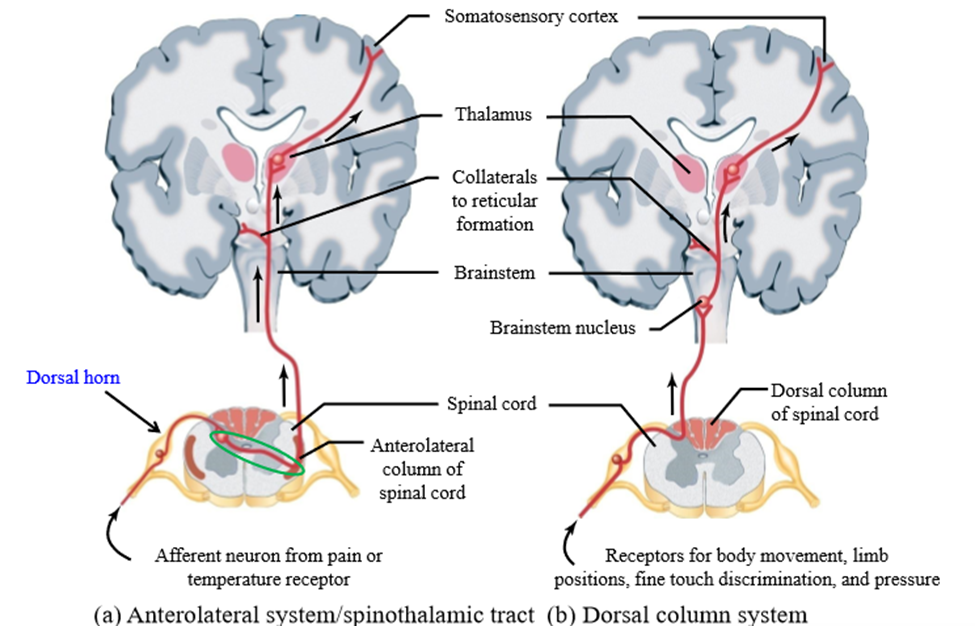
Dorsal system pathway
1. Painful stimulus from right side activates free neuron endings
2. Action potentials generated into mixed peripheral nerve
3. Sensory neuron travels through the dorsal root of spinal cord, and travels up the right side of the spinal cord
4. Sensory neuron synapses with secondary neuron in brainstem
5. Secondary neuron crosses over to left side of brain stem and synapses with cortically projecting neuron
6. Cortically projecting neuron takes information to somatosensory cortex
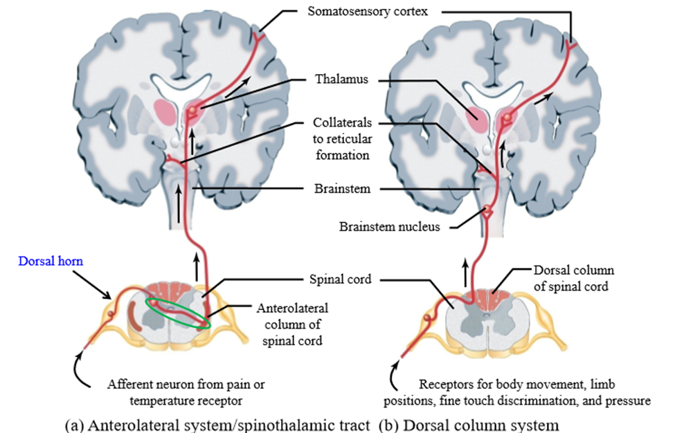
Similarities of anterolateral and dorsal system pathway
Both pathways end up in the brain on the opposite side the stimulus
Difference between the anterolateral and dorsal system pathway
Anterolateral - secondary neuron crosses over in spinal cord
Dorsal - Secondary neuron Crosses over in brainstem
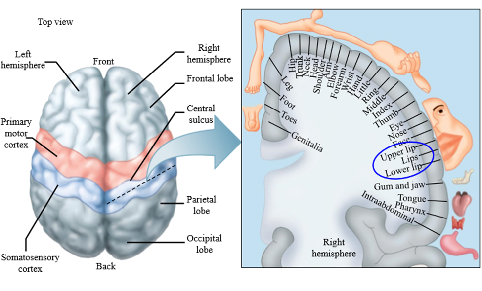
Somatosensory cortex location
Behind the motor cortex and central sulcus
Function of somatosensory cortex neurons
Activate motor cortex neurons, which control movement
Role of motor cortex neurons
Travel down spinal cord via descending systems to motor neurons, activating them based on how we want to move
Somatosensory cortex regions
Each region of the body maps to a region of the somatosensory cortex
How is each body part's amount of room taken up in the somatosensory cortex determined?
Based on how densely innervated the body part is with sensory receptors
High density innervated areas of the body
Fingers, face, lips
Low density innervated areas of the body
Trunk, neck, hips
Photoreceptor depolarization/hyperpolarization
Photoreceptors are depolarized at rest, and hyperpolarized when activated (opposite of most systems)
Components of eyes
Optical component and neural component
Optical component of the eye
Focuses visual image on receptor cells - the front part of the eye
Neural component of the eye
Back part of the eye - transforms visual image into a pattern of graded and action potentials
What light do humans see
In the visible range
When we look at an object, what do we see
Light reflected off the object, and hitting the photoreceptors of our eyes
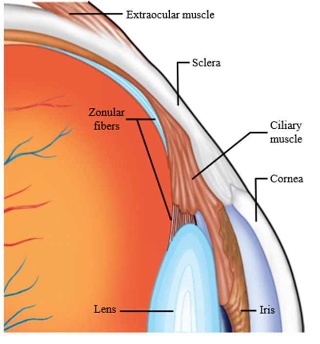
Sclera
White of the eye - membrane surrounding eyeball
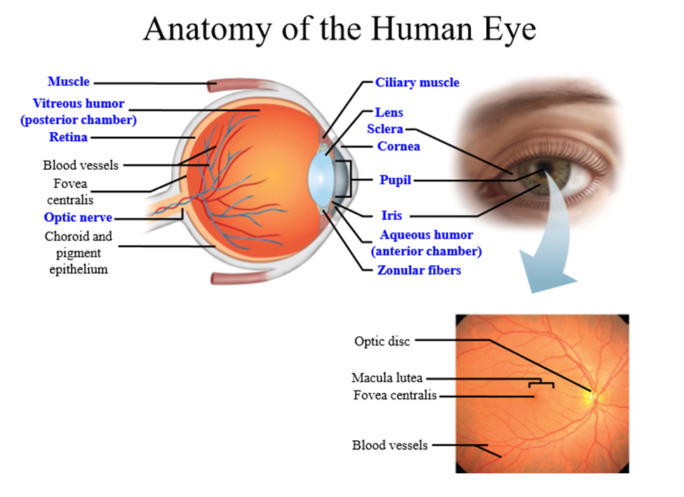
Extarocular muscle
Muscle that is responsible for eye movements, attached to the sclera
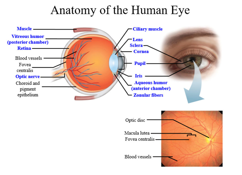
Cornea
Clear tissue at front of the eye that refracts light waves, causing them to converge on photoreceptors
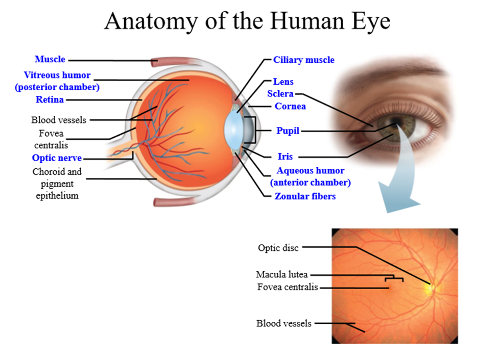
Pupil
Hole that allows light to pass through to photoreceptors
Iris
Colored part of the eye that regulates the size of the pupil - controls amount of light that enters eyeball
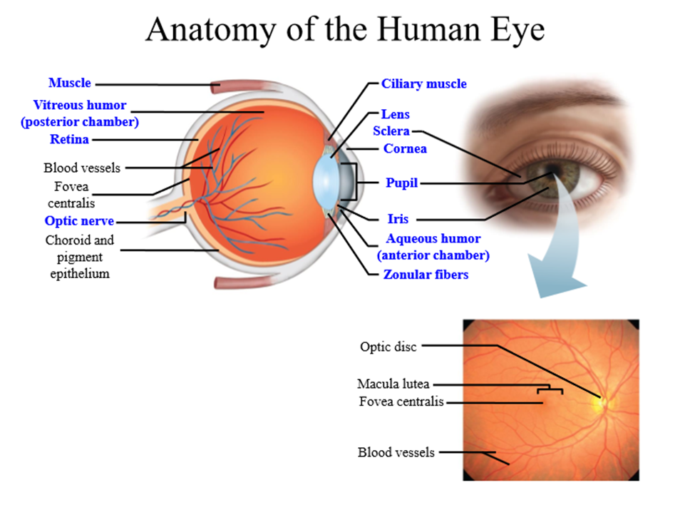
How is the iris innervated
By the autonomic nervous system
- Sympathetic - causes pupil constriction
- Parasympathetic - causes pupil dilation
Lens
Transparent structure that works with cornea to focus the image on the retina. Can change shape based on where viewed object is
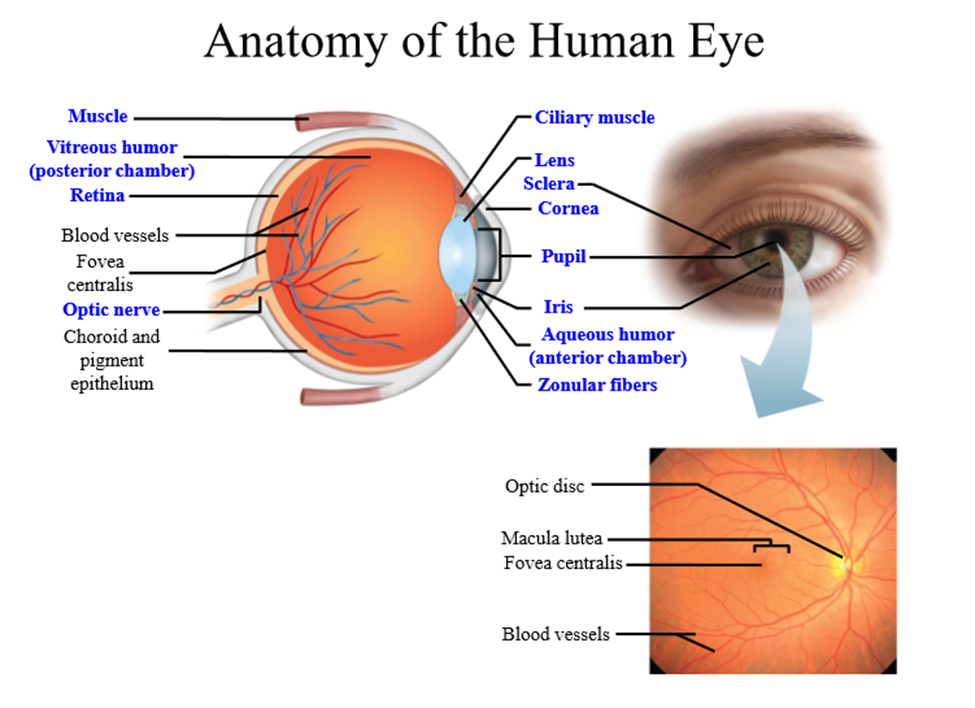
Zonular fibers
Fibers that attach the lens to the ciliary muscles
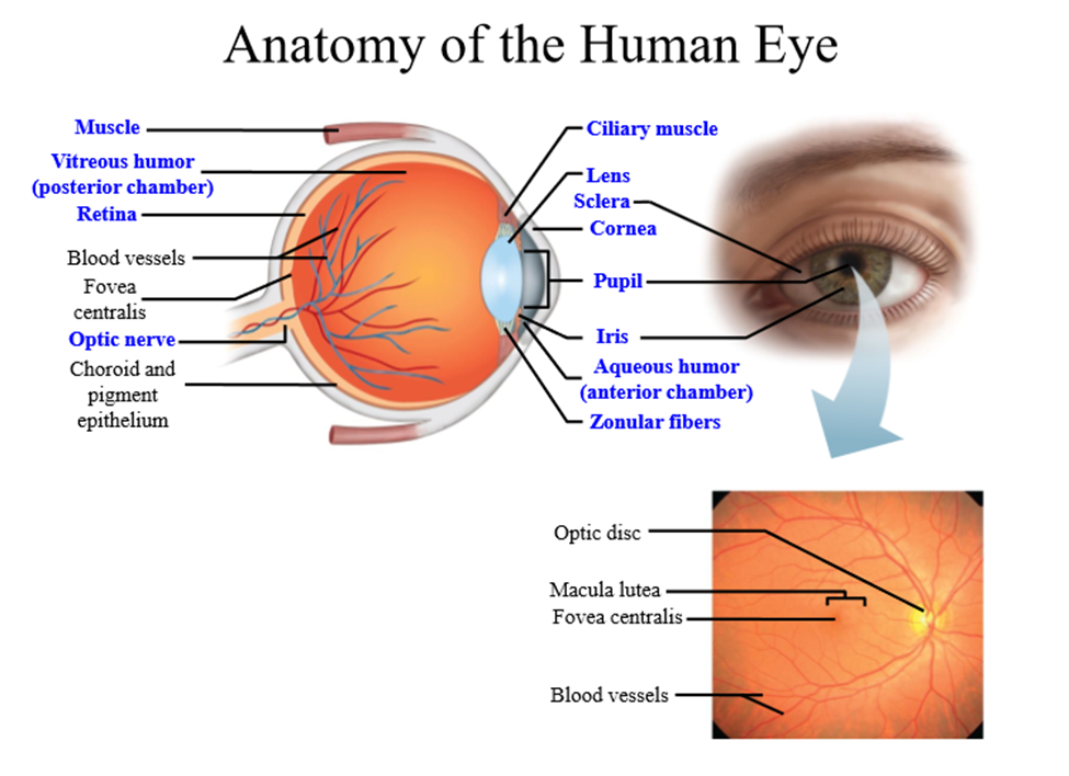
Ciliary muscles
Muscles that contract/relax to change the shape of the lens
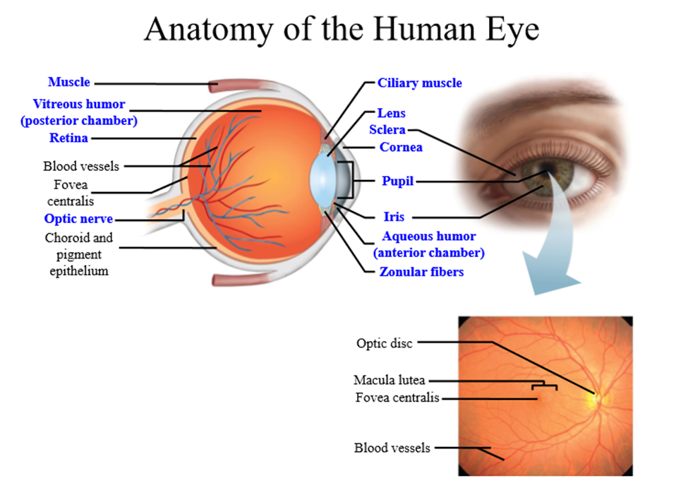
Retina
Light sensitive part at the back of the eye where photoreceptors are found
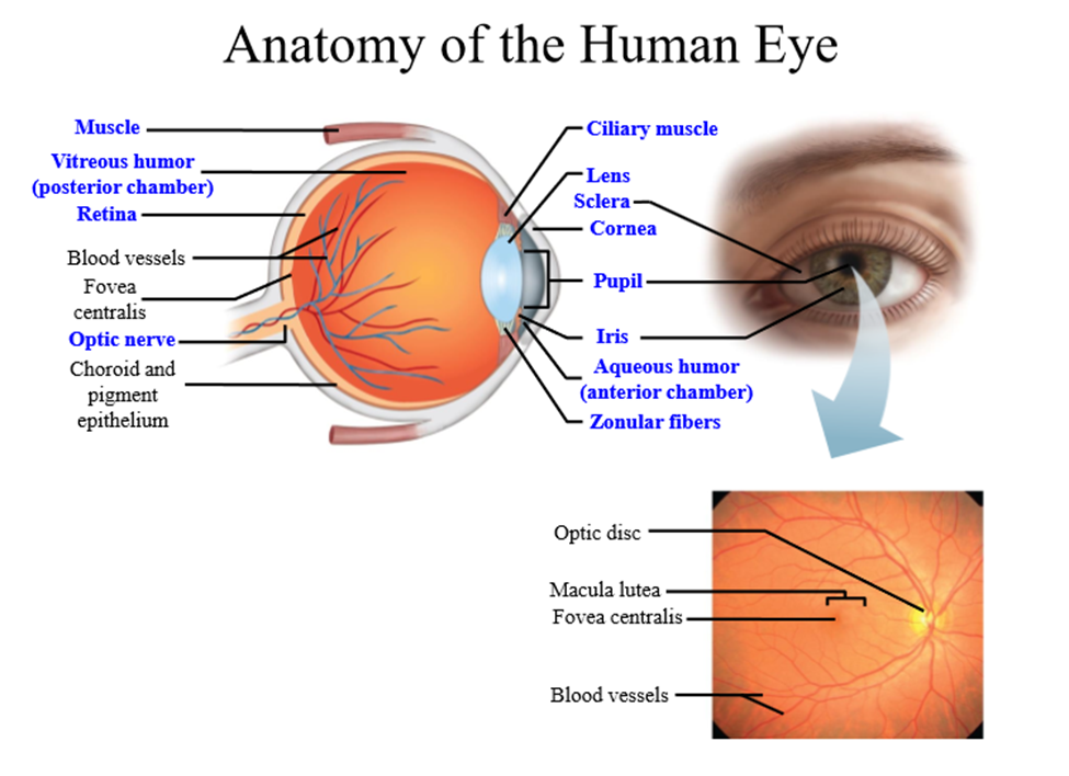
Types of photoreceptors
rods and cones
Rods
Receptors that are monochromatic and activated in low light
Cones
Receptors that are responsible for color vision and activated with more light
Retinal ganglion cells
Take information from the rods and cones to the brain
Optic nerve
Nerve that travels towards the thalamus and cortex
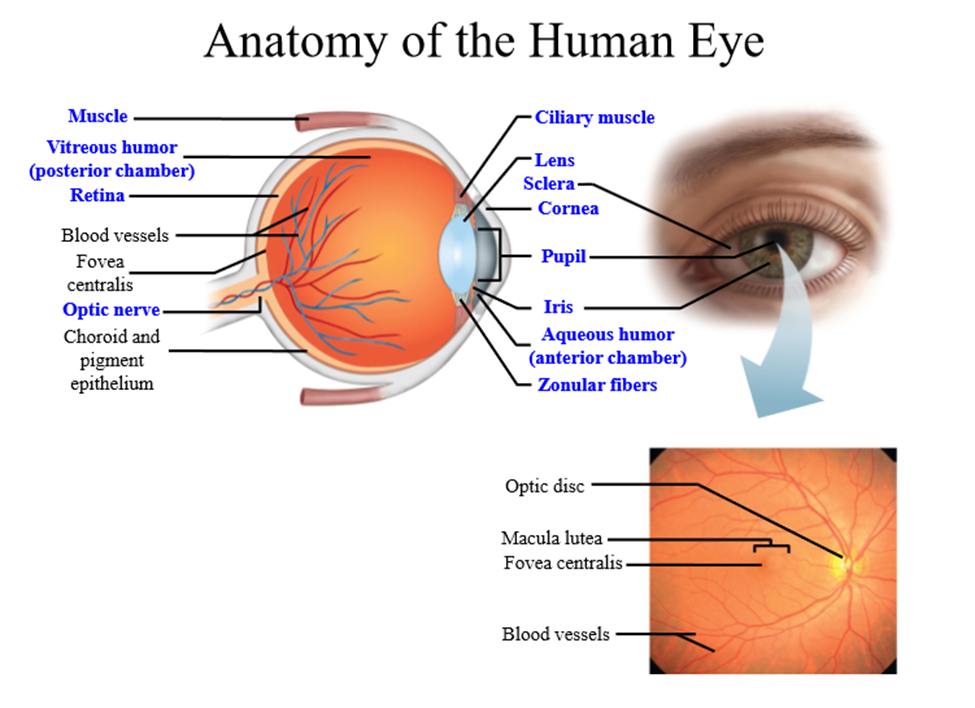
What is the optic nerve made of
axons of retinal ganglion cells
Aqueous Humor
Gelatinous fluid that fills the space between the lens and the cornea
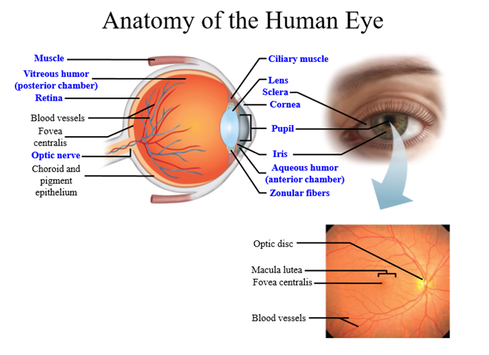
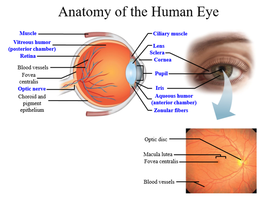
Vitreous humor
Gelatinous fluid that fills the space behind the lens
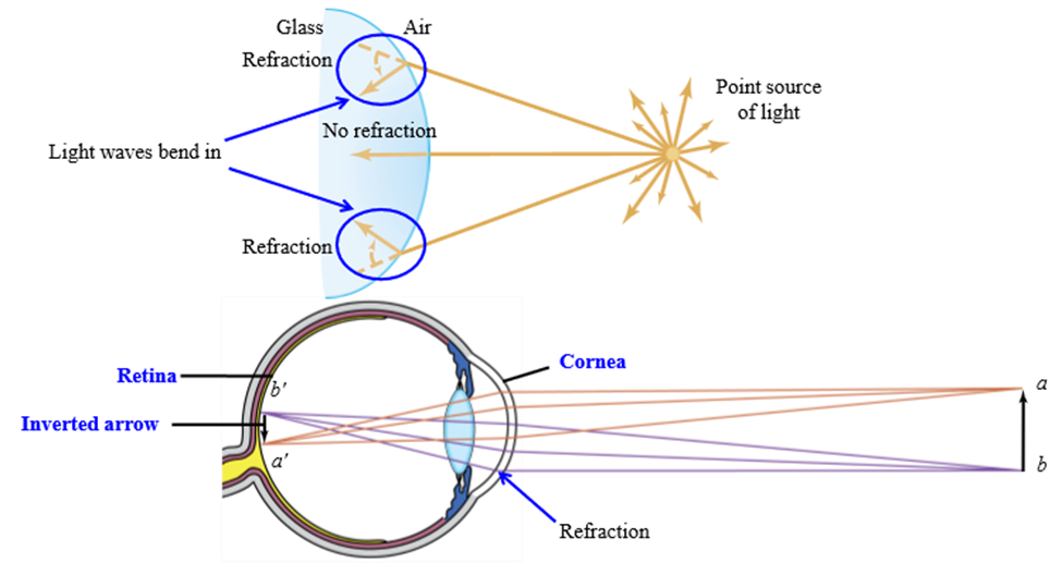
What is refraction
When light travels to a more dense medium (air to cornea), the light waves are bent to hit the retina
How does the image change in the eye
The image is inverted, the brain flips the image around
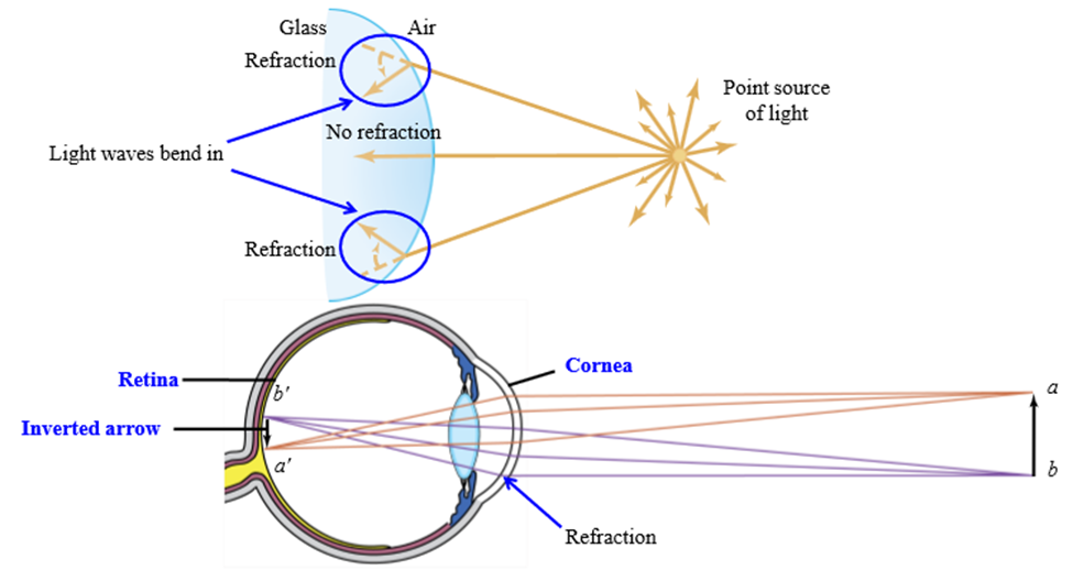
What happens if the amount of refraction is innapropriate in the eye
The image is constructed in front or behind the retina, and the image is not focused
What parts of the eye are responsible for refraction
Cornea refracts light, lens changes shape to focus light on the retina
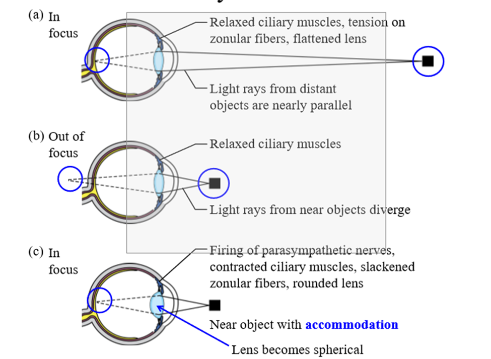
How does the eye work to see an image up close
Ciliary muscle contracts, causing the lens to get fatter and shorter. The lens increases the amount of refraction, allowing the image to focus on the retina
Accommodation
the process by which the eye's lens changes shape to focus near or far objects on the retina
What age do humans lose the ability to accomodate
Around 45 years of age, due to the breakdown of ciliary muscles that control the lens
Presbyopia
Loss of elasticity of the lens, leading to inability to accommodate for near vision. Refraction only comes from the cornea
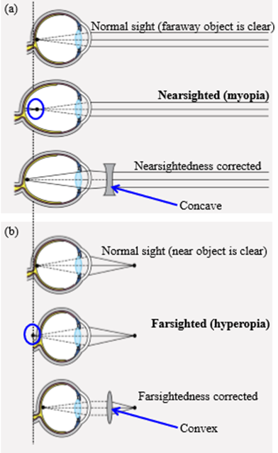
Myopia (near sightedness)
Eyeball is too long, and too much refraction occurs. The image is reconstructed in front of the retina and the image is out of focus
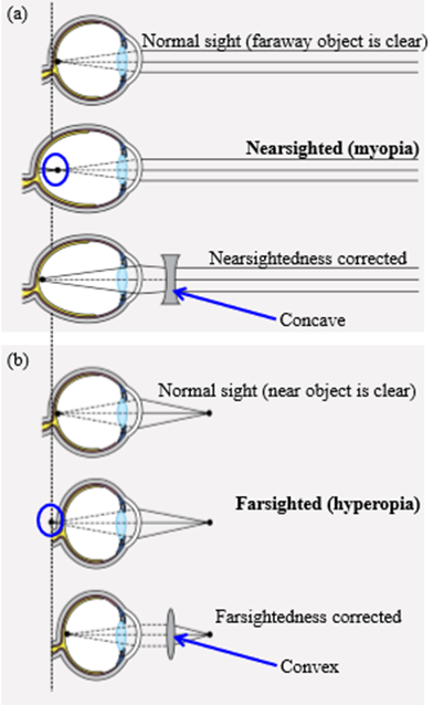
How to fix myopia
Wear lenses with a concave shape - reduces refraction so the image can land on the retina
Hyperopia (far sightedness)
Eyeball is too short, and not enough refraction occurs. The image is reconstructed behind the retina and the image is out of focus