Biopsych
 Call Kai
 Learn
Learn Practice Test
 Spaced Repetition
 Match
Match Flashcards
 Knowt Play
1/122

There's no tags or description
Looks like no tags are added yet.
Name | Mastery | Learn | Test | Matching | Spaced | Call with Kai | Chat |
|---|
No analytics yet
Send a link to your students to track their progress
123 Terms
the nervous system is split into
central nervous system (CNS) and peripheral nervous system (PNS)
compare the function of CNS and PNS
CNS is the main control system for functions including conscious psychological processes whereas PNS transmits information between the CNS and external world.
CNS consists of
brain and spinal chord
PNS consists of
somatic and autonomic
autonomic nervous system is split into
sympathetic and parasympathetic nervous system
CNS: the brain
higher psychological processes
cerebral cortex is the outer layer made up of highly folded grey matter
divided into 2 symmetrical hemispheres the left and right
4 distinct lobes: frontal, parietal, occipital, temporal
Under the cerebral cortex is the more primitive area and is concerned with vital functioning and instinctive behaviour
function of the cerebral cortex
higher cognitive (conscious thought)
emotional, sensory (5 senses)
motor (movement) functions
left hemisphere of the brain
sensory stimulus from right side of the body
motor control of the right side
speech, language and comprehension
analysis and calculations
time and sequencing
recognition of words, letters, numbers
right hemisphere of the brain
sensory stimulus from left side of the body
motor control of the left side
creativity
spatial ability
context/ perception
recognition of faces, places objects
CNS: spinal chord
bundle of nerves, which runs from your brain along a canal in your backbone
sends nerve signals from the brain to the body, and from the body to the brain
also involved in reflex actions, such as the startle response
brain is connected to the spinal cord by the brain stem.
somatic nervous system
responsible for voluntary movement that you do on purpose (e.g. walking).
controls the voluntary movement of skeletal muscles eg biceps
consists of the nerves that carry messages to the eyes, ears, skeletal muscles.
transmits information between the central nervous system and the senses
under conscious control
autonomic nervous system
controls involuntary movement
transmits information from the CNS to the internal organs and from non-skeletal muscles, eg cardiac muscle
differences between SNS and ANS
SNS voluntary ANS involuntary
SNS functions include posture and movement; ANS functions include secretion and control of metabolism
SNS –in vertebrates - includes excitatory neurotransmitters ANS – in vertebrates has both excitatory & inhibitory neurotransmitters
sympathetic nervous system
activated in situations requiring arousal and energy
prepares the body for “fight or flight” responses during stressful situations
prepares the body’s resources for immediate action
produces increased heart and respiratory rate, increasing blood flow to the muscles and pupil dilation to help the body to survive.
parasympathetic nervous system
activated soon after the threat of danger has passed
opposite effect of the SNS and allows for the body to return to homeostasis
person’s heart and respiratory rate decrease to normal levels and blood flow decreases, pupils return to normal size
vital to conserve energy and not to become exhausted
how neurones collect info
through the dendrites and sends info to the cell body
cell body weighs up the info received and makes a binary decision to fire its own electrical charge (action potential)
action potential will travel along the axon to the axon terminal
types of neurone
sensory, motor and relay
neurotransmitters
chemicals that pass from one neuron to another to pass the signal being transmitted
synaptic transmission
Electrical impulses are passed through the axon of a neuron to the axon terminal causing depolarisation of the presynaptic neuron. but cannot go through the synaptic cleft.
Instead, electrical impulse causes calcium channels to open, and positively charged calcium rushes into the cell. This
triggers vesicles with neurotransmitters to move towards the membrane at the axon terminal and release neurotransmitters into the synapse. They then diffuse across the synapse
They then bind to the receptors on the post-synaptic neuron. which triggers the a signal in the post synaptic neuron.
Neurotransmitters can have an excitatory effect on the receiving neuron or an inhibitory effect (making them less likely to fire).
excitatory effect on neurones
making them more likely to fire an impulse
inhibitory effect on neurones
making them less likely to fire an impulse
summation
the charge results in a net effect on the post synaptic neuron
membrane has to reach over a certain voltage (membrane potential) in order to fire.
If the net effect is excitatory (more positive than negative) the neuron will be more likely to fire
if inhibitory (more negative than positive charge) then neuron is less likely to fire.
what happens once the neurotransmitters have bound to receptor sites
a signal has been sent in the post synaptic neuron, the
neurotransmitters unbind from the receptors.
Then either the neurotransmitters travel back into the presynaptic neuron via a reuptake channel, and get repackaged back into vesicles to be used again or are are broken down by enzymes.
sensory neurones (afferent)
located in the PNS
respond to stimulation in sensory receptors for all senses (vision, hearing, smell, taste, touch).
send signals to/ arrives at CNS
long dendrites and short axons, cell body out to the side and not surrounded by dendrites
motor neurones (efferent)
cells in the PNS that send messages from the brain and the spinal cord to the muscles and glands (efferent exits cns)
1 long axon attached to effectors and short dendrites surrounding cell body
help to carry out a response
relay neurones (interneurons)
form connections between other neurons, found in CNS
send signals to other relay neurons, or form links between sensory and motor neurons
short dendrites and short axons
has myelin sheath to insulate axon, speeds up rate of electrical impulse
direction of action potential
dendrites, cell body, axon, axon terminal
reflex arc eg when you touch a hot pan
receptors in your skin sense the stimulus (the hot pan)
Sensory neurons send an electrical impulse towards the CNS from the dendrites, along the axon, to the axon terminal
In the CNS, a relay neuron carries this signal to a motor neuron
A motor neuron now transmits the signal from the CNS to the muscle
you are now able to move your hand away.
function of endocrine system
network of glands across the body that secret hormones (chemical messengers) to regulate vital functions
pituitary gland hormones
Oxytocin, Thyroid Stimulation Hormone (TSH) , ACTH
pituitary gland action
‘Master gland’ as it controls all other glands, TSH signals action in the thyroid, Adrenocorticotrophic Hormone (ACTH) signals action in the adrenal glands
thyroid gland hormone
thyroxine
thyroid gland action
Primarily involved with the regulation of metabolism, such as the conversion of food into energy for the muscles
parathyroid gland hormone
parathyroid hormone (PTH)
parathyroid gland action
acts to increase the concentration of calcium in the blood from kidneys and bone.
pancreas hormone
insulin
pancreas gland action
Promotes the absorption of glucose from the blood into fat, liver and skeletal muscle cells. Insulin lowers blood glucose levels.
medulla adrenal gland hormones
Adrenaline & Noradrenaline
adrenal glands action
Responsible for reacting to threat via the fight or flight response eg increased heart rate
ovaries hormones
Oestrogen and progesterone
ovaries gland action
Responsible for the development and regulation of the female reproductive system and secondary sex characteristics.
testes hormone
testosterone
testes action
key role in the development of male reproductive system such as the testes and prostate, as well as promoting secondary sexual characteristics eg increase muscle/ bone mass, growth of body hair
pineal gland hormone
melatonin
pineal gland action
regulates the sleep-wake cycle
fight or flight response
a threat is detected by sensors in the eye via hypothalamus and our sympathetic nervous system is activated at the same time as endocrine
hypothalamus signals to the pituitary gland to activate endocrine system
pituitary releases ACTH which travels through blood stream and is detected by cells in the adrenal gland
which stimulates adrenaline to be released
fight or flight symptoms
increase breathing/ heart rate to increase O2
pupils dilate to increase light entering eye
inhibition of digestion so more blood for muscles
increased sweating to regulate temp
parasympathetic response to fight or flight
After a few minutes, the parasympathetic branch of the ANS is activated, and the body returns to normal by establishing homeostasis.
Heart rate and respiratory rates decrease, adrenaline secretion slows down, the feeling of butterflies subside and sweating stops.
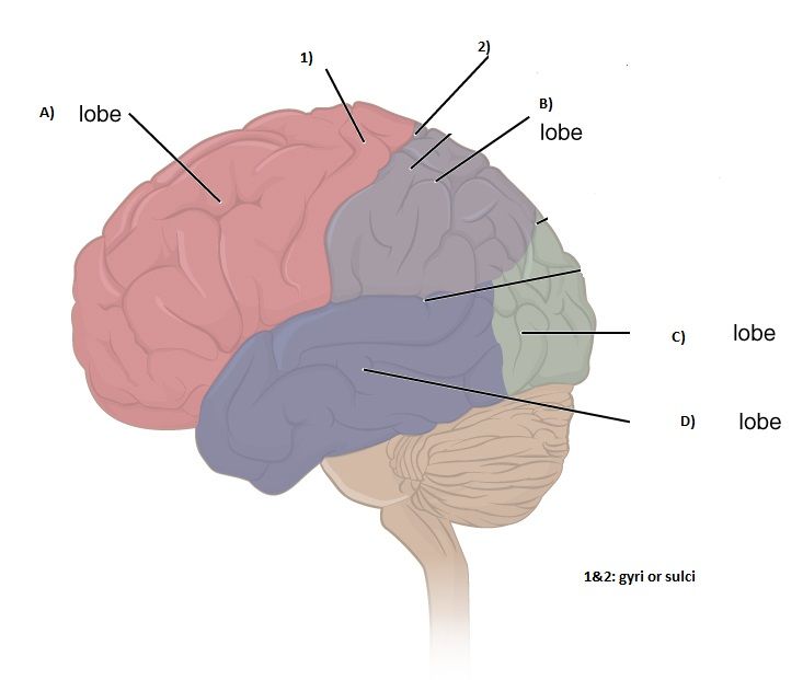
label each lobe ABCD
A= frontal
B= parietal
C= occipital
D= temporal
motor cortex lobe
frontal lobe
motor cortex function
regulates movement, damage may result in loss of control over movements
somatosensory cortex lobe
parietal lobe
somatosensory cortex function
processes sensory info such as touch, the more somatosensory area devoted to a certain body part, the more sensitive it will be
visual cortex lobe
occipital lobe
visual cortex function
receives and processes visual info, damage results in blindness
auditory cortex lobe
temporal lobe
auditory cortex function
analyses speech, damage results in hearing loss
language centers
broca’s area and wernicke’s area
Brocas area lobe
temporal lobe
Brocas area function
responsible for speech production, damage results in inability to produce words and sentences called broca’s/ expressive aphasia
Wernicke’s area lobe
temporal lobe
Wernicke’s area function
responsible for speech comprehension, damage results in inability to speak in coherent sentences or make sense, called wernicke’s/ receptive aphasia
localisation has research evidence supporting strength AO3
Petersen et al used brain scans to demonstrate how Wernicke’s area was active during a listening task and Broca’s during a reading task
in the listening task, participants were required to understand whats being said (wernnicke) whereas in reading they were required to produce the words (Broca’s)
support localisation as evidence that specific areas have specific functions
strength of case study supporting theory of localisation AO3
clive wearing sustained damage to hippocampus through a viral infection and damaged his semantic and episodic memory. (couldn’t remember if wife visited etc) but no damage to procedural memory (could still play piano)
supports localisation as suggests that the hippocampus may be involved with semantic or episodic memory whereas another part of the brain may be involved with procedural memory. If the function was more holistic we would see more severe effects on all areas of his memory
However, as this is a case study, the sample size is only of one person, meaning that the results cannot be generalised to the wider population. Therefore, this only provides supporting evidence, not definitive proof of the localisation of memory function.
research that challenge the theory of localisation AO3
Lashley suggests higher cognitive process such as learning and decision making are not localised but distributed holistically (all throughout the brain).
Lashley taught rats to solve a maze. This utilised their frontal cortex. He then removed between 10-50% of areas of the cortex in rats, and observed whether they were still able to solve the maze.
No particular area was shown to be more important in terms of the rats’ ability to complete the maze, they could still solve it (just a little slower) when they had the lesions.
This suggests the process of learning, specifically spatial navigation is not localised, but requires every part of the cortex. This seems to suggest learning is too complex to be localised to just one area, supporting a more holistic and multifunctional theory in regards to the function of the brain
law of mass action
Lashley put forward that behavioural function is spread widely across cortices as opposed to being localised
equipotentiality theory
proposed by Lashley which suggests basic motor and sensory functions are localised but higher mental functions aren’t
claimed that intact areas of the cortex could take over responsibility for specific cognitive functions following brain injury
does plasticity change localisation
theory of plasticity challenges the idea of localisation because it suggests that if a certain area is damaged, other areas can take over their function (showing flexibility of functions).
Evidence shows that when the brain has become damaged through illness or accident and a particular function has been compromised or lost, the rest of the brain appears to be able to reorganise itself to recover the function.
For example, if a stroke victim had damage to their right motor cortex they still may be able to recover movement in their left hand side, because the left motor cortex would step in to do the job of the right side which is damaged
plasticity
ability of the brain to change and develop as a result of our experience and learning
hemispheric lateralisation
specialised areas associated with language are found in one of the hemispheres rather than both eg ability to produce and understand language, for most people, is controlled by the left hemisphere
brain as contralateral
for movement, sensation and vision, which means that the left hemisphere controls the right hand side of the body, and the right hemisphere controls the left hand side of the body
corpus callosum
bridge fro 2 hemispheres that allows both sides of the brain to communicate with each other. Information processed by the left hemisphere (eg: moving my right hand), is passed to the right hemisphere via the corpus collossum and vice versa
left hemisphere functions
sensory stimulus from right side
motor control of right side
speech, lang, comprehension
analysis and calculating
time snd sequencing
recognition of words, number, letters
right hemisphere functions
sensory stimulus from left side
motor control of left side
creativity
spatial ability
context/ perception
recognition of faces, places, objects
better a visual motor tasks
split brain researcher and aim
roger sperry
Sperry devised a way of being able to test hemispheric lateralisation using visual and tactile tasks. This involved using a piece of equipment called a ‘T-scope’ which allowed each hemisphere to be tested in isolation of the other. Participants had to focus on a particular spot on the board (a fixation point), this allowed their left and right hemispheres to be tested separately.
quasi exp, 11 participants, epileptics whoo couldn’t be treated with drugs
asked patients to: describe what they see, do tactile task, do drawing task, composite task (splitting wordsr4)
sperry’s split brain experiment hypothesis
Split brain patients will not be able to verbally describe an image or word that is presented to their left visual field while controls will have no difficulty.
This is because this information is processed by the RIGHT hemisphere, which does not contain a language center.
As the corpus callossum in the split brain patients is severed, the two hemispheres of the brain cannot communicate with each other
sperry’s split brain experiment method
First the participant being asked to focus on the ‘fixation point’ and then an image or word was projected for 1/10th s (to stop info travelling to other visual field) to one or both visual fields (to the left or to the right).
To test for non-verbal processing, this equipment also enabled the participants to be able to pick up or match objects that were out of the participant’s sight.
The participants were then asked to describe the object, image, or word that had been presented to them. In some variations of the study, they were asked to draw what they had seen or touched.
In a ‘normal’ brain, the corpus callosum would immediately share information between both hemispheres giving a complete picture of the visual world. However, presenting the image to one hemisphere of a split-brain patient meant that information could not be conveyed from that hemisphere to the other.
sperry’s split brain experiment results
When a picture/word was projected to the right visual field (left hemisphere), the patient could easily describe what had been shown. However, when the picture/word was projected to the left visual field (right hemisphere), the patient could not describe what had been shown and typically reported that there was nothing there. This supports hemispheric lateralisation showing that lang is processed in the left hemisphere as the patients could only describe what they had seen when it was projected to the right visual field
Although the patients could not describe what had been shown to their left visual field, they were able to use their left hand to point to a matching object or picture. This shows that the right hemisphere has processed the information and controls the left hand, but obviously cannot verbalise what was shown.
If two words/pictures were projected at the same time, one on either side of the visual field (e.g. ‘a dollar sign’ on the left and ‘a question mark’ on the right), the patient would say that they saw a question mark but when asked to draw (with their left hand) what they saw, they would draw a dollar sign. The patients were not aware that they had drawn a different object or picture to the one they said they had seen. This suggests the two hemispheres were working separately from each other and that drawing ability is dominant in the right hemisphere.
An object placed in the patients right hand (the patient could not see it just feel it) it could be easily described or named in speech or writing, whereas, if the same objects were placed in the left hand, the patient could only make wild guesses. This also supports hemispheric lateralisation as it shows the left hemisphere is dominant for speech and writing
sperry’s split brain research experimental and involves the use of specialised equipment strength AO3
use of this equipment, such as the T-Scope, allows for the image or word to be projected for 1/10th of a second to one or both visual fields This meant that the split-brain patients would not have time to move their eyes across the image and so the visual information would only be processed by one visual field (and 1 hemisphere) at a time, increasing validity of the study.
Furthermore, the research uses highly standardised procedures such as giving the same tasks to each participant, and using standardised equipment.
The same procedure has been used on a number of split-brain patients and the results on the left hemisphere being dominate for language has been found to be consistent, increasing the reliability of the task.
sperry’s split brain research control group were people with no history of epileptic seizures weakness AO3
they could be seen as an inappropriate group to use as a comparison. E:
As the split brain patients suffered from epilepsy, it could be argued that it may have caused unique changes in the brain which could have influenced the results, so a more appropriate control group would have been people who had a history of epilepsy but had not had the split-brain procedure.
Moreover, Small sample sizes are used in split brain research meaning it is difficult for the results on hemispheric lateralisation to be generalised to the wider population.
However, as commissurotomy is a rare procedure, there is a limited amount of ‘split brain’ patients available for investigation therefore small sample sizes are unavoidable.
sperry’s split brain research
conducted in a highly controlled lab environment, so could be argued to lack mundane realism.
means that the data gathered from the split brain research came from the patients being testing under artificial conditions.
In real life a severed corpus callosum can be compensated for by the unrestricted use of two eyes.
herefore, the research findings cannot be generalised to how split brain patients function in everyday tasks. However, for the purposes of testing one hemisphere at a time the controls were essential.
theory of hemispheric lateralisation research evidence AO3
Sperry conducted experiments on split brain patients who had undergone the severing of their corpus callossum as a treatment for severe epilepsy. This meant that the two hemispheres of their brains could no longer communicate with each other.
Sperry found that when an image or word was presented to the left visual field, split brain patients could not verbally describe what they had seen. This is because they were processing the information with their right hemisphere, which does not contain a language center. Because of the lack of corpus callossum, their brain could not send the message across to the left hemisphere to process in the language center to allow the participant to verbalise what they had seen. This was compared to a control group with an intact corpus callossum, and no history of epilepsy.
These results support, as it suggests that the two hemispheres of the brain are working separately from each other to process info, has been very useful for investigating and demonstrating lateralisation of function. leading to a significant improvement in our understanding of the role of each hemisphere and brain processes associated with each hemisphere.
theory of hemispheric lateralisation: Modern neuroscientists suggest that the differences in function may be overstated AO3
the actual distinction between the each hemisphere is less clear and more complex.
In a neurotypical brain the two hemispheres are in constant communication when performing everyday tasks, and many of the behaviours typically associated with one hemisphere can be effectively performed by the other when the situation requires it.
This suggests that the theory may not be useful in explaining behaviour outside of a niche context of split-brain patients.
Perhaps a more holistic explanation of brain function is a more realistic way of understanding how the two hemispheres interact with one another.
theory of hemispheric lateralisation:prompted a theoretical and philosophical debate about the degree of communication between the two hemispheres in everyday functioning and the nature of consciousness AO3
Some theorists have suggested that the 2 hemispheres are so functionally different that they represent a form of ‘duality’ in the brain – that in effect we are all ‘two minds"‘.
In contrast, other researchers have argued that, far from working in isolation, the two hemispheres form a highly integrated system and are both involved in most everyday tasks.
This lack of agreement from different areas of the scientific community suggests that more research is needed to understand fully the nature of lateralization
Functional recovery
describes how the brain can recover after trauma and brain injury
synaptic pruning
As we age, connections that we don’t use are deleted and connections that we use a lot are strengthened
brain plasticity researcher and aim
Maguire wanted to test whether being a cab driver in London has affected brain structure
brain plasticity method
The posterior hippocampus is a part of the brain is associated with spatial and navigational skills in humans and other animals.
Part of a London taxi driver’s training involves taking a test known as ‘The Knowledge’, which assesses their ability to recall the names and locations of the streets in the city of London.
Maguire scanned (MRI scans) the brains of 16 right-handed London taxi drivers and compared them to a matched control group (bus drivers – who follow the same route so do not need the same dynamic spatial and navigational skills).
brain plasticity results
Maguire found that London taxi drivers had a significantly greater volume of grey matter in the posterior hippocampus than the matched control group.
Their posterior hippocampus was larger, the longer they’d been a taxi driver, because they were using it to learn the street names and routes.
there was a positive correlation between how great the volume of grey matter was and how long they had been in the job
brain plasticity conclusion
results of the study suggest that the learning the drivers undertake as part of their training alters the structure of their brains.
However, Maguire’s research is correlational so we cannot conclude causality; claiming that the length of time they worked as taxi drivers CAUSED them to develop a larger posterior hippocampus.
It could be that they chose to be taxi drivers because they were so good at spatial awareness, (maybe because their hippocampus was naturally bigger!)
brain plasticity study strengths
Control group allows us to say that there is a significant difference between taxi drivers and others (same mean age and all right handed)= good internal validity.
Use of scientific, objective measurements (MRI)
An attempt to study a real-world phenomena (natural experiment).
Good mundane realism = external validity
brain plasticity study weaknesses
No female taxi drivers used in sample – generalisability is poor due to beta gender bias (the assumption that findings from males can be applied to everyone).
Issue of correlational research – ability to prove cause and effect. We can’t be 100% sure that the difference is due to the ‘knowledge’, they weren’t tested before. They could have been taxi drivers because of their already existing difference.
HOWEVER the further positive correlation found between length of time in the job (amount of experience) and size of hippocampus means that causation is very likely
study supporting plasticity
Mechelli et al found a larger parietal cortex in the brains of bilingual people, compared to non bilingual people.
structural changes for brain recovery to form new connections
Axonal sprouting: The growth of new nerve endings which connect with other undamaged nerve cells to form new neuronal pathways.
Reformation of blood vessels where blood flow is increased to the affected area.
Recruitment of homologous (similar) areas on the other side of the brain to take over specific tasks
how does brain recovery work
brain is able to reorganise and rewire itself by forming new synaptic connections close to the area of damage. Secondary neural pathways that would not usually be used to carry out certain functions are activated to enable functioning to continue, often in the same way as before. Support for this comes from structural changes that are known to take place in the brain.
research supporting functional recovery
case study of EB by Danelli. she investigated EB, a 17yr old Italian boy who had his entire left brain hemisphere removed at 2yrs old. (Due to non-cancerous tumour).
EB case study
17yr boy had his left brain hemisphere removed at 2yrs old. (Due to benign tumour).
By 5, his language fluency improved due to intensive rehabilitation and by 17, using various brain scans, although there were minor problems with his grammar, in his everyday life, EB’s language appeared virtually normal.
Suggests lang abilities can still function after severe trauma, eg removal of left hemisphere.
Evidence suggests that the person’s level of educational attainment will influence how well the brain recovers after trauma. Schneider (2014) found that the more time brain injured patients had spent in education, (known as their cognitive reserve) the greater their chances of a disability-free recovery.
This suggests that the cognitive reserve could be an important factor in brain recovery after trauma.
factors that impact functional recovery
Education: Schneider’s cognitive reserve is the term for her finding that for the more years spent in education the higher the chance of disability free recovery.
Age: The younger the person when the brain damage occurs, the greater their chance of disability free recovery.
Sex: Women tend to have higher connectivity between the two hemispheres (denser neurons in the corpus callosum), which means that women have a higher chance of disability free recovery.
negative consequences of plasticity
Prolonged drug use, has been shown to result in poorer cognitive functioning as well as an increased risk of dementia in later life.
60-80% of amputees are known to develop phantom limb syndrome. This is the continued experience of sensation in the missing limb. These sensations are usually unpleasant and painful and are thought to arise from cortical reorganisation in the somatosensory cortex that results from the limb loss.
Therapies can help people to ensure that rewiring is beneficial to their function.
Research evidence shows that the brain is plastic and can recover from injury AO3
PLASTICITY: Maguire’s well controlled research
FUNCTIONAL RECOVERY: Danielli’s case study of EB (shows the extent of reorganisation possible)
This evidence presents compelling evidence that the brain is highly adaptive to the way it is being used, this is evidence for plasticity and functional recovery.