clinical chem test 1
 Call Kai
 Learn
Learn Practice Test
 Spaced Repetition
 Match
Match Flashcards
 Knowt Play
1/134

There's no tags or description
Looks like no tags are added yet.
Name | Mastery | Learn | Test | Matching | Spaced | Call with Kai | Chat |
|---|
No analytics yet
Send a link to your students to track their progress
135 Terms
routine void
no patient prep needed
MOST CONTAMINATED
midstream “clean catch” (CC)
used for culture
procedure: clean area w antiseptic, discard initial portion of urinary stream, and collect midstream portion into a clean container
avoids vaginal discharges/contamination
catheterized collection technique
usually used for pts having difficulty voiding
insert sterile catheter thru urethra into bladder, urine flows into bag
collect by aspiration or directly from port NEVER ACCEPT URINE FROM BAG
things accumulate
urine in bag can be old - only good for 2 hrs

suprapubic aspiration collection
use of syringe and needle inserted thru the abdominal wall and directly into bladder
not usual, LEAST contaminated

collection containers
use clean, dry container w lid
sterile container for culture
pedi-bags for infants
LABELED PROPERLY on container, not just lid
necessary for proper labeling of urine specimen
NAME
DATE
PT ID NUMBER
TIME OF COLLECTION
random collection
collected at any time
adv: convenient for patiend
disadv: may be diluted; false picture of health
1st morning collection
patient collects urine as soon as they wake up
adv: most concentrated; PERFERRED METHOD
disadv: none
post-prandial collection
collected 2-3 hrs after eating
adv: can be used for detection of carbohydrate metabolism disorders
disadv: patient responsible for collecting on time
24-hour collection
1st void is discarded; collect all urine for 24 hrs
adv: allows us to quantitate substances (protein, glucose, hormones)
disadv: pt responsible for collecting on time, may forget
timed collection
collected at a particular time of day
ex: urine collection between 2-4 PM
adv: allows us to test substances w/ diurnal variation (different concentrations at different times of day, like cortisol being higher in the morning)
disadv: pt responsible for collecting on time
handling of urine specimen
delivered to laboratory immediately (ASAP)
a delay of MORE THAN 2 HRS requires a PRESERVATIVE
if no preservative and OVER 2 hrs - DISCARD AND RECOLLECT
changes in unpreserved urine
chemical
physical
microscopic
#1 way to preserve urine
refrigeration
gives up to 8 HRS to work w/ urine (follow lab policy)
prevents bacterial proliferation
slows/inhibits enzymatic processes that break down cell components
what changes occur in urine stored at room temp?
rapid bacteria multiplication
urea splitting bacteria INCREASE PH IN URINE (ammonia)
increase in urine pH → decomposition of casts
glucose is used up by bacteria (false lower/negative reading)
blood cells and casts deteriorate over time
preservatives - boric acid
most commonly used
keeps bacteria stable
preserves formed elements, INTERFERES W PH READING
used to preserve urine for culture and sensitivity testing
preservatives - chloroform
inhibits bacterial growth
changes CELLULAR SEDIMENT
may be used for aldosterone testing
preservatives - chlorohexidine
inhibits bacterial growth
useful glucose preservative
affects BILIRUBIN and UROBILINOGEN testing
preservatives - formalin
preserves urinary sediment
may PRECIPITATE PROTEIN and give FALSE POSITIVE result for REDUCING SUBSTANCES
preservatives - preservative tablets
release formaldehyde (not used anymore - toxic to humans)
interferes w REDUCING SUBSTANCES at high concentrations
INCREASES S.G.
preservatives - toluene
preserves ketones, proteins, reducing substances
not effective against BACTERIA PRESENT
floats on surface of urine and is flammable
preservatives - thymol
rarely used
interferes w ACID PRECEPITATION test for protein, but NOT STRIP TEST
TESTS THAT REQUIRE SPECIMEN TO BE UNPRESERVED
pregnancy test
molecular methods
GCE and chlamydia tests
PCR- related tests
physical changes in unpreserved urine
color: changes due to oxidation or reduction of substances (darker)
clarity: falsely decreased due to bacterial growth + solute precipitation (cloudier)
odor: increased due to bacterial proliferation + decomp. of urea to ammonia (stinkier)
unpreserved urine - glucose
DECREASES
it is metabolized by bacteria or yeast
unpreserved urine - ketones
DECREASES
ketones volatize
unpreserved urine - bilirubin
DECREASES
photo-oxidation and hydrolysis
unpreserved urine - urobilinogen
DECREASES
oxidation of urobilinogen
unpreserved urine - pH
INCREASES
bacterial decomposition and conversion of urea to NH3
unpreserved urine - nitrites
INCREASES
bacteria proliferation (up to a certain point)
unpreserved urine - RBCs, WBCs, casts
DECREASES
degeneration by enzymes
urinalysis components
physical characteristics - quickest/easiest part
chemical characteristics - dipstick
microscopic examination - centrifuge → supernatant/sediment → microscope
usually omitted if physical and chemical exam meet normal findings
physical characteristics noted
color
clarity
odor
concentration
volume received
color
urochrome and UROBILIN (lipid soluble pigments)
formed in large intestines
gives urine its characteristic color
other possible pigments
influence of hydration
pale to dark yellow
intensity correlates w/ concentration
darker = more concentrated
oxidation can cause color change
colorless - pale yellow urine
diluted urine
normal
pale/straw to dark yellow urine
normal
dark amber urine
bilirubin
also causes yellow foam
orange urine
bilirubin
medication
light red/pink urine
blood, hemoglobin, myoglobin
menstruation
beets, rhubarb, blueberries artificial dyes
red-brown uria
hematuria
menstruation
rhabdomyolysis (myoglobin)
prophyrinuria
meds
black/brown/tea urine
homogentisic acid
melanin
antibiotics
senna laxatives
green/blue urine
biliverdin
pseudomonas bacterial infection
amitriptyline (antidepressant); cimetidine (ulcer/acid reflux med.)
food dyes
clarity
overall visual appearance
degree of transparency
normal urine = clear
indicates hydration and healthy UT
cloudiness/turbidity
due to contaminants
pathogenic and nonpathogenic sources
clarity and color usually go hand in hand
levels of clarity
clear - no visible particulate matter
slightly cloudy - some visible particles, background not obscured
cloudy - visible particles, background slightly blurred
turbid - cannot view background through urine

substances causing turbidity - PATHOLOGIC
RBCs
WBCs
bacteria
yeast
trichomonas
renal epithelial cells (should stay in kidneys)
abnormal crystals
pus
substances causing turbidity - NON PATHOGENIC
mucus
squamous epithelial cells
normal crystals
radiographic media
sperm
fecal contamination
powder
lotion
foam caused by SHAKEN urine
normal urine foam will dissipate
stable foam = ABNORMAL
stable WHITE = moderate/large amount of albumin (protein)
stable YELLOW = bilirubin is present
note: foam usually not reported, used as support presence/absence of indicated substances
differentiation of acidic urine
amorphous urates
radiographic contrast media
differentiation of alkaline urine
amorphous phosphates
carbonates
odor
not reported
can give clues on abnormal substances
normal = faintly aromatic
ammonia (pungent) = bacteria = old urine or uti
sweet/fruity = ketones
maple syrup = congenital metabolic disorder
mousy/musty (in infants) = phenylketonuria
concentration and specific gravity
amount of solutes present in sample
SG of urine consists primarily of urea and electrolytes (Na, K)
varies w diet, health, hydration status
useful for assessing renal function - kidney’s ability to conserve water as needed
color is crude indicator

low specific gravity
hyposthenuria
range: less than 1.010
excess fluid status - can indicate renal failure
possible causes:
diabetes insipidus
kidney failure
pyelonephritis
malignant hypertension
excess glucose and protein
high specific gravity
greater than 1.025
suggests dehydration
possible causes:
volume loss
diabetes mellitus
diarrhea
CHF
shock
syndrome of inappropriate ADH
trauma, stress, drugs
note: IV radiography dyes/drinks give false elevaiton
specific gravity : diabetes mellitus vs insipidus
BOTH: HIGH URINARY VOLUME
Mellitus
deficiency of insulin (excess glucose excreted in urine)
INCREASED specific gravity (glucose = dense molecule)
Insipidus
deficiency ADH
DECREASED specific gravity (kidneys not concentrating urine; more water
osmolality
another measure of concentrating/diluting ability of kidneys
better than specific gravity; unaffected by high molecular weight solutes
changes = DIRECTLY PROPORTIONAL TO AMOUNT OF SOLUTE
normal reference range = 500-850 mOsm/kg water
direct methods for specific gravity measurements
urinometer
falling drop method
NOT DONE ANYMORE
indirect methods for specific gravity measurements
reagent strip method
refractometer
urinometer
replaced by more accurate methods
direct method - measures SG at room temp
no longer recommended
requires CORRECTION for temp, glucose, protein
a min. of 15 mL of urine needed
weighed glass float w long, narrow calibrated stem
falling drop method
time rate of fall of drop of urine through temperature controlled column of silicone based oil
rate proportional to mass of solutes present
more precise and accurate than refractometer and urinometer
ALREADY TEMP CORRECTED
CORRECT ONLY FOR PROTEIN AND GLUCOSE
refractometry
measure refractive index of a solution
refractive index = ratio of velocity of light in air to velocity of light in sltn
based on refractive index of light
quality control
check with water daily - 1.000
5% NaCl - 1.022
reagent strips
varioud configurations
blood
pH
ketones
glucose
protein
specific gravity
bilirubin
urobilinogen
nitrite
leukocyte esterase
pH principle
normal urine pH: 4.5 - 8.0
double indicator system
enables pad to produce readings over natural range of urine pH - SENSITIVE TO H+ IONS
methyl red = acid indicator
bromthymol blue = alkaline indicator
we read a combined color change

run over effect
due to excess urine after dipping - “leaching”
can falsely get an acidic reading
to fix: dab dipstick on paper towel
why do we avoid testing old urine?
causes false pH increase do to ammonia formation by bacteria
acidic urine can be caused by
diet (high protein, cranberry ingestion)
sleep
acidifying drugs
diarrhea (water loss = bicarb loss)
metabolic acidosis
respiratory acidosis
UTI w acid producing bacteria (E.coli)
alkaline urine can be caused by
diet (vegetarian; low carb)
alkaline drugs
acute and chronic renal failure
metabolic alkalosis
respiratory alkalosis
UTI w alkaline producing organism - pseudomonas
protein in urine
normally, only small amnt of low weight protein is filtered in glomerulus
ALBUMIN and HMWP should NOT be in urine
Tamm-Horsfall protein = normal
excreted by tubules, not contained in plasma
forms matrix of urinary casts
SSA test is used for confirmation - detects albumin, globulins, and bence-jones proteins
protein - reagent strip
detects ALBUMIN only
pad principle:
based on “protein error of indicators”
point of color change of some pH indicators is different in presence of protein from that observed in absence of protein
TETRABROMPHENOL BLUE - most common indicator
no protein = buffered at pH 3 = yellow
protein = H+ acceptor
presence of protein = error occurs in behavior of indicator = color changes to blue/green

protein - false positive
highly buffered alkaline urine
prolonged exposure of pad to urine = “leaching”
container cleaning compounds (bleach contamination)
urine w blood
protein - false negative
dilute urines
proteins other than albumin present (dont get picked up by dipstick)
protein in urine - PATHOLOGIC CAUSES
glomerular nephritis (inflammation of glomeruli - decreased filtration)
malignant hypertension (kidney damage)
kidney disease
pyelonephritis (UTI) - bacterial infection
renal vascular disease
protein in urine - PHYSIOLOGIC CAUSES
usually happens in short amount of time
physical stress
exercise
heat/cold exposure
fever
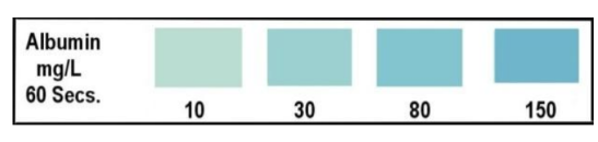
albuminuria
presence of albumin in the urine (aka microalbuminuria)
DETECTS EARLY SIGNS OF KIDNEY PROBLEMS IN DIABETICS
may be positive due to UTI
tested w sensitive techniques
immunoassay and tablet protein error of indicator test
tests to measure
albumin to creatinine ratio in random urine
24 hr collection of urine
orthostatic/postural proteinuria
occurs following time in VERTICAL POSITION only
benign cause
more frequent in young adults
confirmed w timed study:
empty bladder before bed
collect specimen immediately after rising
collect another specimen after in vertical position for several hours
NEGATIVE in the MORNING and POSITIVE on specimen collected LATER IN THE DAY
bence-jones protein
abnormal immunoglobin light chains in urine
present in MULTIPLE MYELOMA
heat precipitation test - screening procedure
bence jones protein is present when precipitation appears between 40-60oC (test = 40-60 precipitate → 100 disappears → back to 40-60 precipitate again)
confirm w immunoelectrophoresis

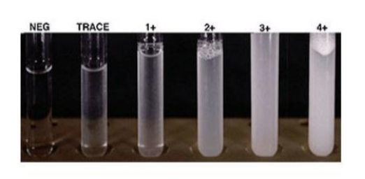
SSA protein precepitation
confirmatory test for proteins in urine
3% sulfosalicylic acid = detects albumin, globulins, bence-jones proteins
added to small and equal volume of urine
acidification causes precipitation of proteins in urine
semi-quantitated visually or more precisely using photometry
glucose
normally contained in ultrafiltrate and reabsorbed in the proximal tubule
NEVER NORMAL IN URINE
presence in urine: glucosuria
amount in urine depends on level in blood, rate of glomerular filtration, degree of tubular reabsorption
renal threshold exceeds 160-180 mg/dL

glucose pad principle
double enzyme rxn
enzyme Glucose Oxidase coupled w peroxidase
1st rxn: glucose in urine reacts w enzyme. glucose is oxidized forming gluconic acid and H2O2
2nd rxn: H2O2 reacts w chromogen in presence of peroxidase enzyme. causes visible color change proportional to amnt of glucose in urine
specific for glucose

glucose false positive
none in sample
contamination
strong oxidizing agents: bleach, peroxides, cleaning agents
glucose false negatives
low temp
high SG
vitamin C (inhibits enzyme)
aspirin in rly high amounts
high ketones
high bacteria
clinitest
test for reducing substances
sugars occasionally found in urine (galactose, lactose, fructose, maltose)
copper reduction test
used to screen for other reducing substances
TETS DONE ON ALL INFANTS
early detection of galactosemia
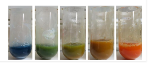
clinitest principle
reducing sugars will reduce copper sulfate (CuSO4) to cuprous oxide (Cu+) in the presence of heat and strong alkaline solution
blue → green → yellow → orange
clinitest false positive
nalidixic acid
cephalosporins
ascorbic acid (vitamin C)
urinary preservatives
formalin
formaldehyde
clinitest false negatives
technique errors
“pass through” effect can cause erroneous results
5 drops of urine normally tested nut high glucose levels can pass through quickly and return to blue
USE 2 DROP TECHNIQUE and diff chart
ketones
chemicals when the body breaks down fat for energy (fatty acid metabolism)
fat used when glucose (carbs) not available
ketone bodies produced in urine:
2% acetone
78% beta-hydroxybutyric acid
20% acetoacetic acid (aka diacetic acid)
only type of ketone detected by strip test
ketone pad principle
utilizes sodium nitroprusside and an alkaline buffer
ketone bodies in urine (acetoacetic acid/diacetic acid) react w sodium nitroprusside to produce magenta/purple color

ketones false positives
urine highly pigmented (dark colored red urine)
both high SG and low pH
phenylketones (red-orange color)
confirm questionable results w tablet test
ketones false negatives
controls containing acetone
breakdown by bacteria - old urine
volatilization of ketones (time) - old urine
acetest
used for ketones
contains sodium nitroprusside, glycine, alkaline buffer, and lactose
lactose allows for better color differentiation
can be used on serum, urine, plasma, and whole blood
10x more sensitive to diacetic acid than acetone

blood
strip detects hematuria, hemoglobinuria, myoglobinuria
correlates w urine appearance, microscopic exam, appearance of patient’s plasma, and plasma chemical test
CAN COEXIST - must confirm and differentiate
blood pad principle
based on the peroxidase activity of hemoglobin and myoglobin
catalyze a rxn between peroxide and a chromogen
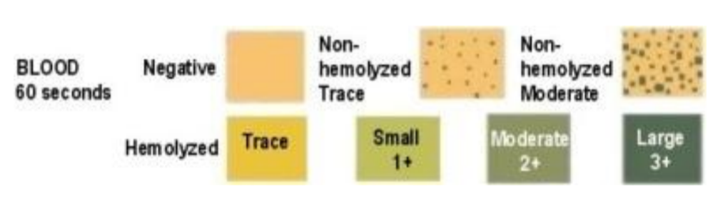
free hemoglobin/myoglobin = uniform color change
intact/whole RBCs = speckled pattern
color intensity = proportional to concentration
hemoglobin rxn
H2O2 + chromogen → oxidized chromogen + H2O2
hemoglobin acts as peroxidase
blood false positives
presence of oxidizing contaminants (bleach)
high bacterial content - bacterial peroxidases
menstrual blood
povidone-iodine (betadine)
blood false negatives
high levels of ascorbic acid (vit. C)
high SG
proteins
nitrites
formalin (preservatives)
blood - hematuria
RBCs in the urine
may result from bleeding at any point in urinary system
GLOMERULONEPHRITIS, TUMORS, TRAUMA, RENAL CALCULI
PYELONEPHRITIS, TOXIC CHEMICALS, DRUGS, UTIs
early indicator of renal disease
RED CELLS DISINTEGRATE IN DILUTE URINE (less than 1.010 SG)
if low RBC quantity present, only detect chemically and microscopically
can cause color change → pink/red, red/brown, smoky
blood hemoglobinuria
intravascular hemolysis (red cell components lyse into plasma)
hemolysis w kidney, lower urinary tract, or in sample
transfusion reactions
hemolytic anemia
severe burns
haptoglobin = protein that binds free hemoglobin
when haptoglobin in plasma saturated → see hemoglobin in urine
some hemoglobin absorbed into tubular cells of the kidneys as hemosiderin
blood - myoglobinuria
released after acute destruction of muscle fibers (rhabdomyolysis)
injury
strenuous exercise in untrained individual
convulsions
electric shock
can damage kidneys
rare
SERUM NORMAL
urine is red/brown to black

blood hemoglobin vs myoglobin
both will produce positive reagent strip blood results
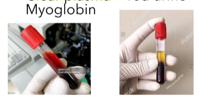
1st macroscopic differentiation
red plasma + red urine = hemoglobinuria
clear plasma + red urine = myoglobinuria
myoglobin screen principle
hemoglobin is precipitated by ammonium sulfate
mix 2.8 g ammonium sulfate in 5ml of urine
let stand for 5 mins then filter
test filtrate w strip (positive = pink/myoglobin ; negative = clear/hemoglobin)
bilirubin
formed by breakdown of hemoglobin in the RES (reticuloendothelial system)
normally, NO DETECATABLE AMOUNT SHOULD BE DETECTED
if present = SIGN OF LIVER OR BILIARY SYSTEM DISEASE
only water soluble (unconjugated bilirubin) can be excreted by urine

bilirubin pad principle
coupled rxn of a diazonium salt w bilirubin in an acid medium
produces tannish-pink/purple color
often hard to read
bilirubin rxn:
bilirubin + diazide → azobilirubin
