The orbit & adnexa
1/28
There's no tags or description
Looks like no tags are added yet.
Name | Mastery | Learn | Test | Matching | Spaced |
|---|
No study sessions yet.
29 Terms
What is the bony orbit ?
A protective ( pyramid shaped ) space within the skull which contains the eyeball, eye muscles, nerved and blood vessels.
How many bones make up the bony orbit ?
7
Name the bones in the bony orbit:
frontal
Sphenoid
Lacrimal
Ethmoid
Maxilla
Zygomatic
Palatine
What is the ocular adnexa ?
They are the supporting and protective structures that are attached to the eyeball.
This includes:
eyelids
Eyelashes
Lacrimal drainage system
The function is to protect the eye and to keep the cornea moist and clean.
The bony orbit has four walls. Name them:
medial
Lateral
Floor
Roof
Describe the structure and function of the orbital roof:
Structure:
thin and fragile ( some parts can be absorbed in old age due to osteoporosis)
Composed of the frontal bone
Composed of the lesser wing of sphenoid.
Function: separates the eyeball from the frontal lobe
What are the greater and lesser wings of sphenoid and what is the difference between them ?
they are two pairs of wings that extend from the spheniod bone.
Lesser wings= smaller, part of the roof
Greater wings= larger part of lateral wall
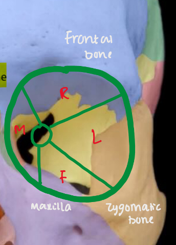
Describe the structure and function of the orbital floor:
Structure:
Weakest wall ( prone to a blow out fracture)
Composed of the maxilla, zygomatic and palatine bone
Function:
Supports eyeball
The inferior oblique muscle originates from the orbital floor, its crucial for eye movement.
Describe the structure and function of the medial wall:
Structure:
Largely composed of the ethmoid bone
Maxilla
Sphenoid
composed of lacrimal bone*
* smallest bone of face, located in the medial wall of each eye orbit. Contains lacrimal sac, for tears.
Describe the structure and function of the lateral wall:
Structure:
thickest wall
Composed of zygomatic bone
Composed of greater wing of sphenoid
Function:
strongest of the four orbital wall, which protects eye from external forces
wall contains Whitnall’s tubercle which serves as an attachment site for ligaments such as the lateral recuts muscle.
What are the openings of the orbit ?
There are 6 openings which allow the passage of nerves and blood vessels to come to and from the orbit.
Name the 6 openings in the orbit:
1) optic canal
2) Superior orbital fissure
3) Inferior orbital fissure
4) Ethmoidal foremen
5) Zygomaticfacial & zygomatictemporal
6) Nasolacrimal canal
What structures are found in the orbital space ?
muscles ( 6 extraocular & levator palpebrae superiosis)
Nerves ( CN 2,3,4,6)
Vessels ( ophthalmic artery and vein)
Lacrimal system ( gland, ducts, sac)
Connective tissues ( orbital septum and tendons capsule)
What is the ocular adnexa ?
The structures that surround and support the eye.
This includes the eyelids and lacrimal system.
Describe the eyelids and its function:
fold of skin that covers the globe.
Shields eye from injury
Sustains the tear film
Supplies oxygen to the cornea when lid is closed.
Eyelid is also known as the palpebral fissure.
What is epicanthal folds ?
Fold of skin at the medial/ nasal side of a persons eye.
What is lagophthalmos ?
when the eyelid doesn’t close properly during sleep
Can lead to eyes becoming dry
Can cause epithelial damage of cornea and cause a loss of transparency
How many eyelid muscles are there ? And name them all:
4 muscles
Orbicualris oculi
Levator palpebral superiosis
Mullers muscle
Tarsal plate muscle
Describe the orbicularis oculi:
it is an eyelid muscle
Attached nasally and laterally by ligaments
Causes the eyelid to close
Theres two main divisions:
Orbital part- causes the eyelid to tightly close ( force-full blinking)
Palpebral part- gently closes the eyelid ( normal blinking)
Innervated by cranial nerve 7 ( facial nerve)
Describe the levator palpebrae superiosis:
eyelid muscle
Causes the upper eyelid to retract and be raised.
Innervated by cranial nerve 3 ( oculomotor nerve) * damage to CN3 - causes the upper eyelid to droop.
Describe Muller’s muscle:
eyelid muscle
Assists the levator palpebrae superiosis in keeping the upper eyelid elevated.
Innervated by the sympathetic nervous system.
Describe the tarsal plate muscle:
eyelid muscle
Provides rigidity and structure to the lids
Secretes lipid layer of tears- which prevents tears from evaporating.
This is important during eyelid eversion
Describe the tarsal gland ( meibomian):
embedded inside the tarsal plate of both the upper and lower eyelids
Secretes lipids
To prevent the tears from evaporating too quickly.
What are the glands of Zeis
located near the eyelash hair follicles
Secrets sebum ( oil) to keep the eyelashes from becoming dry and brittle.
Way to remember* - Z ( zobia)= hair follicles
Describe the glands of Moll:
Located near the eyelash follicles
Acts as a sweat gland by producing sweat to help the tear film.
Describe the conjunctiva:
thin, transparent mucous membrane that keeps the eye moist and protected from microorganisms
at the medial edge there is a condensed bit of tissue called the caruncle.
there are three main sections to the conjunctiva:
Palpebral conjunctiva- this lines the inner surface of the eyelids
Bulbar conjunctiva- covers the scalera of eyeball
Fornix- the fold where the eyelid and eyeball meet ( acts like a junction)
Describe the tear film and its 3 layers:
covers the anterior surface of the eye
Outermost layer = lipid layer
Inner layer= aqueous layer
Innermost layer= mucous layer
Lipid layer
contains cholesterol and fatty acids- prevents the evaporation of tears. Keeps the tear surface smooth by providing lubrication for eyelid movement
Aqueous layer
thickest layer- provides nutrients such as salts, glucose and proteins to cornea.
Mucous layer
allows the tear film to stick to the corneal surface
Describe the lacrimal drainage system:
the lacrimal glands which is situated above the eyelid produces tears
The tears get drained from the upper and lower puncta.
The upper canaliculi merge to form the common canaliculus and tears move through that passage.
The valve of Hasner is at the beginning of the canaliculus and prevents the back flow of tears towards the eye.
The tears then move to the lacrimal sac which is compressed when blinking.
Tears are then drawn into the lacrimal apparatus as the sac re-expands ( lacrimal pump)
The nasolacrimal duct connects the lower end of the lacrimal sac with inferior meatus.
Another valve guards the opening and prevents air from entering the sac during blowing of the nose.