posterior abdominal wall, kidneys and adrenal glands
 Call Kai
 Learn
Learn Practice Test
 Spaced Repetition
 Match
Match Flashcards
 Knowt Play
1/109

There's no tags or description
Looks like no tags are added yet.
Name | Mastery | Learn | Test | Matching | Spaced | Call with Kai | Chat |
|---|
No analytics yet
Send a link to your students to track their progress
110 Terms
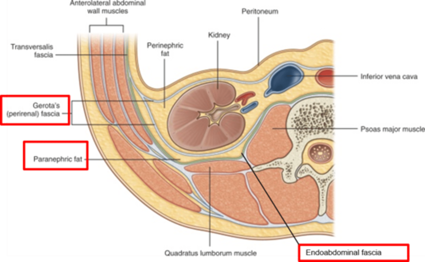
What does the endoabdomicnal facia of the posterior abdominal wall lie between? What is it continuous with?
lies between the parietal peritoneum and the muscles and is continuous with the transversalis fascia
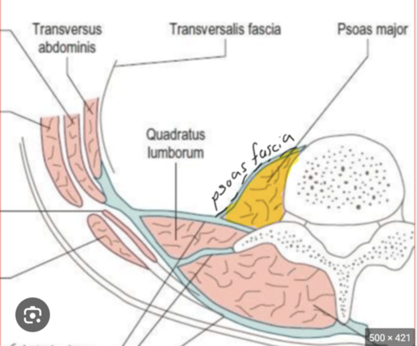
what does the psoas fascia of the posterior abdominal wall surround? what is it attached medially to?
surrounds the psoas major muscle and is attached medially to the lumbar vertebrae and pelvic brim
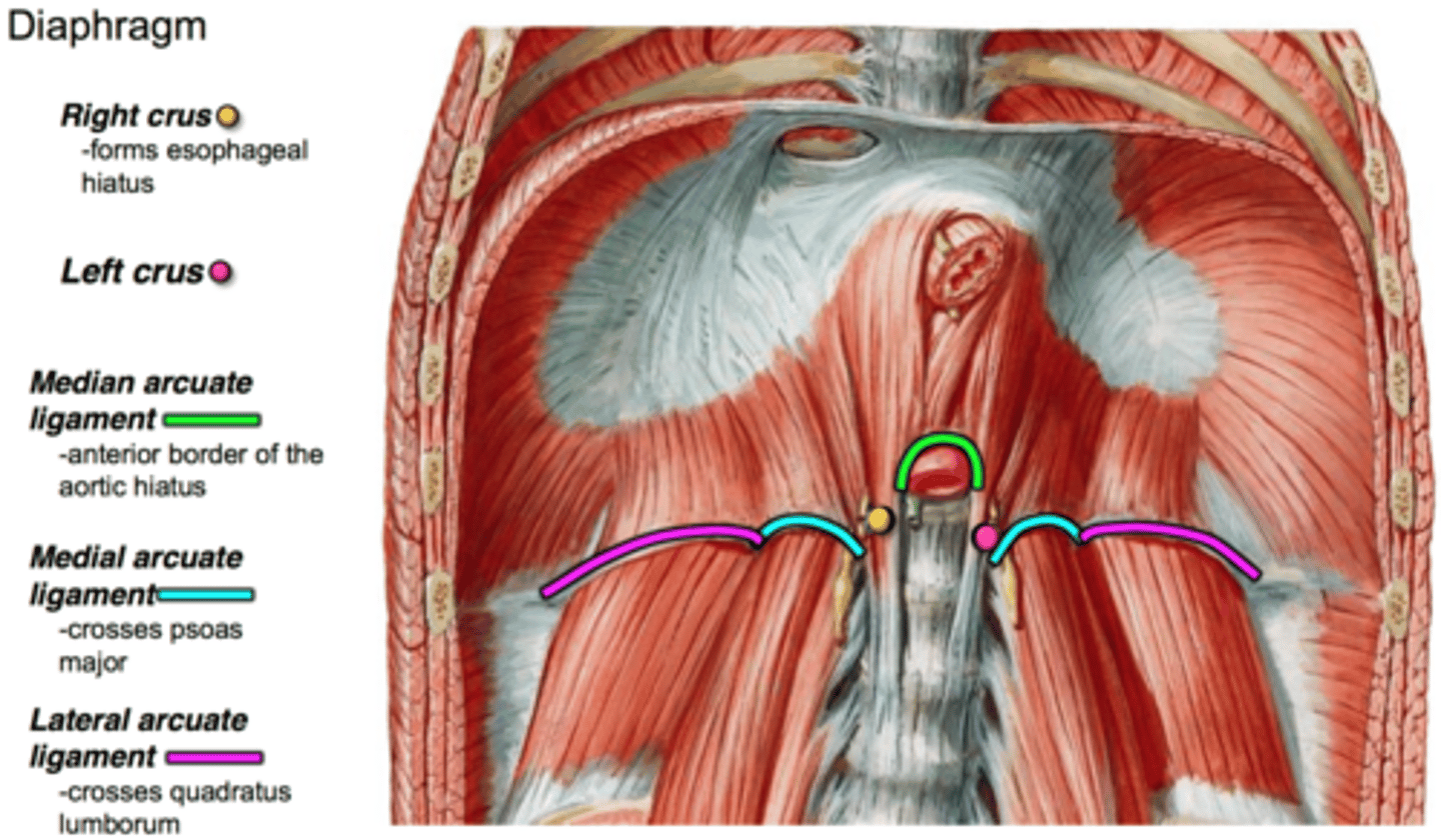
what does the psoas fascia thicken to form? what fascias is it continuous with?
thickens to form the medial arcuate ligament and is continuous with the quadratus lumborum and thoracolumbar fascia
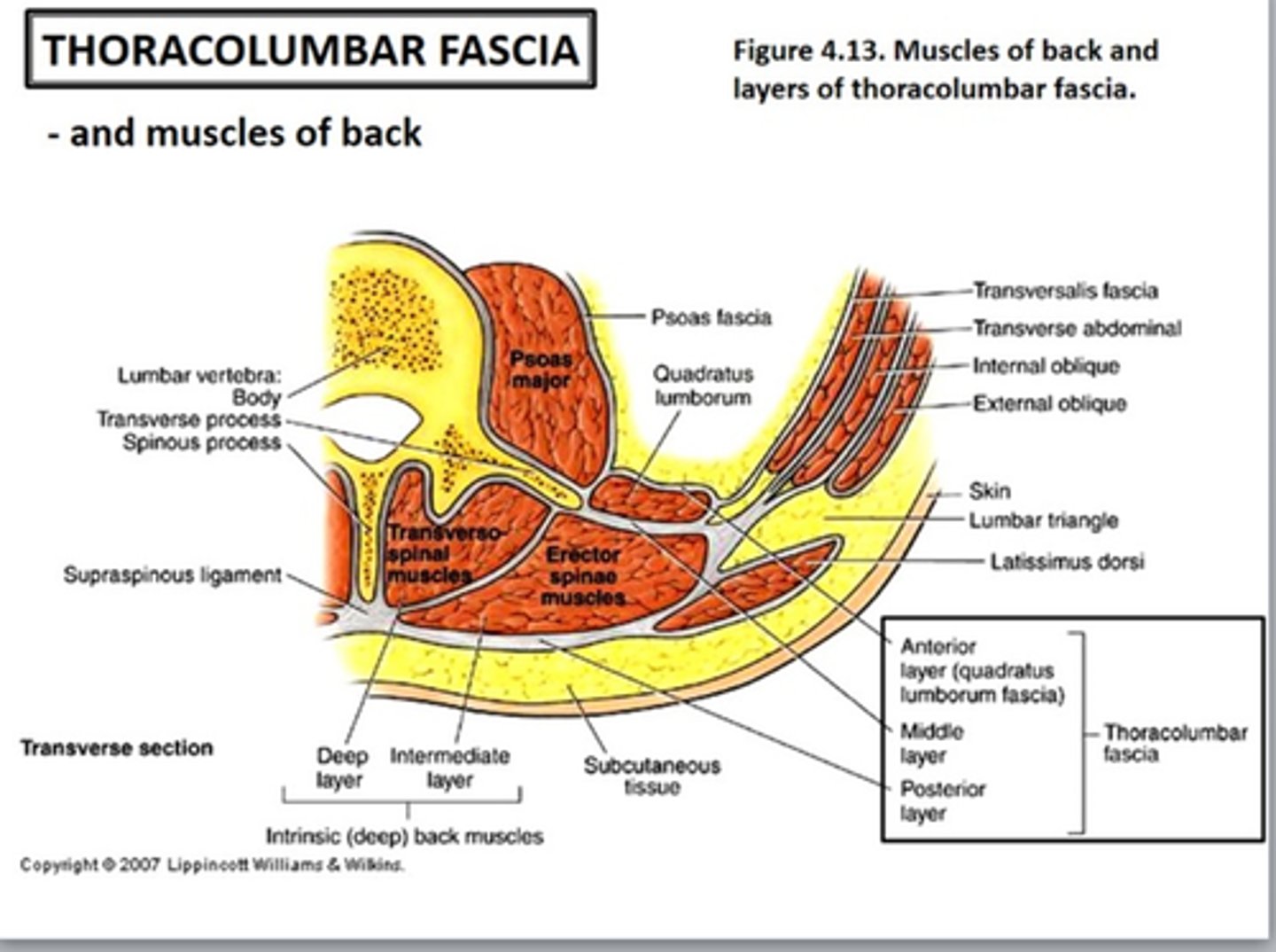
what is the thoracolumbar fascia? what is it attached to? what does it envelope?
extensive fascia that is attached to the vertebral column and envelopes the deep back muscles (in a similar manner to the rectus sheath)
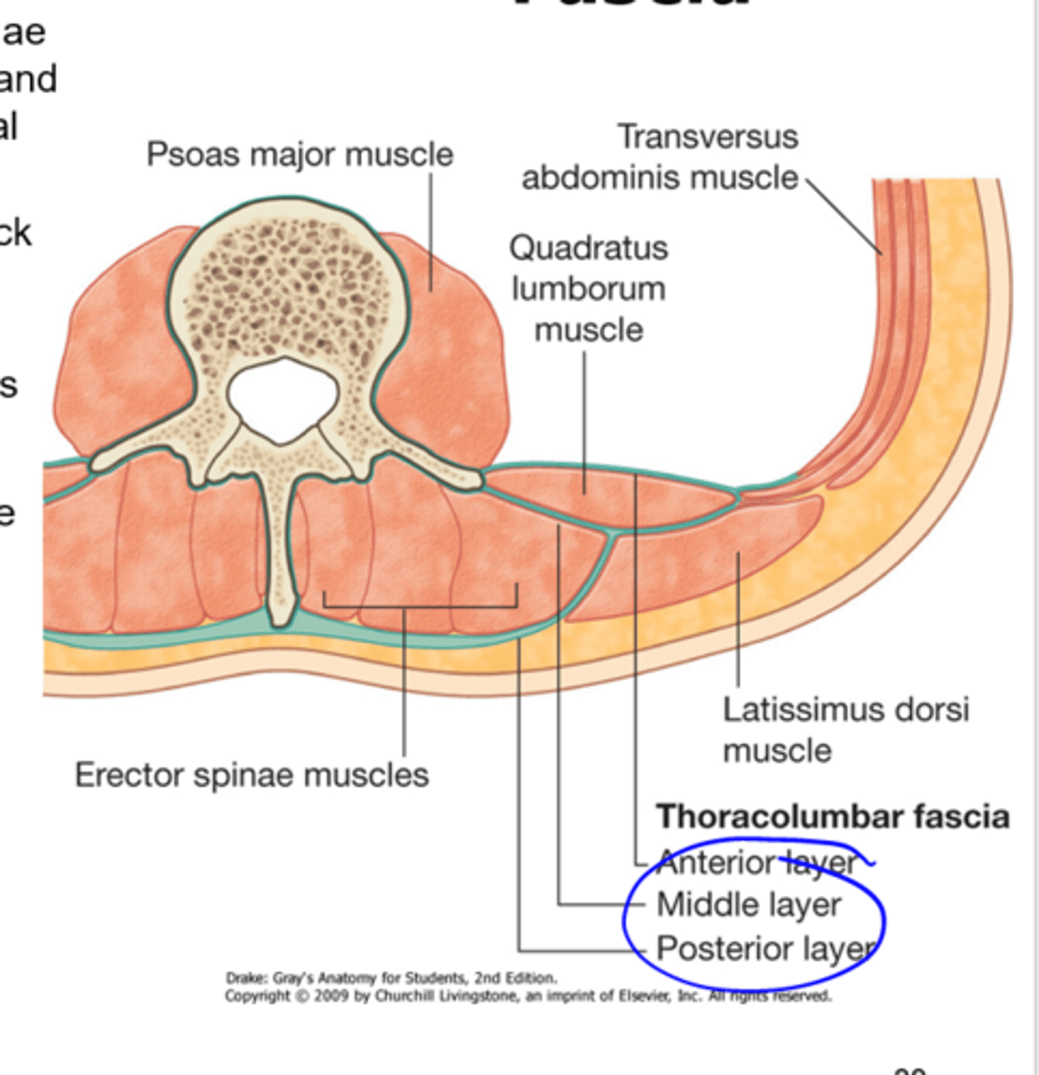
the thoracolumbar fascia is thinner in what area compared to the lumbar area?
thoracic
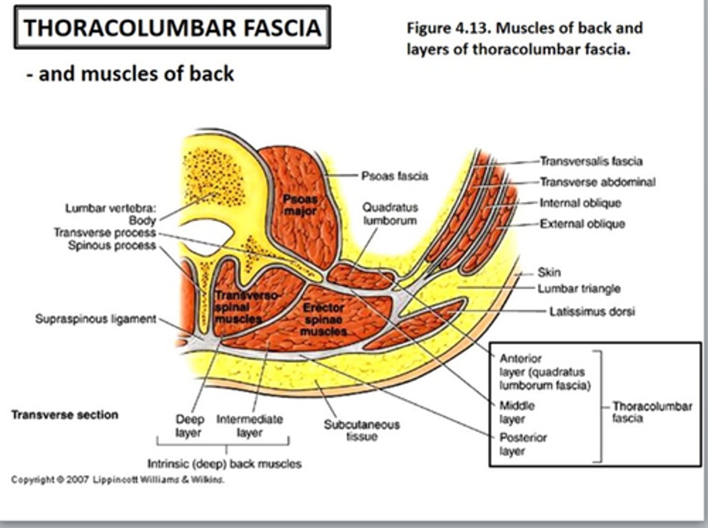
the thoracolumbar fascia in the lumbar region attach laterally to what muscles?
in the lumbar region, attaches laterally to the Internal oblique, transverse abdominis, and latissimus dorsi muscles
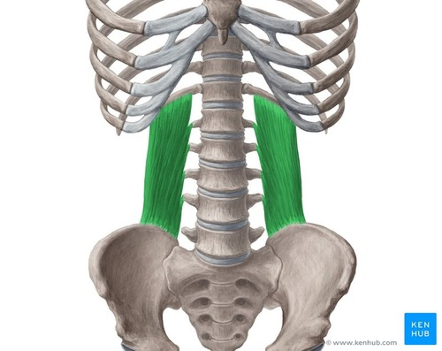
what layer is the quadratus lumborum facia? what muscle does it cover?
anterior layer of the thoracolumbar fascia that covers the quadratus lumborum muscle
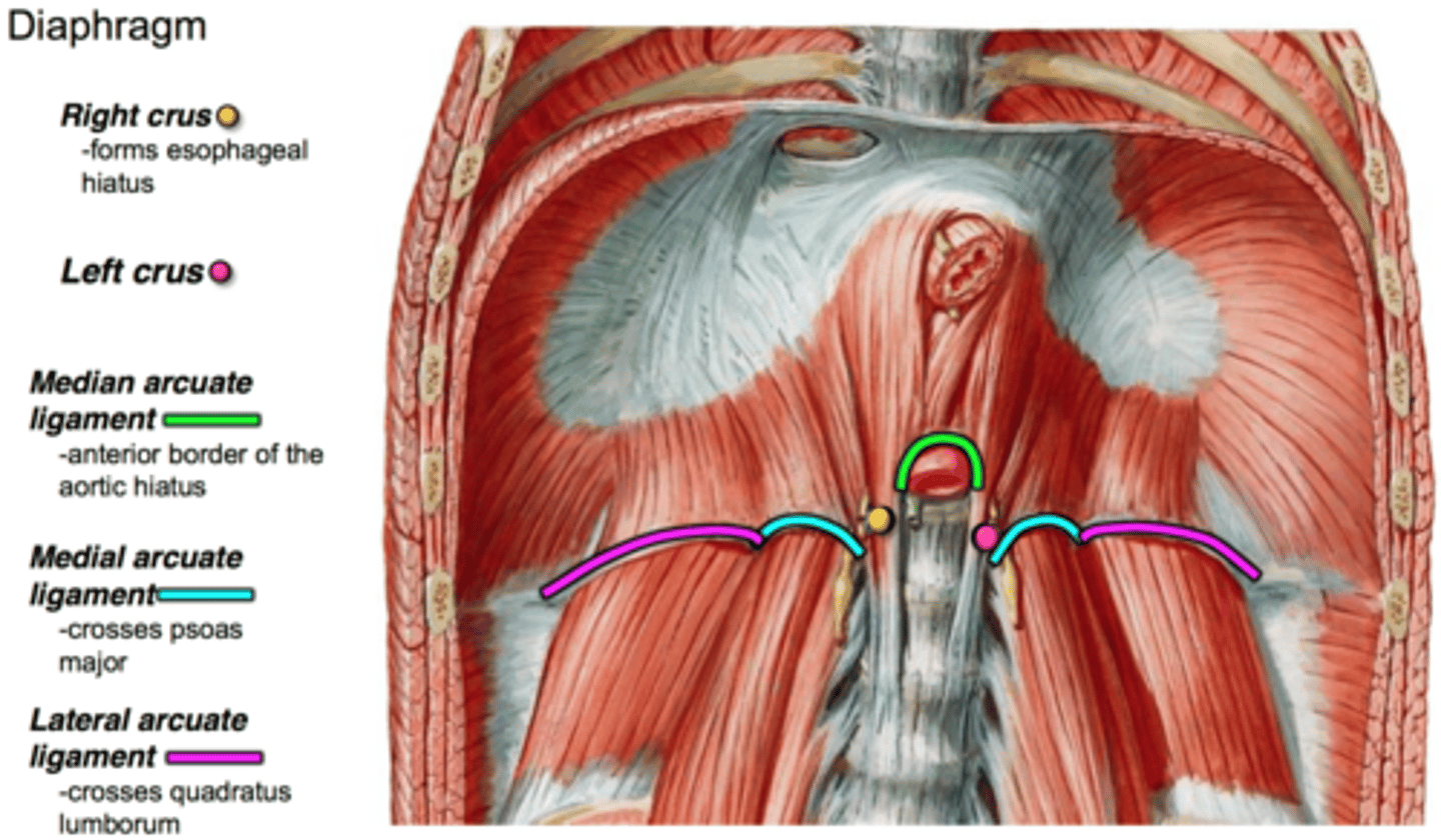
what does the quadratus lumborun fascia thicken to form?
thickens to form the lateral arcuate ligament
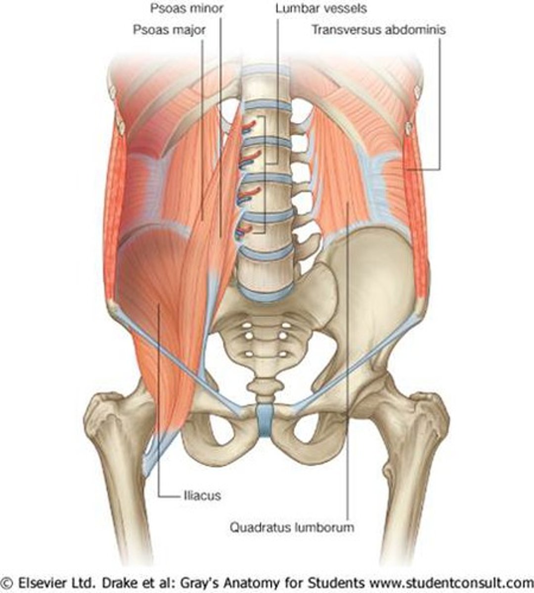
what are the muscles of the posterior abdominal wall?
psoas major, Iiacus, and quadratus lumborum
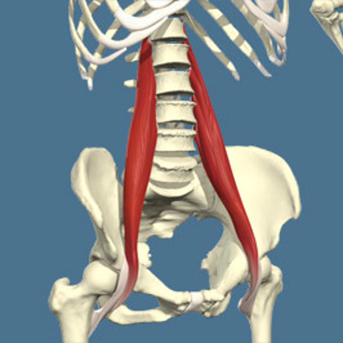
what is the psoas major muscle lateral to?
lateral to lumbar vertebrae
what is the function of the psoas major muscle?
flexes thigh or trunk
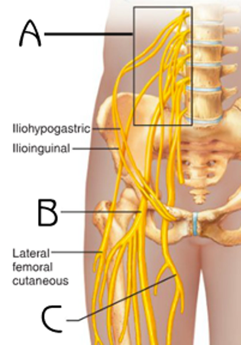
what is the psoas muscle innervated by?
anterior rami of L1 - L3
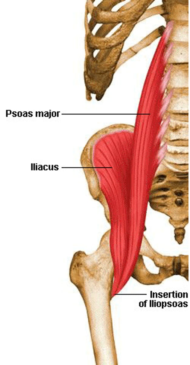
what is the Iliacus muscle lateral to? the tendons join those of the posas major to form what?
lateral to inferior part of psoas major and its tendons join those of the psoas major to form the iliopsoas (chief flexor of the thigh)
what is the function of the Iliacus muscle?
flexes thigh and stabilizes the hip joint
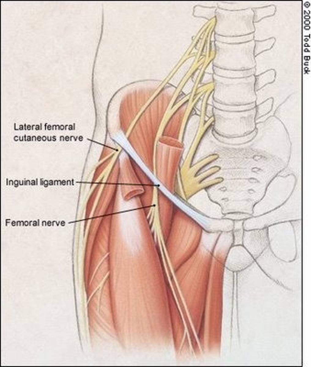
what is the Iliacus muscle innervated by?
femoral nerve (L2 - L4)
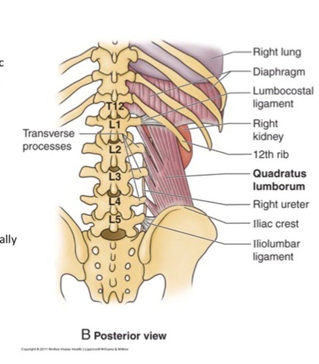
what is the quadratus lumborum muscle lateral to? what does it form?
lateral to superior part of psoas major and forms the kidney bed
what is the function of the quadratus lumborum muscle?
extends and laterally flexes vertebral column
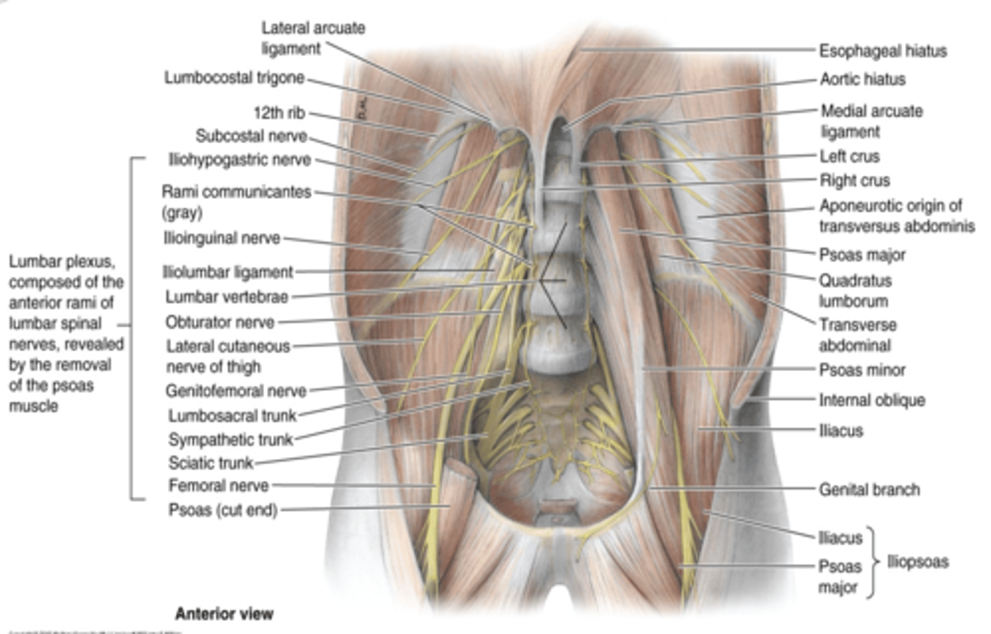
what is the quadratus lumborum muscle innervated by?
subcostal nerve and L1 - L4 nerves
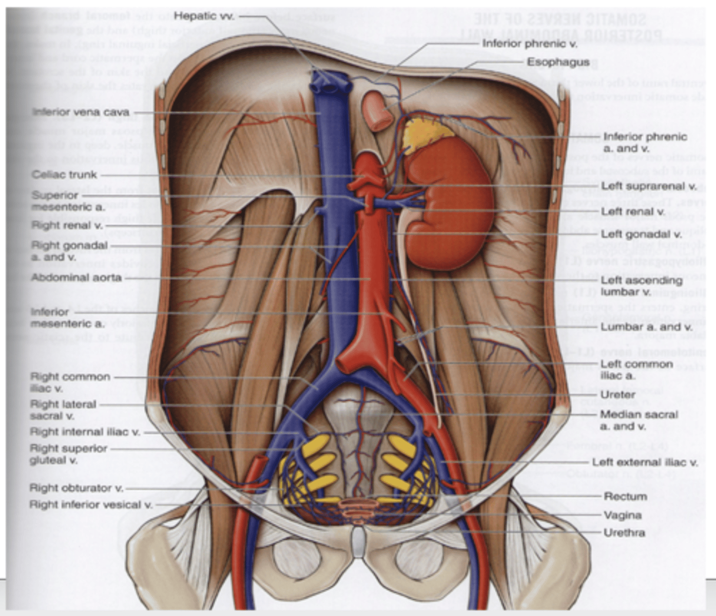
where does the abdominal aorta begin at and end? what does it divide into?
begins at aortic hiatus (level of T12) and ends at the level of L4, where divides into right and left common iliac aa
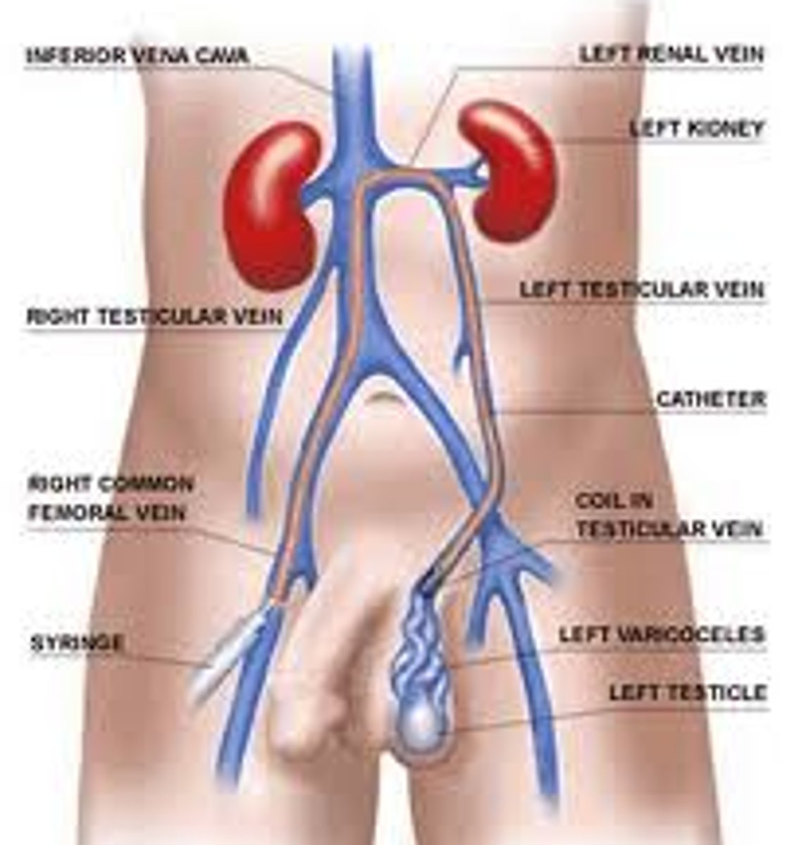
what is the largest vein in the body?
IVC
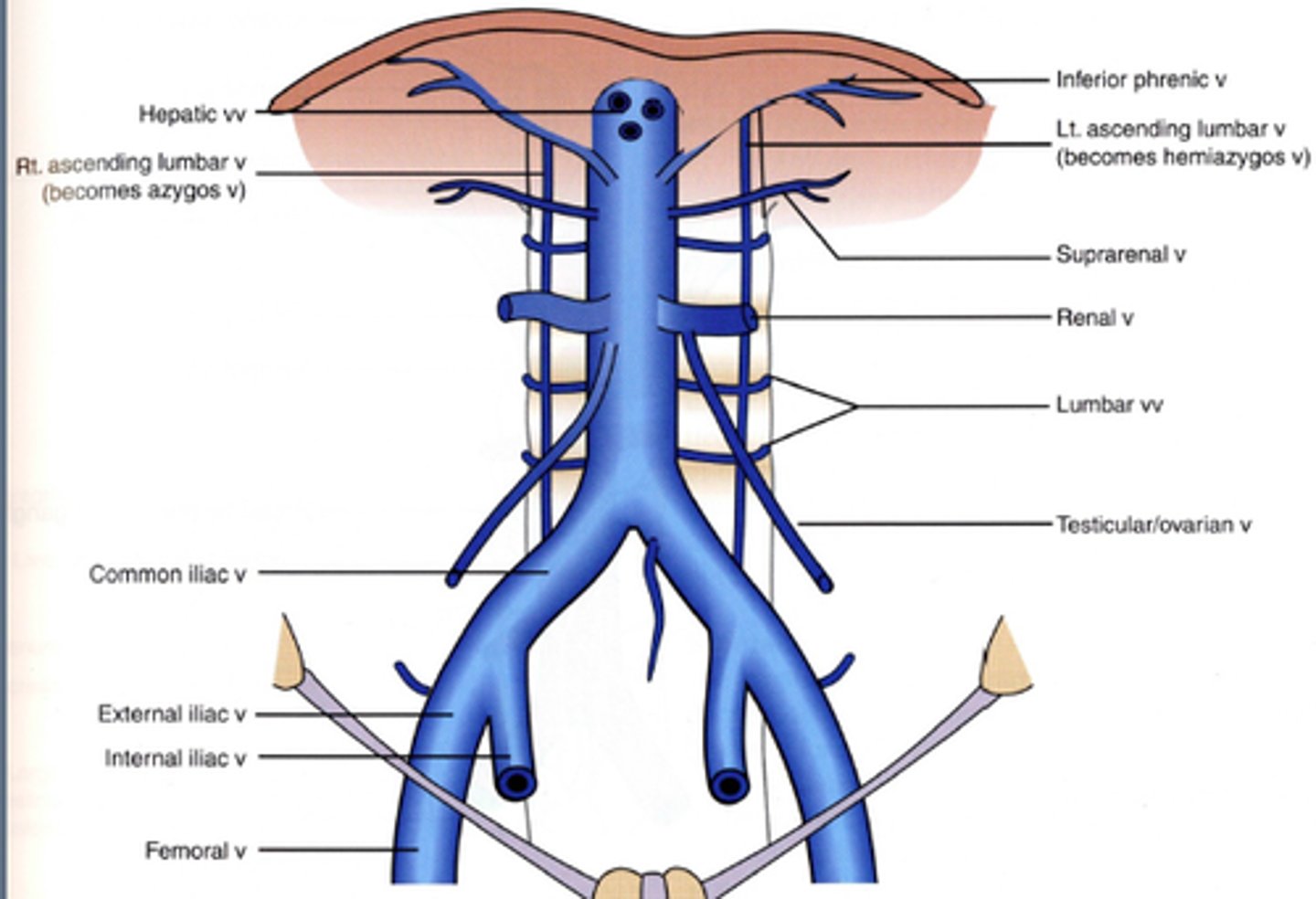
where does the IVC begin anterior to? by the union what enters the caval opening at the level of what vertebrae?
begins anterior to L5 by the union of the common iliac vv. inferior to aortic bifurcation > enters caval opening and thorax at level of T8
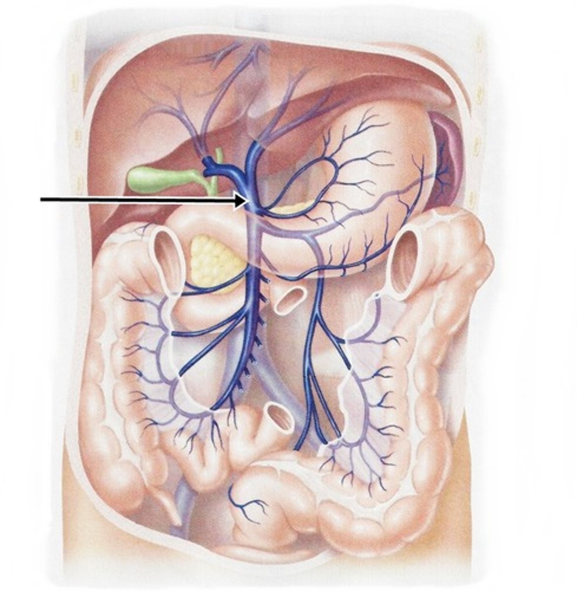
what does the hepatic vein drain of the IVC?
drains the liver
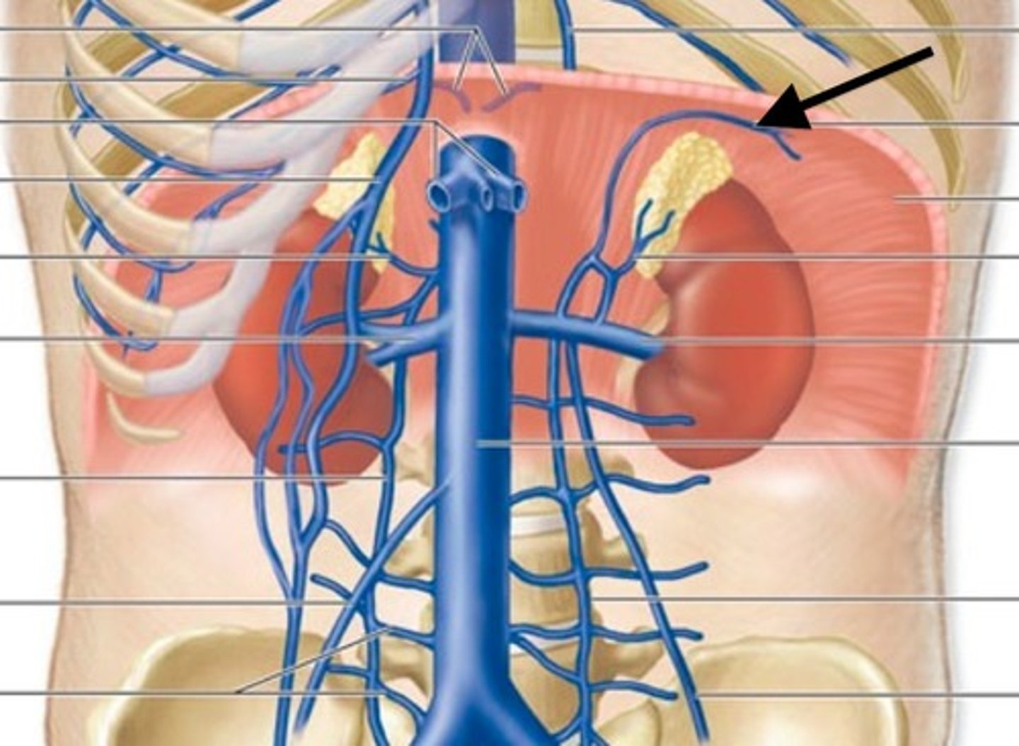
what does the inferior phrenic vein drain of the IVC?
drains inferior surface of diaphragm
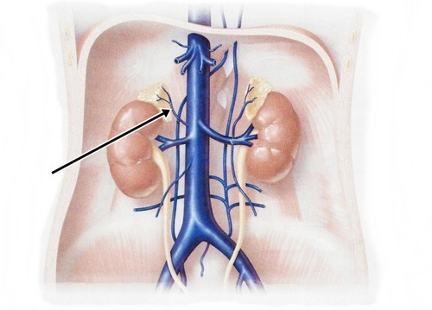
what does the right suprarenal vein drain of the IVC?
drains right suprarenal gland
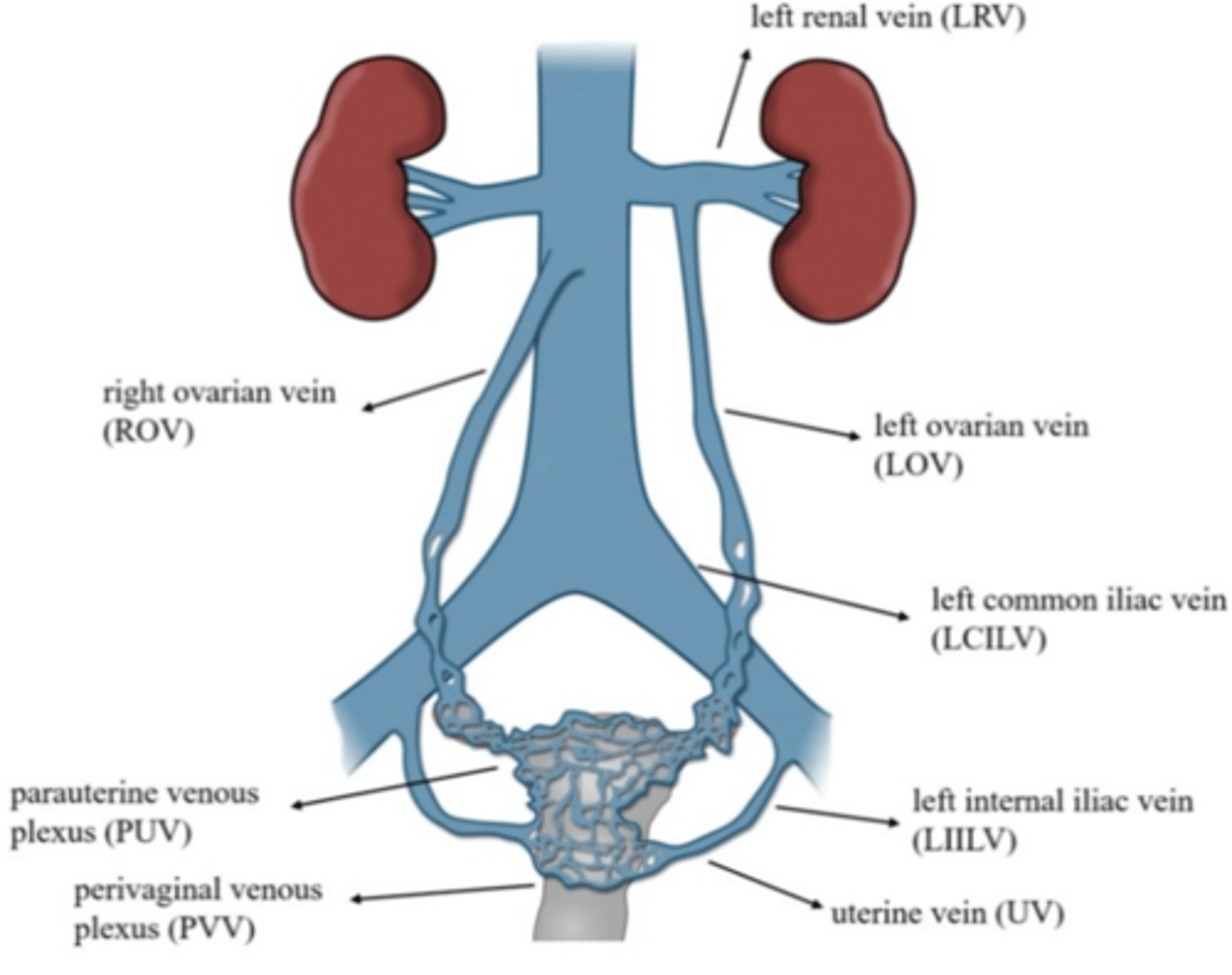
what does the right goadal vein drain of the IVC?
drains R. testis or R. ovary
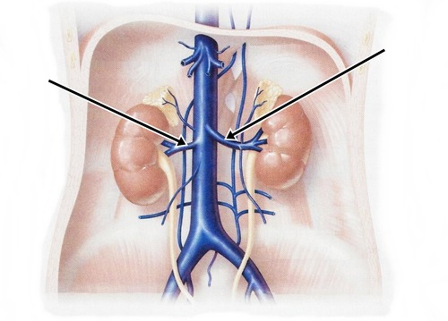
what does the renal vein of the IVC drain?
drain kidneys and the left renal also drains:
left gonadal v.: drain left testis or left ovary
left suprarenal v.: drain left suprarenal gland
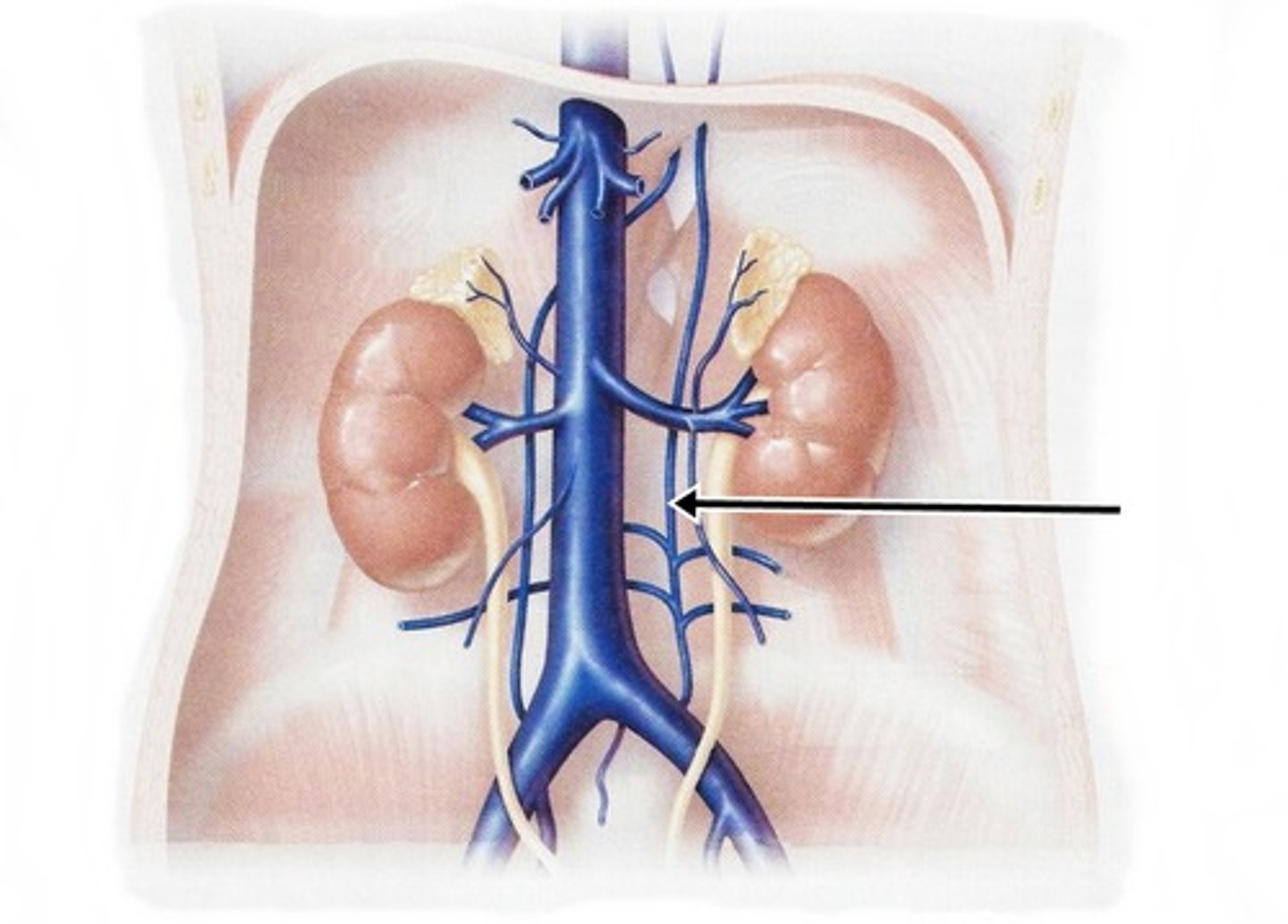
what does the lumbar vein of the IVC drain?
drain the abdominal wall
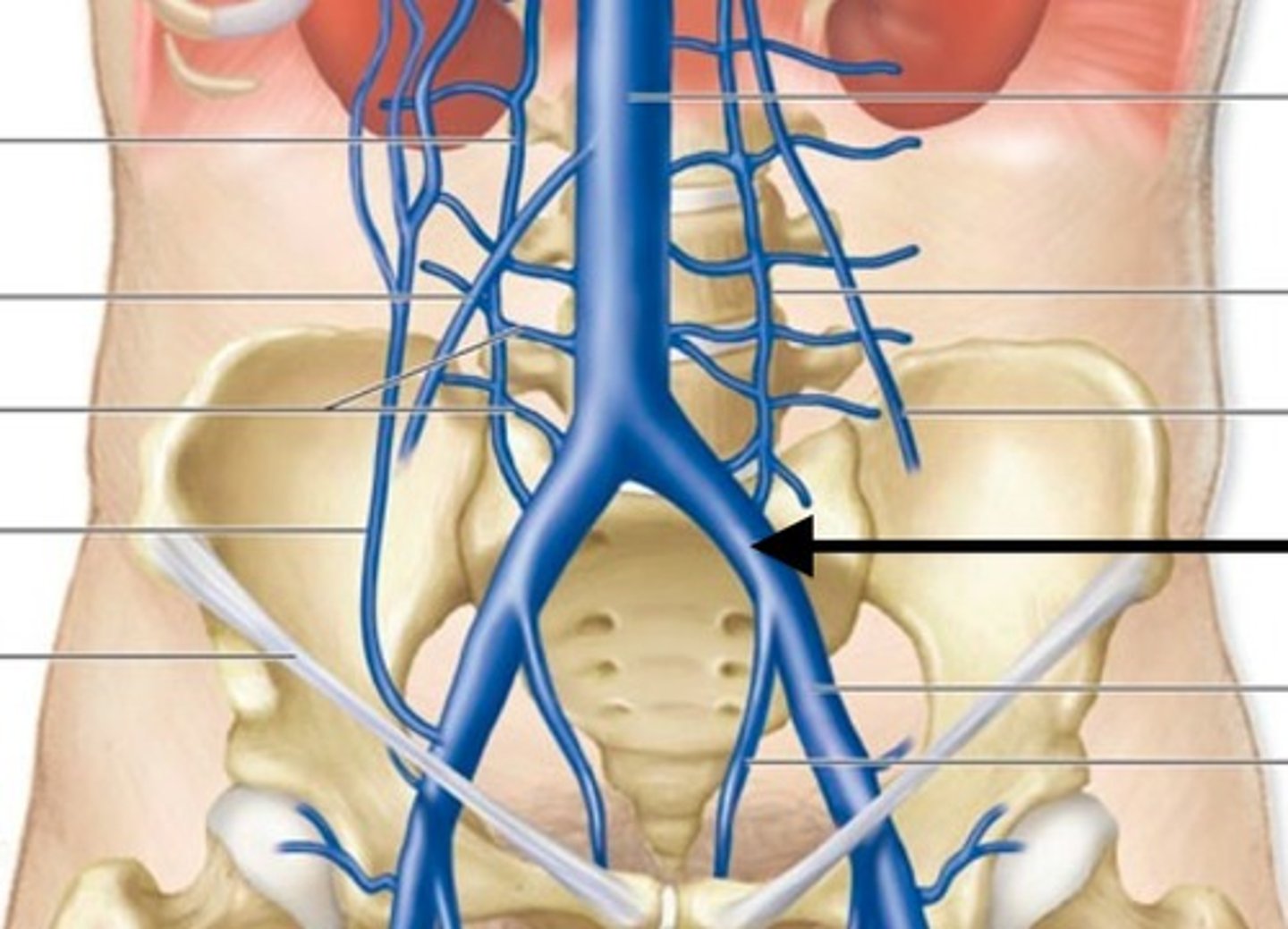
what does the common iliac vein of the IVC drain?
drain the legs
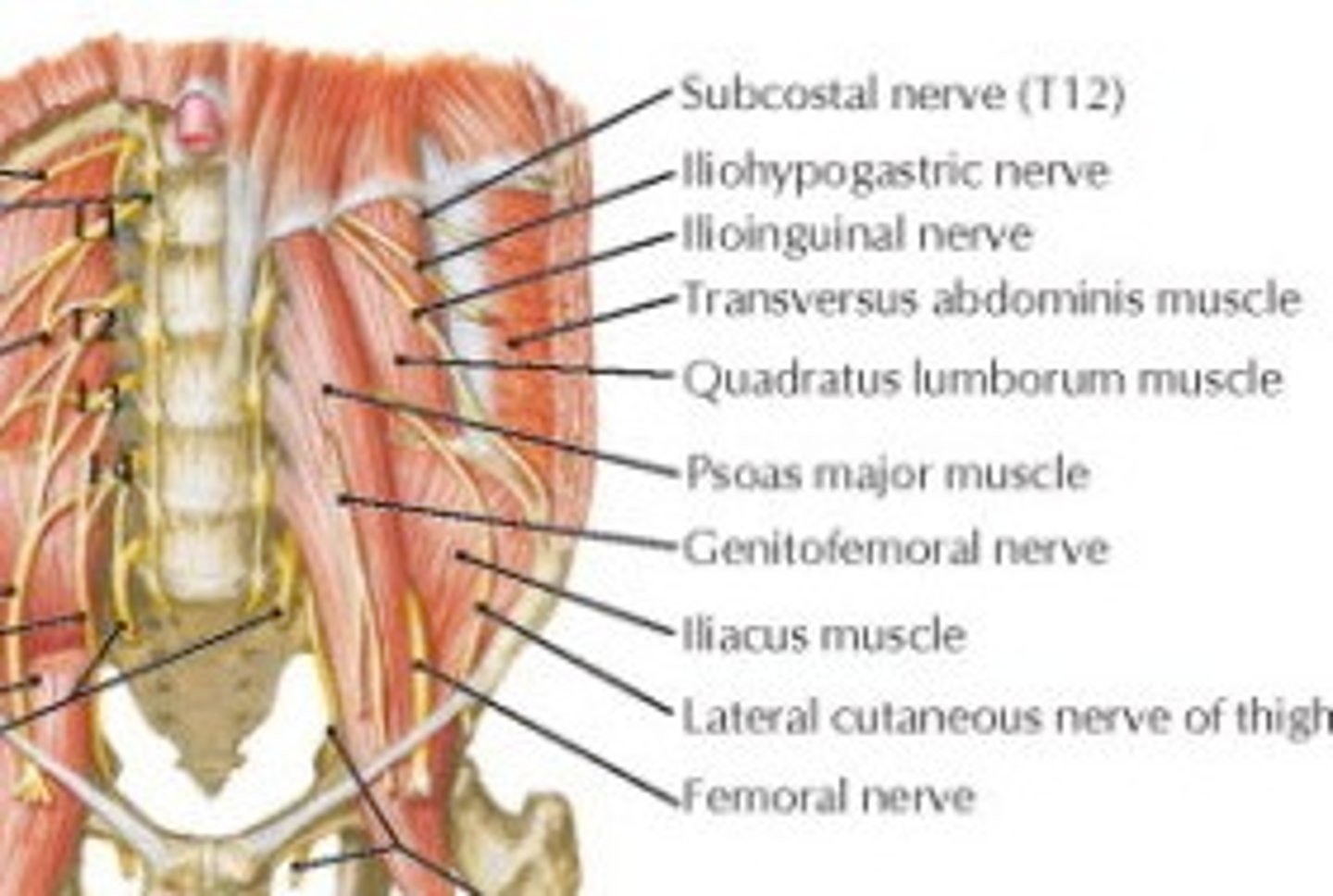
what is the course of the subcostal nerve (T-12)?
runs inferolaterally on the anterior surface of the quadratus lumborum muscle.
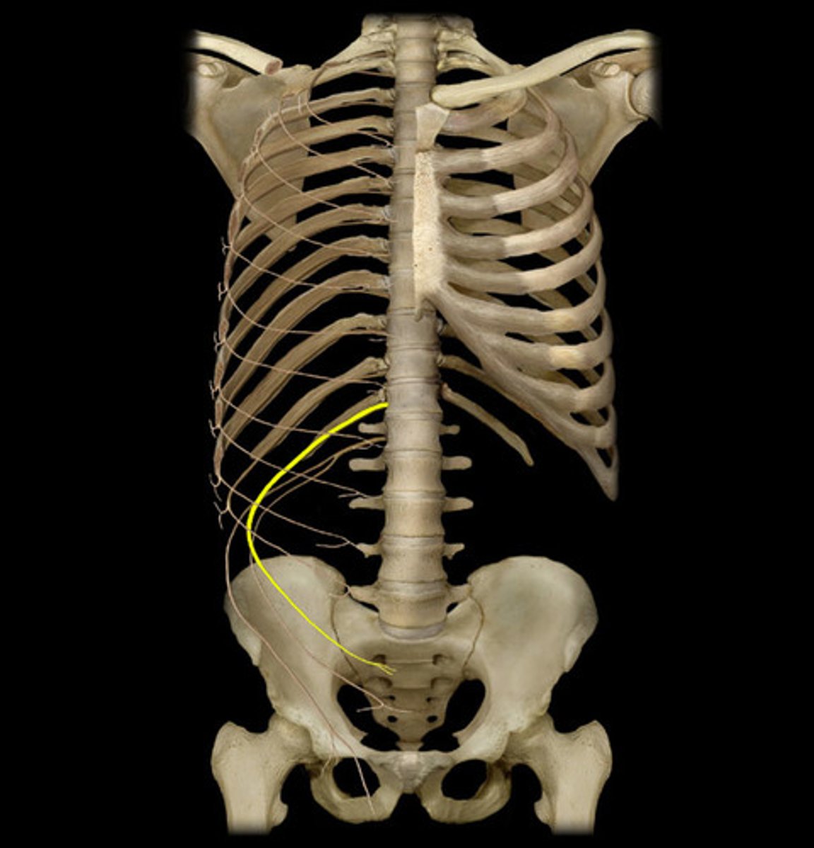
what does the subcostal nerve (T-12) innervate?
muscles of anterolateral abdominal wall and skin on side of hip
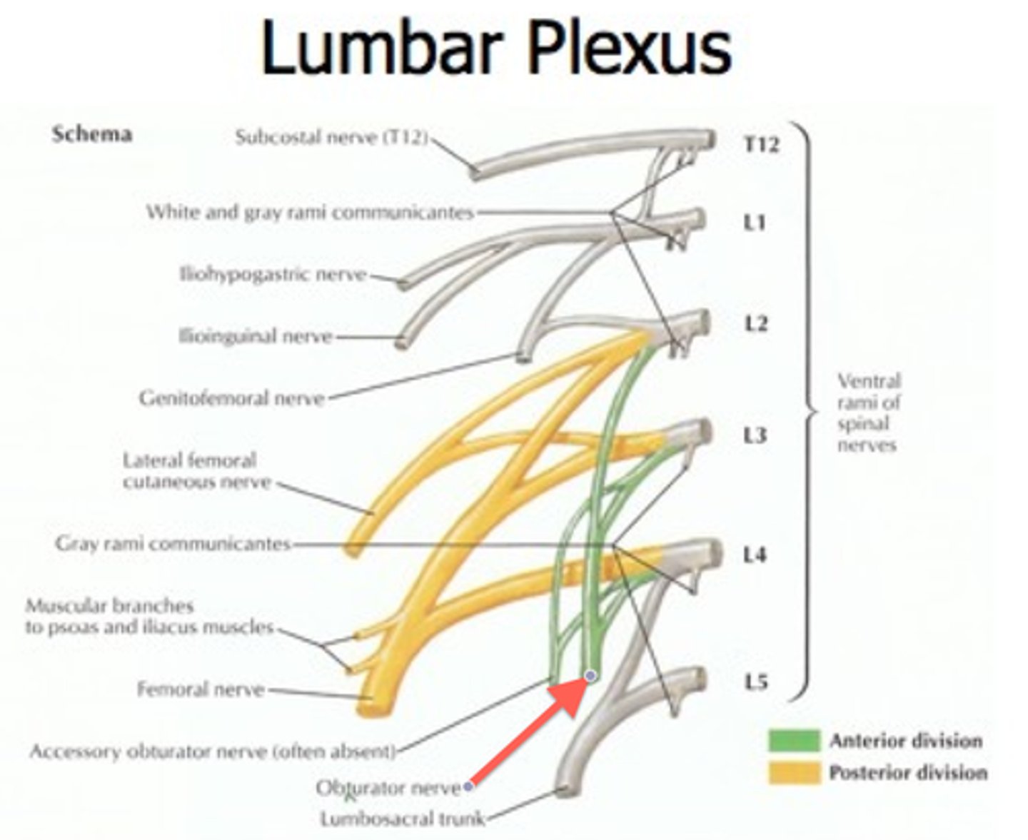
what nerves belong to the lumbar plexus?
1. Iliohypogastric nerve (L1
2. Ilioinguinal nerve (L1)
3. Genitofemoral nerve (L1 - L2)
4. Lateral cutaneous nerve of the thigh (L2 - L3
5. Femoral nerve (L2 - L4)
6. Obturator nerve (L2 - L4)
7. Lumbosacral trunk (L4 - L5)
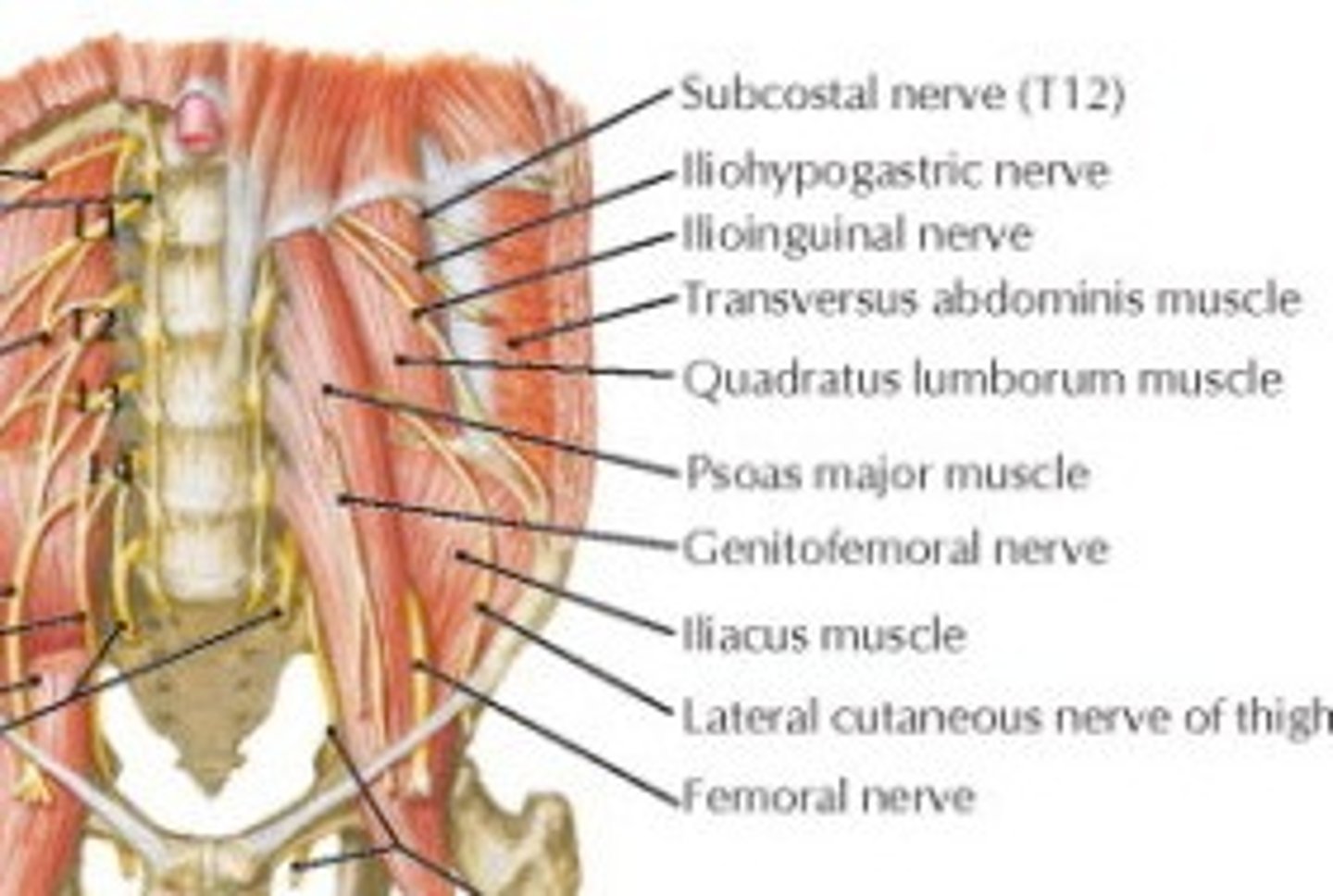
where does the Iliohypogastric nerve (L1) of the lumbar plexus run?
runs inferolaterally on the anterior surface of the quadratus lumborum muscle
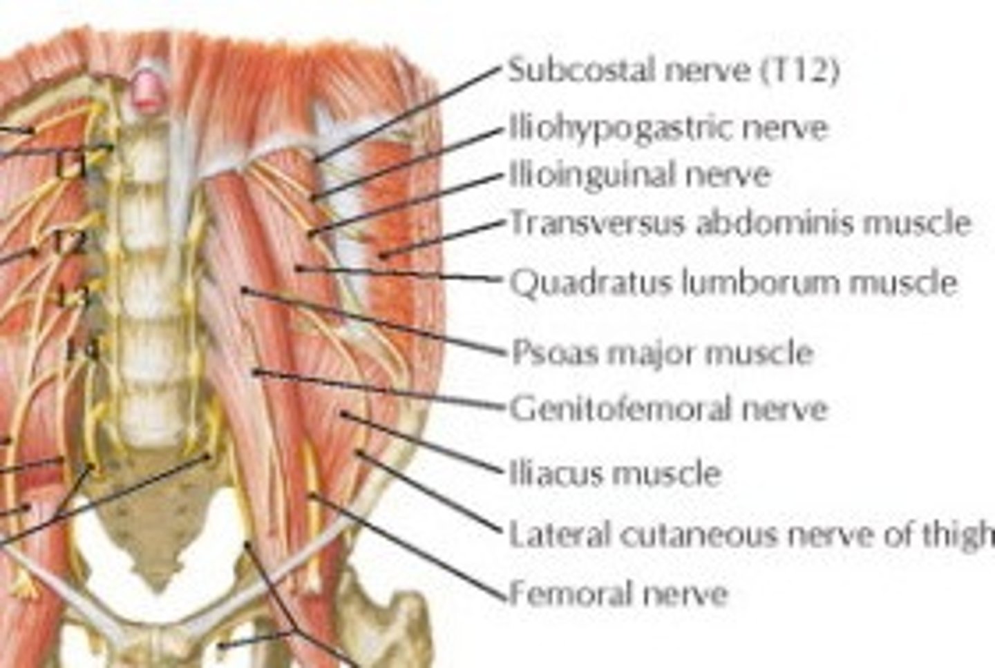
what does the Iliohypogastric nerve (L1) of the lumbar plexus innervate?
internal oblique and transverse abdomius muscles, and skin above pubis
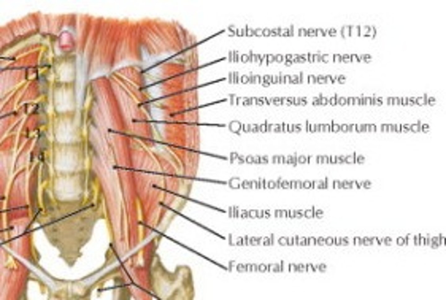
what is the course of the Ilioinguinal nerve (L1) of the lumbar plexus?
runs inferolaterally on the anterior surface of the quadratus lumborum muscle
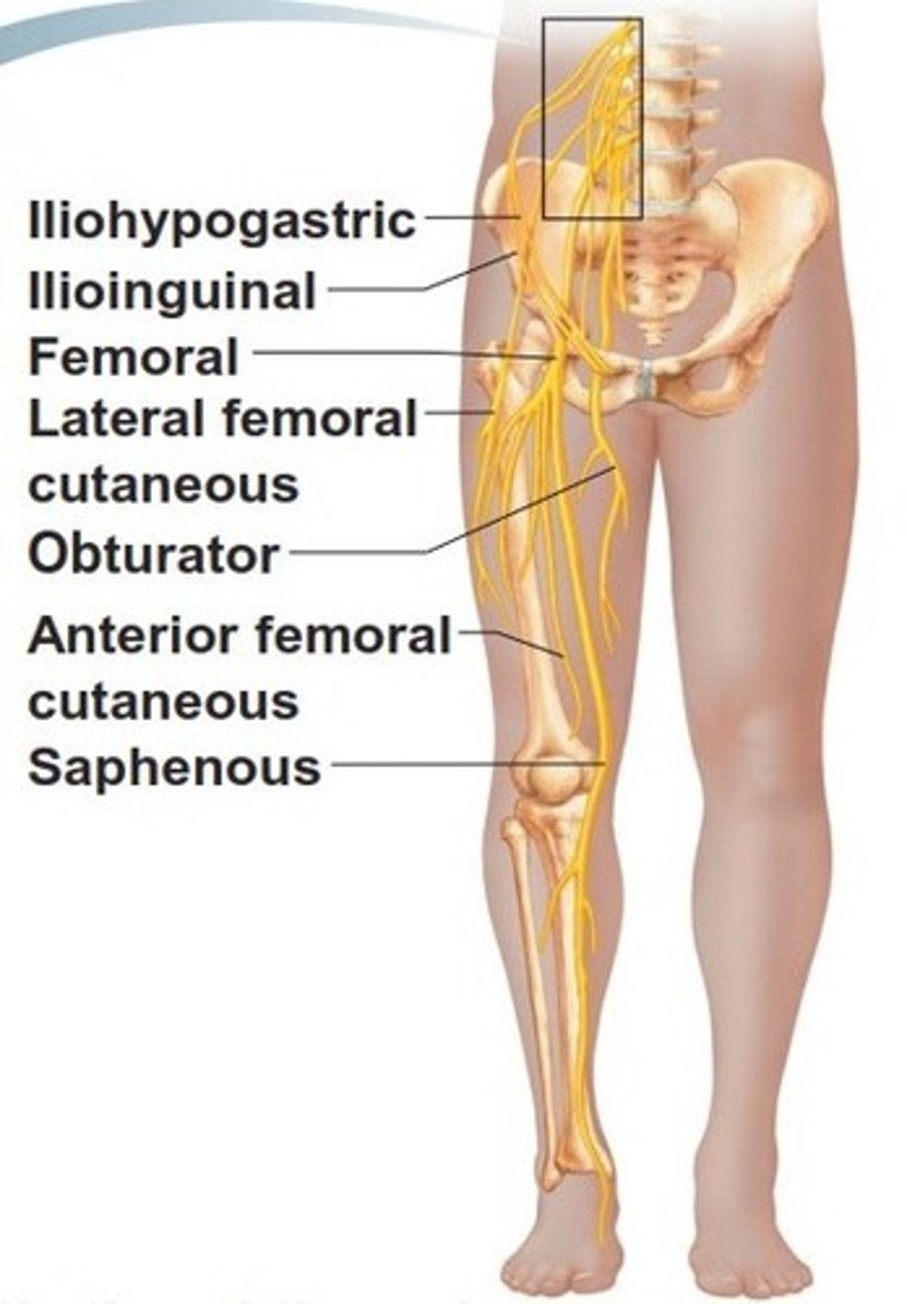
what does the Ilioinguinal nerve (L1) of the lumbar plexus innervate?
internal oblique and transverse abdominus muscles, and skin on root of penis (or mons pubis), and anterior scrotum (or labium majus)
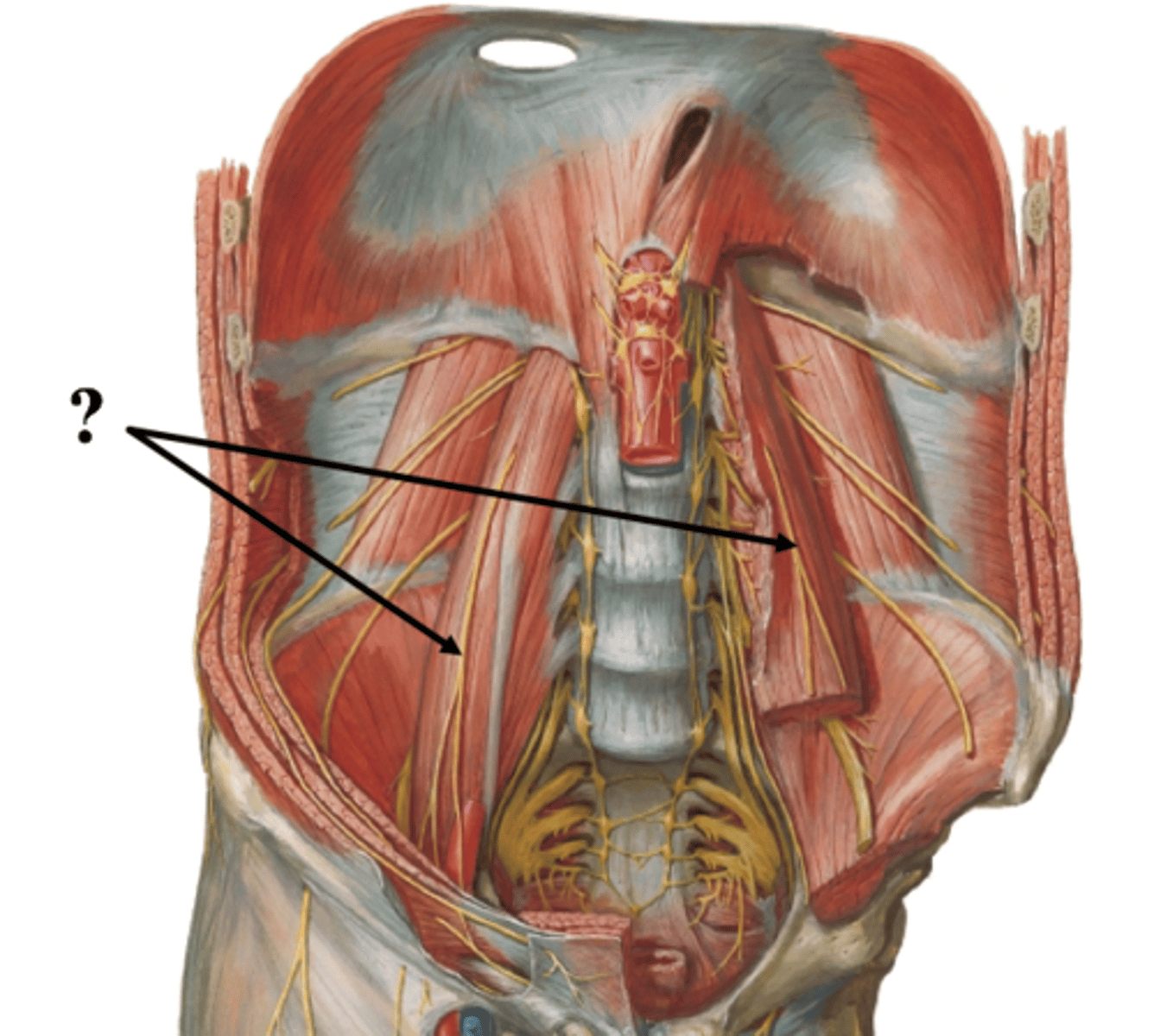
the genitofemoral nerve (L1 – L2) of the lumbar plexus pierces what muscle and runs inferiorly on its anterior surface?
pierces the psoas major and runs inferiorly on its anterior surface
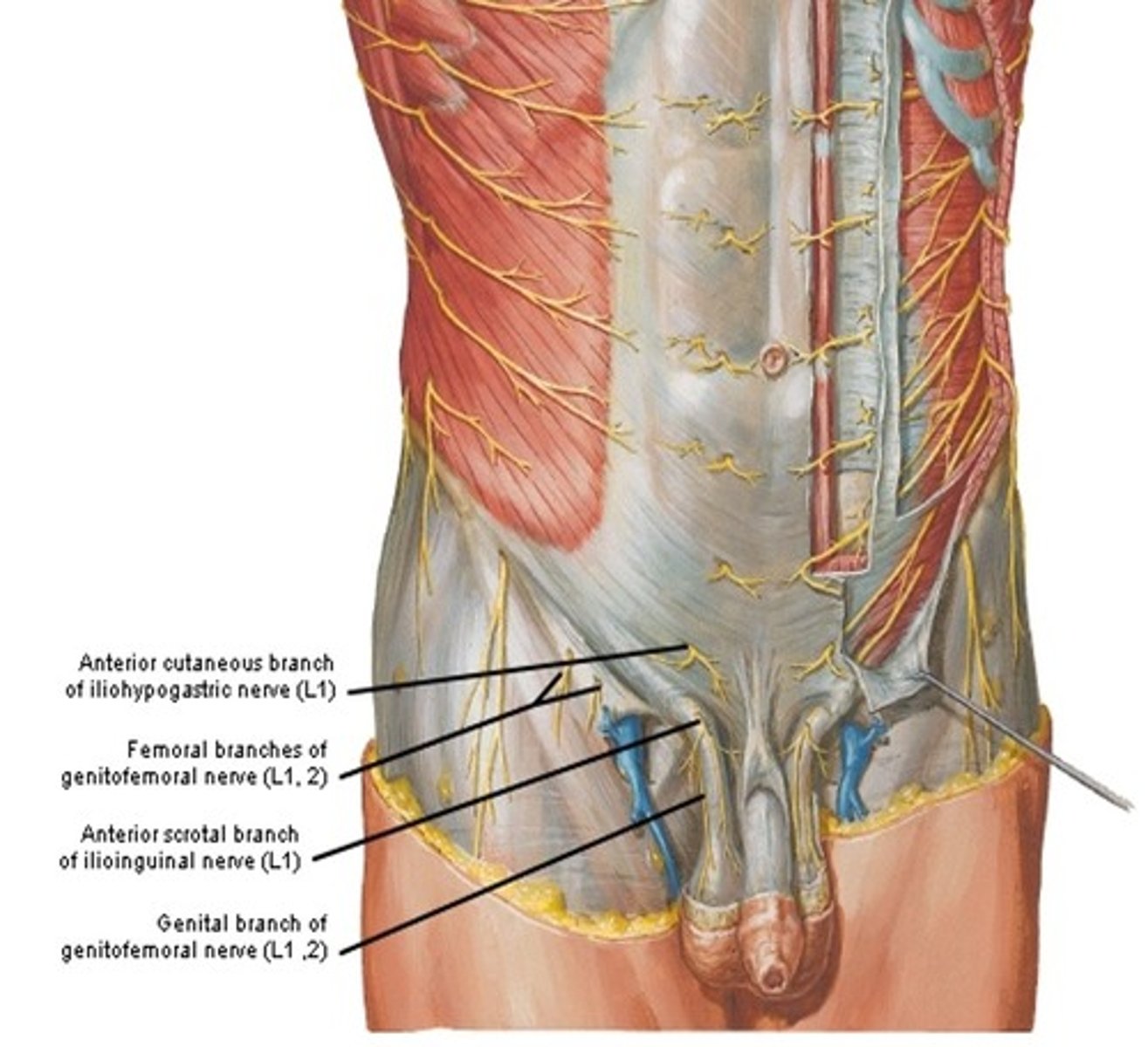
the femoral branch of the genitofemoral nerve (L1 – L2) innervates what?
skin on anterior thigh
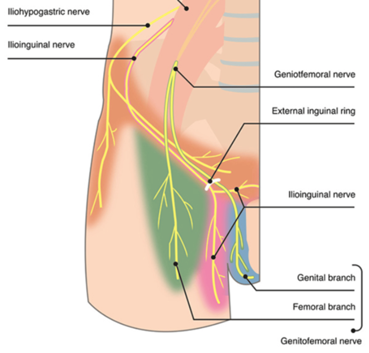
the genital branch of the genitofemoral nerve (L1 – L2) innervates what (males V.S females)
males: cremaster muscle and skin of scrotum
females: skin on mons pubis and labia majora
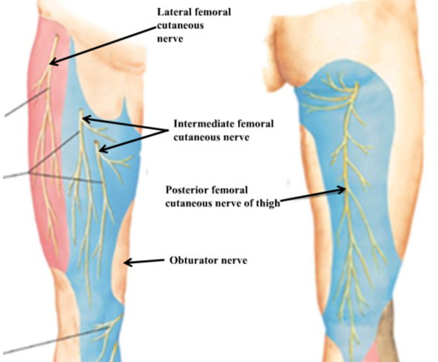
what is the course of the lateral cutaneous nerve of the thigh (L2 – L3) of the lumbar plexus?
runs inferolateral on the iliacus and enters the thigh deep to the inguinal ligament
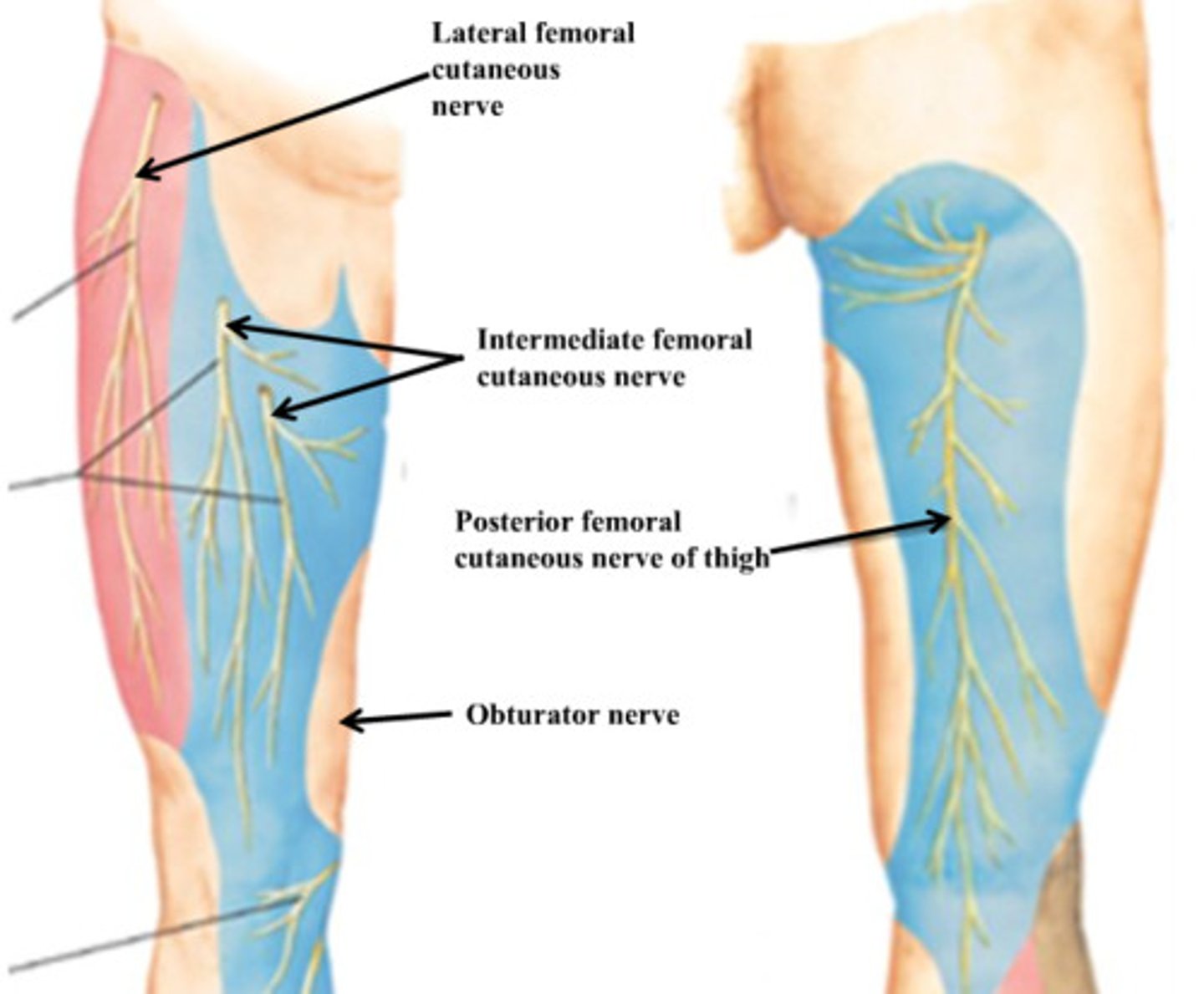
what does the lateral cutaneous nerve of the thigh (L2 – L3) of the lumbar plexus innervate?
skin on anterolateral surface of thigh
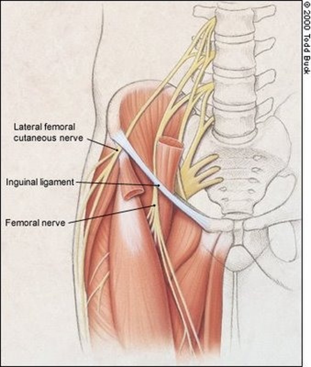
what is the course of the femoral nerve (L2 – L4) of the lumbar plexus?
emerges from lateral border of psoas major and passes to the thigh deep to the inguinal ligament
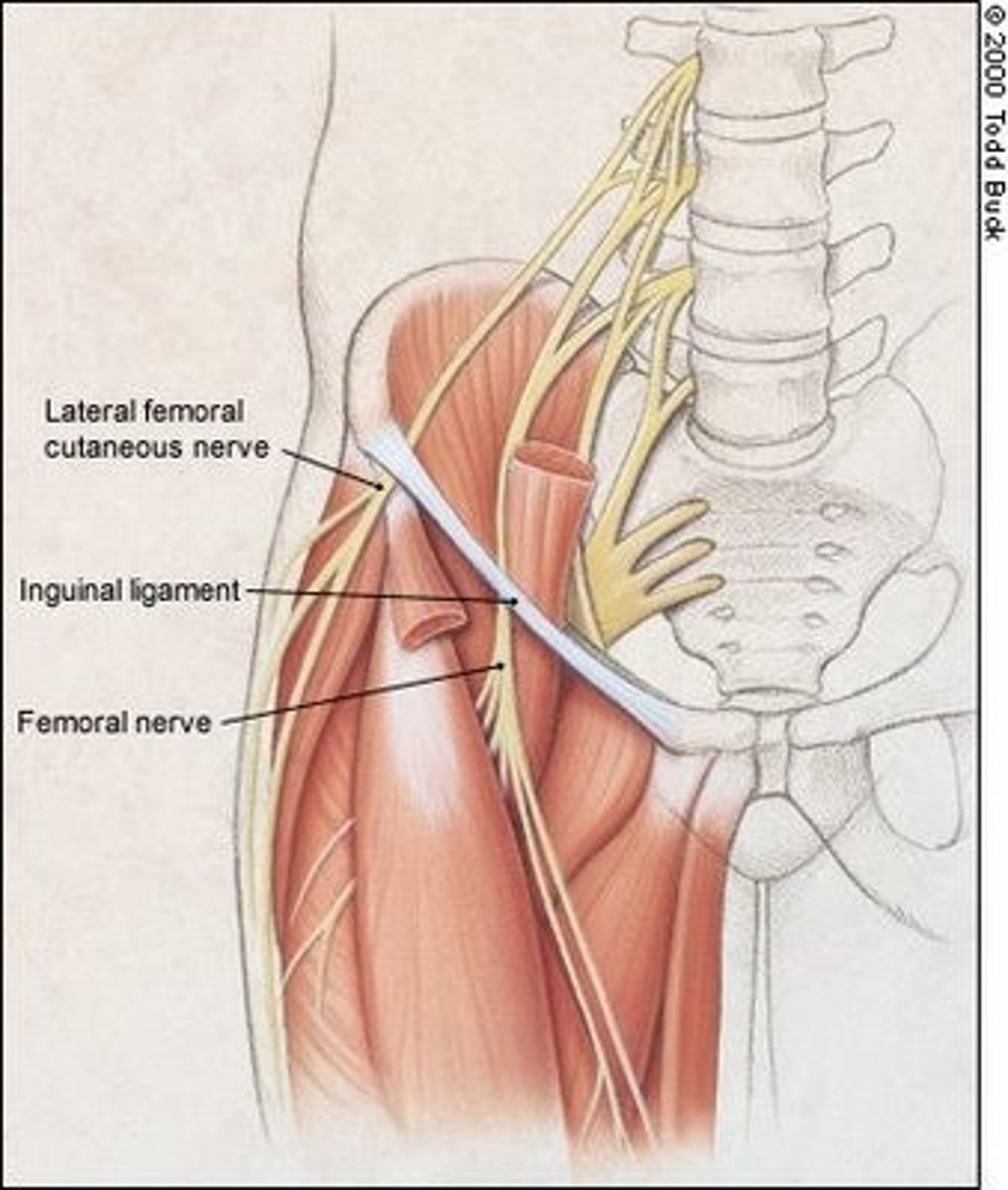
what does the femoral nerve (L2 – L4) of the lumbar plexus innervate?
iliacus muscle, and muscles of anterior compartment of thigh (flexors of the hip and extensors of the knee)
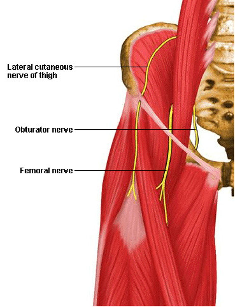
what is the course of the obturator nerve (L2 – L4) of the lumbar plexus?
emerges from medial border of psoas major and passes through the obturator foramen to the medial thigh
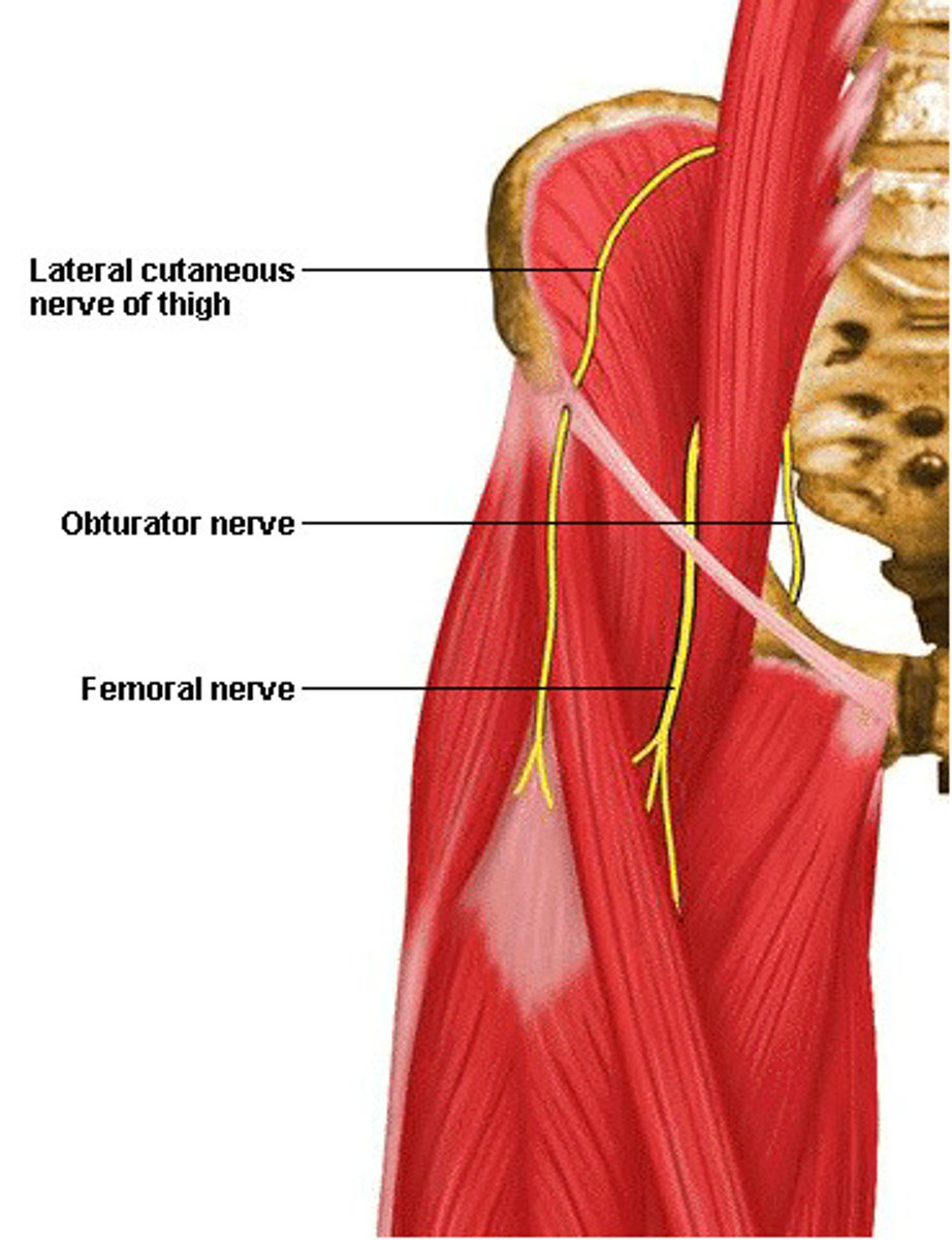
what does the obturator nerve (L2 – L4) of the lumbar plexus Innervate?
muscles of medial compartment of thigh (hip adduction)
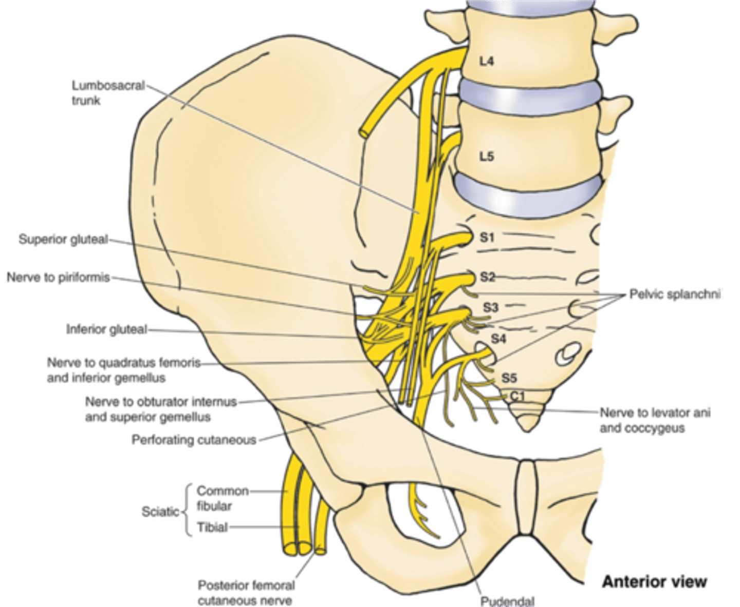
what is the course of the lumbosacral trunk (L4 – L5) of the lumbar plexus?
descends into the pelvis to contribute to the sacral plexus
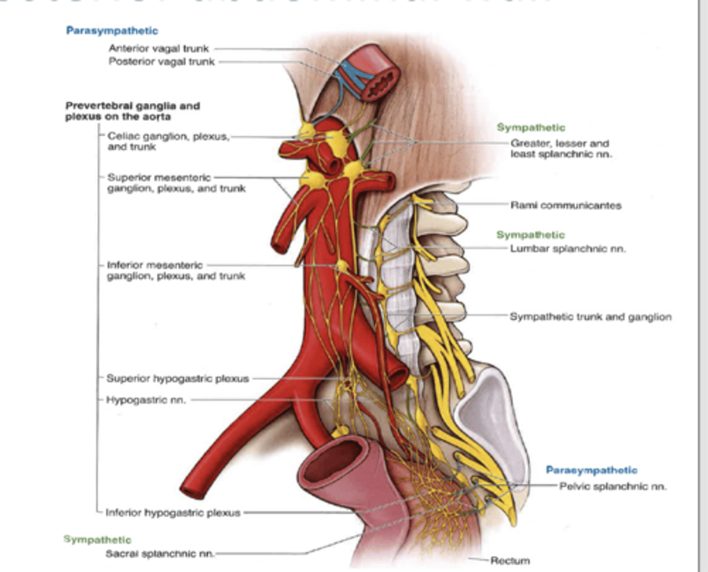
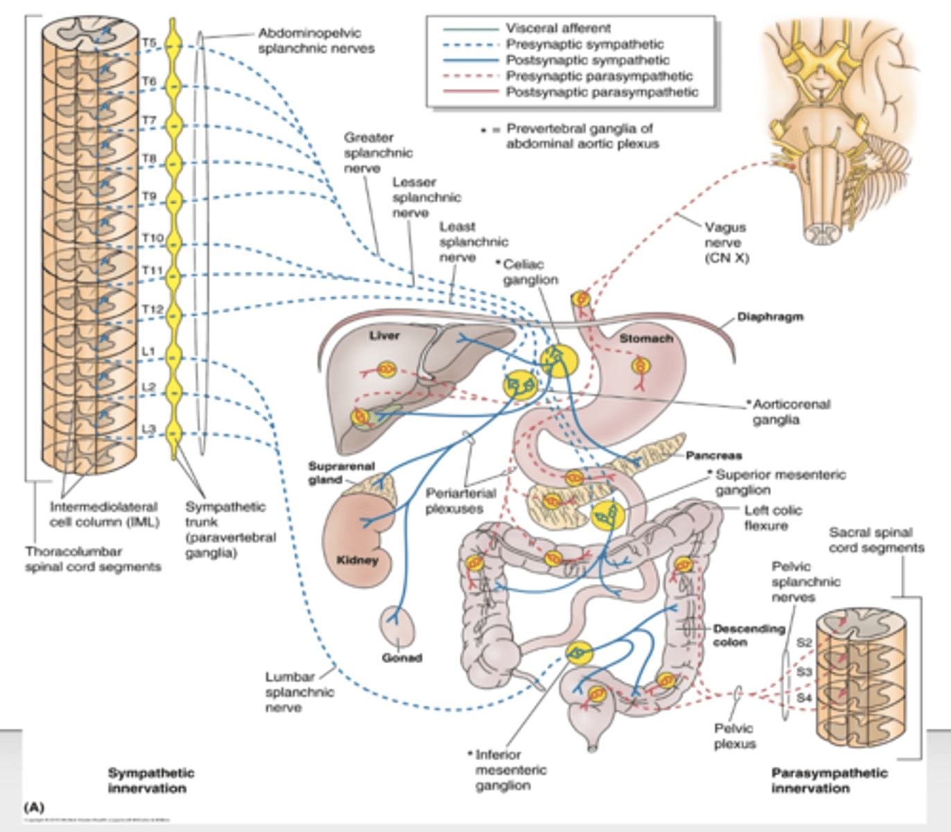
what are the nerves of sympathetic innervation to the posterior abdominal wall?
1. lower thoracic splanchnic nerves
2. lumbar splanchnic nerves (L1 - L3)
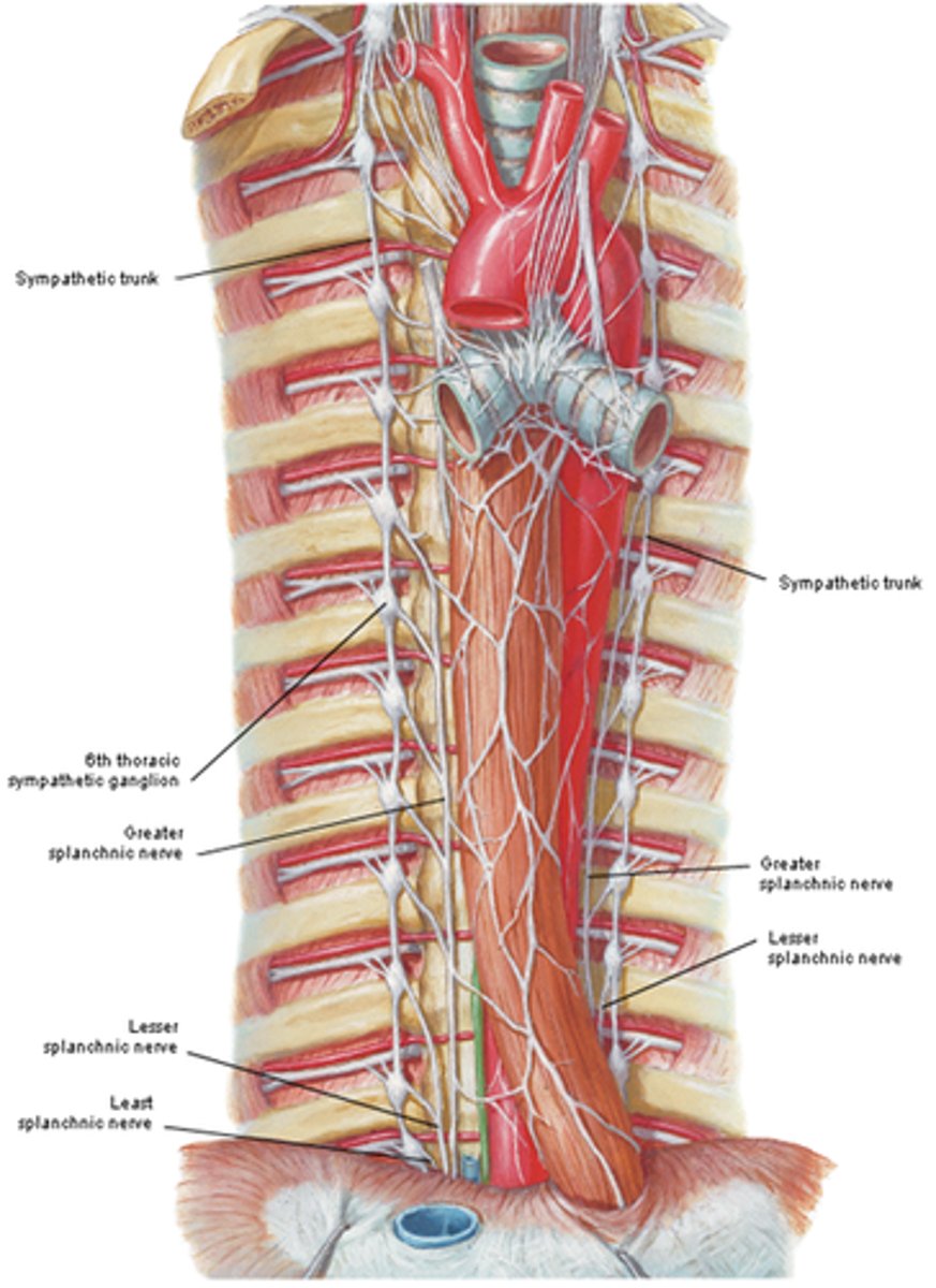
what nerves make up the lower thoracic splanchnic nerves?
greater (T5 – T10), lesser (T10 – T11), and least (T12) splanchnic nerves
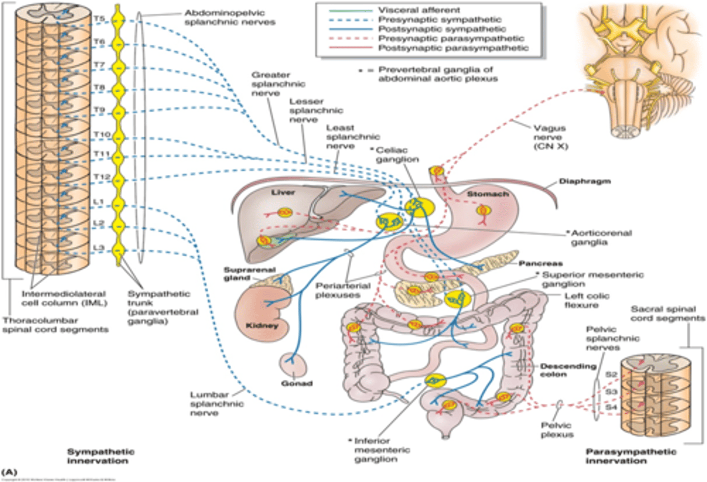
what do the lower thoracic splanchnic nerves convey presynaptic fibers to?
celiac, superior mesenteric, and aorticorenal SP ganglia
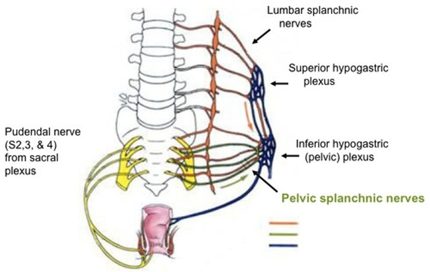
where do the lumbar splanchnic nerves (L1-L3) orginates from?
originate from medial portion of abdominal sympathetic trunk
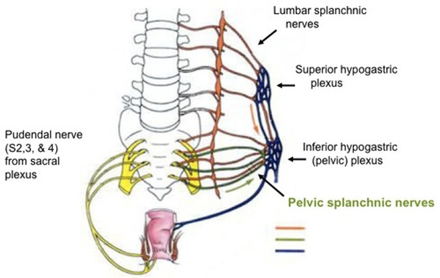
where does the lumbar splanchnic nerves (L1 – L3) convey presynaptic fibers to?
inferior mesenteric and superior hypogastric plexuses
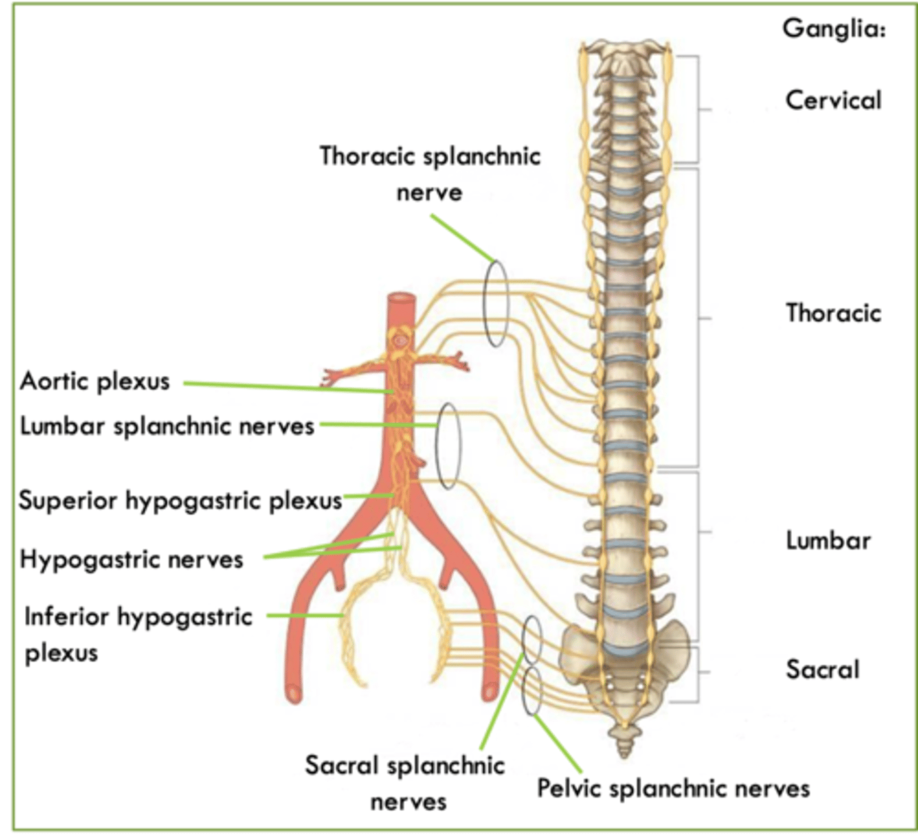
what is the parasympathetic innervation of the posterior abdominal wall?
1. anterior and posterior vagal trunks
2. pelvic splanchnic nerves
what do the anterior and posterior vagal trunks innervate?
innervate the GI tract as far down as the left colic (splenic) flexure
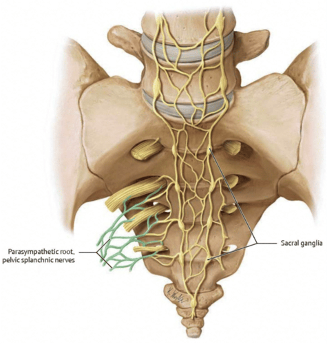
where do the pelvic splanchnic nerves originate from?
originates from anterior rami of spinal nerves S2 – S4
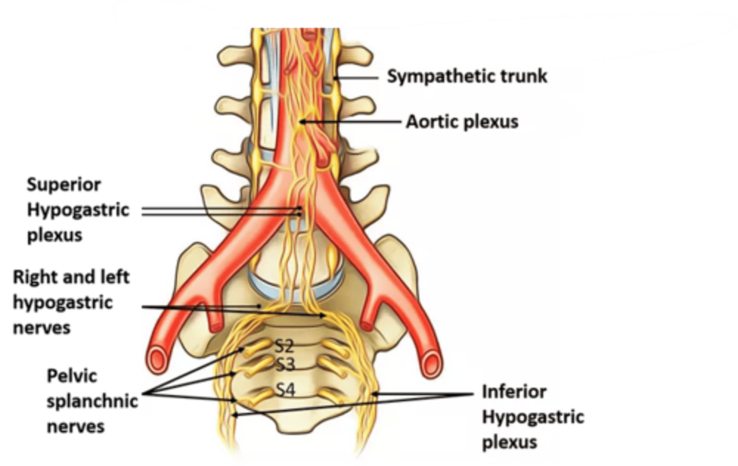
what does the pelvic splanchnic nerve convey presynaptic fibers to? what does it supply?
convey presynaptic fibers to: inferior hypogastric plexus, and supplies the GI tract inferior to the left colic flexure
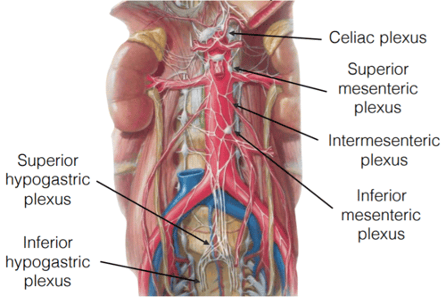
what are the abdominal automonic plexus comprised of? what does it surround?
comprised of nerve networks of SP and PSP fibers, surrounding the abdominal aorta and its major branches
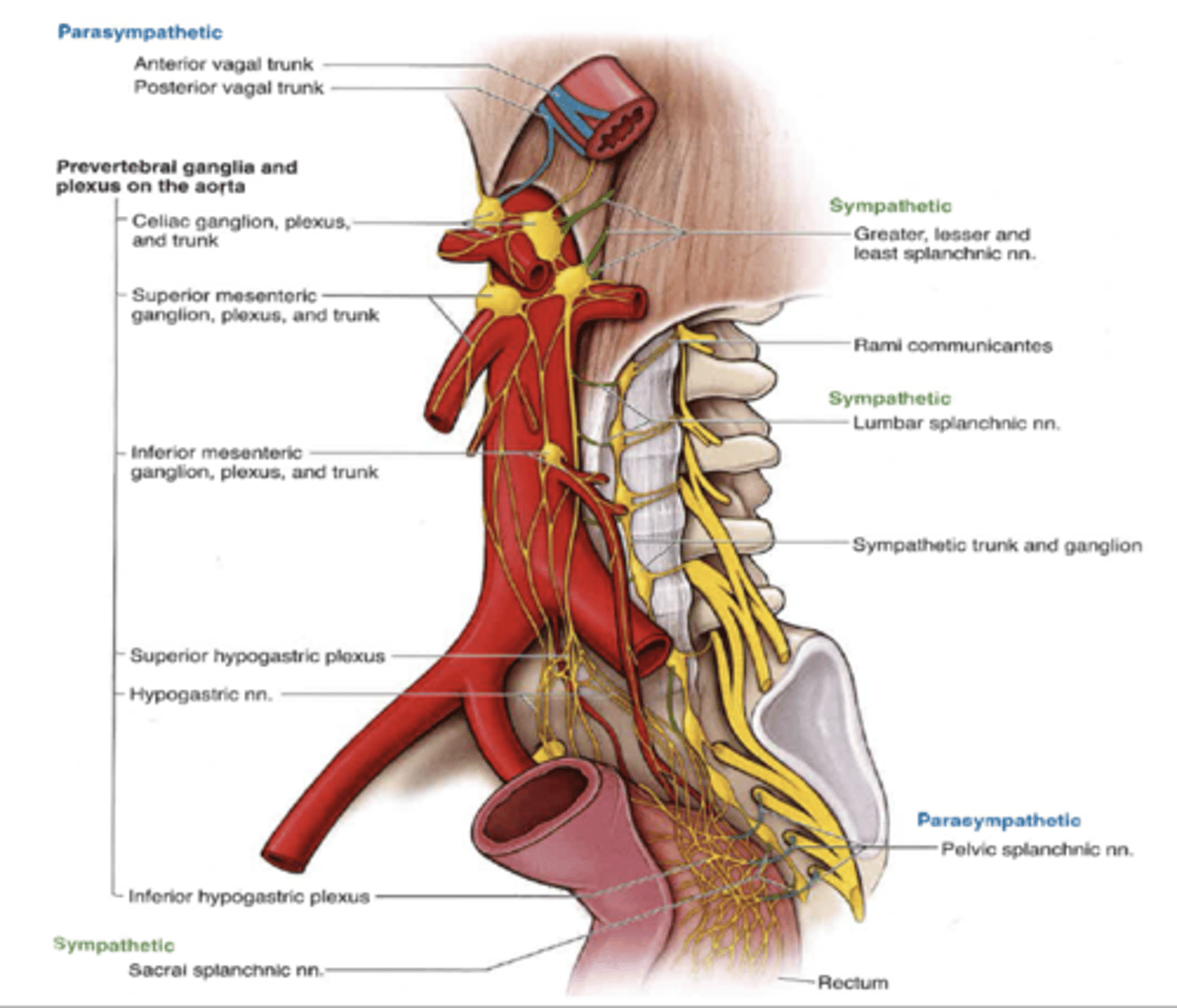
what abdominal autonomic plexus are interconnected?
1. celiac
2. superior mesenteric
3. inferior mesenteric plexuses
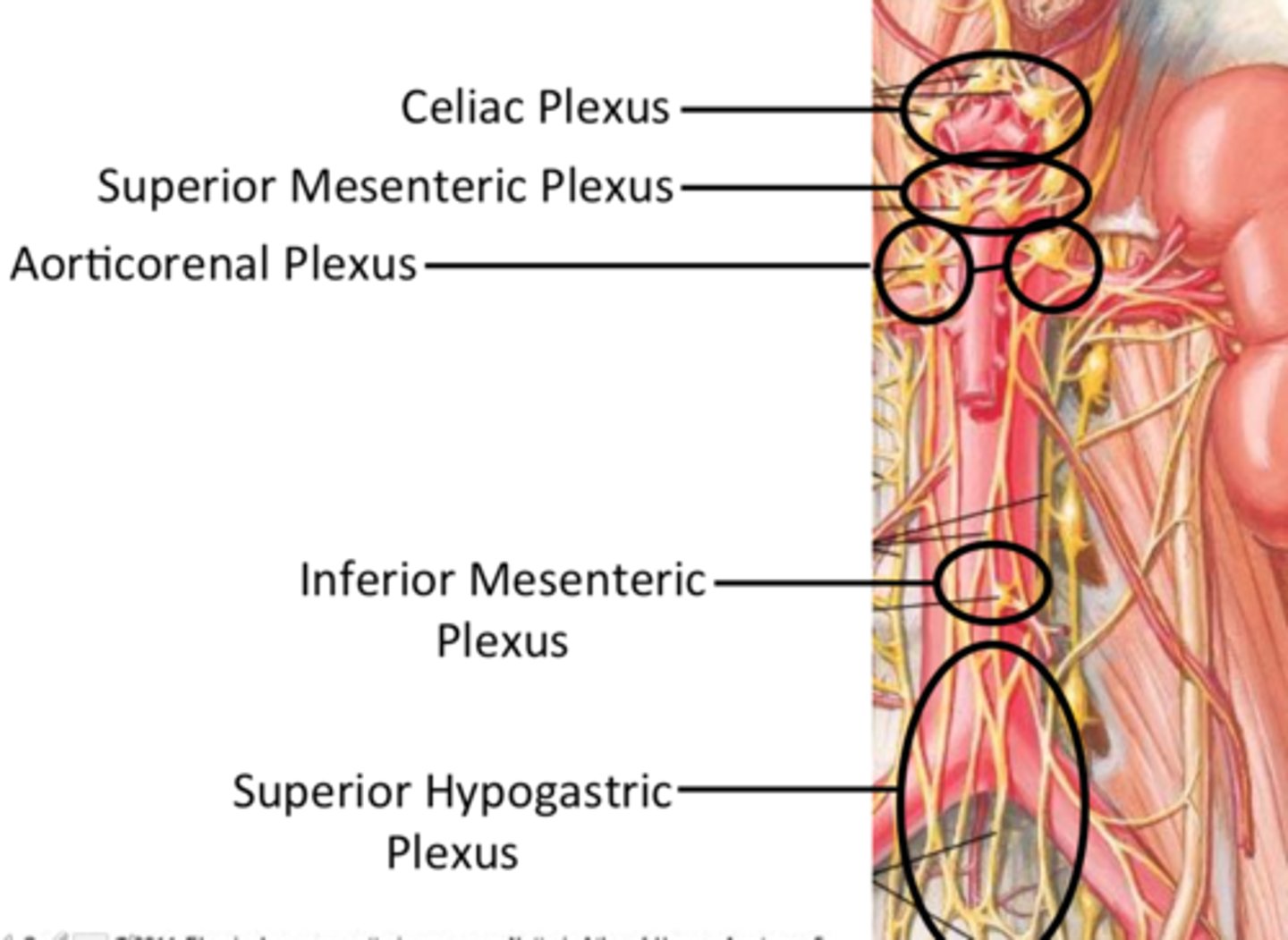
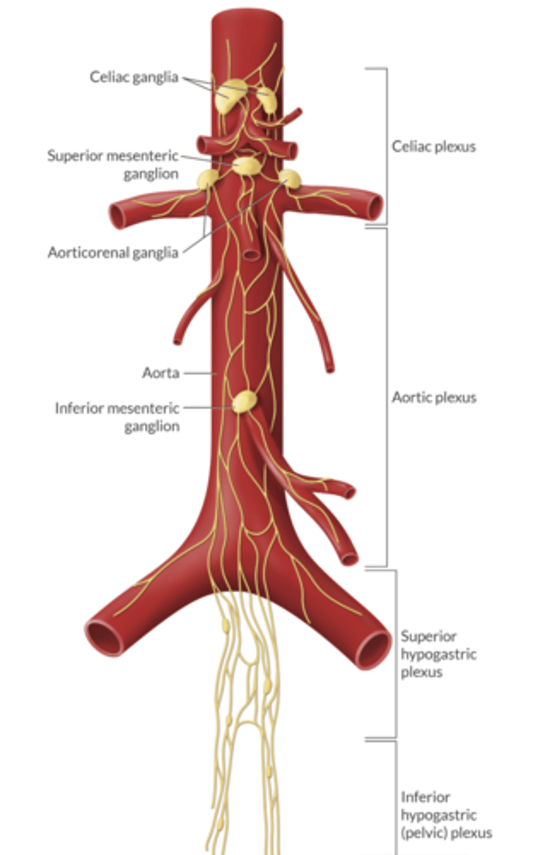
where is the location of the celiac ganglia and plexus?
around origin of celiac trunk
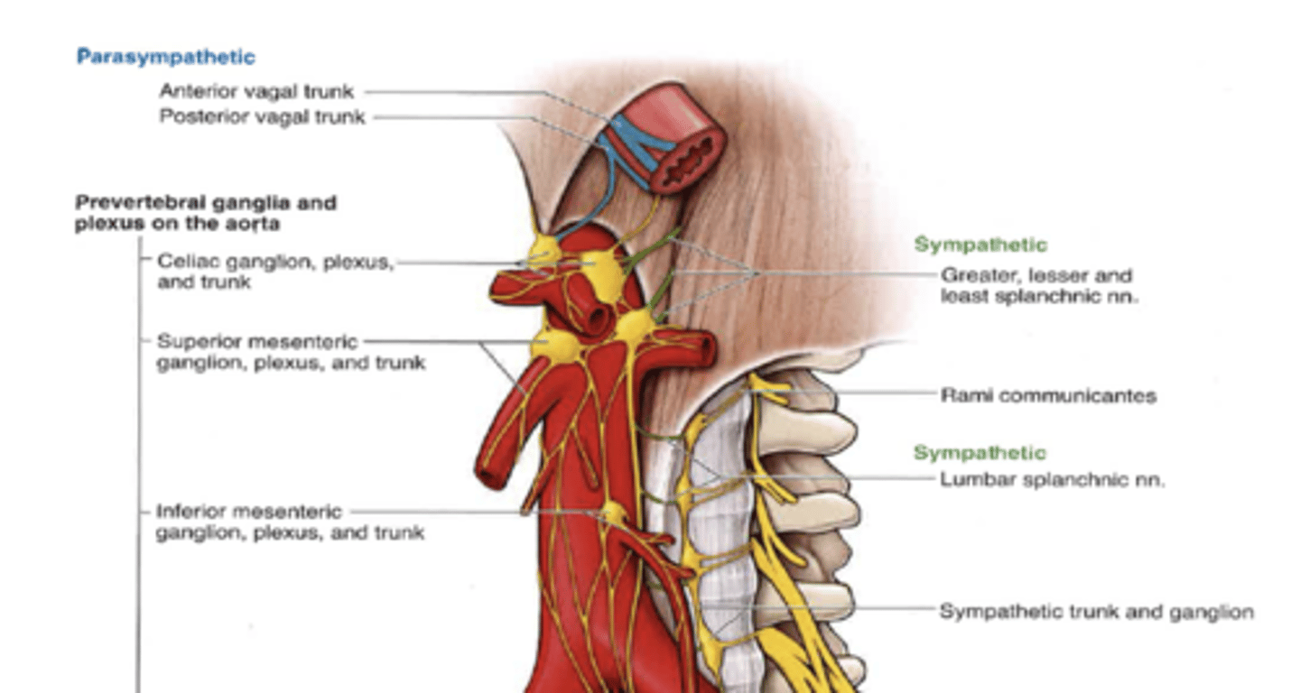
what are the sympathetic nerves of the celiac ganglia and plexus
greater & lesser splanchnic nerves
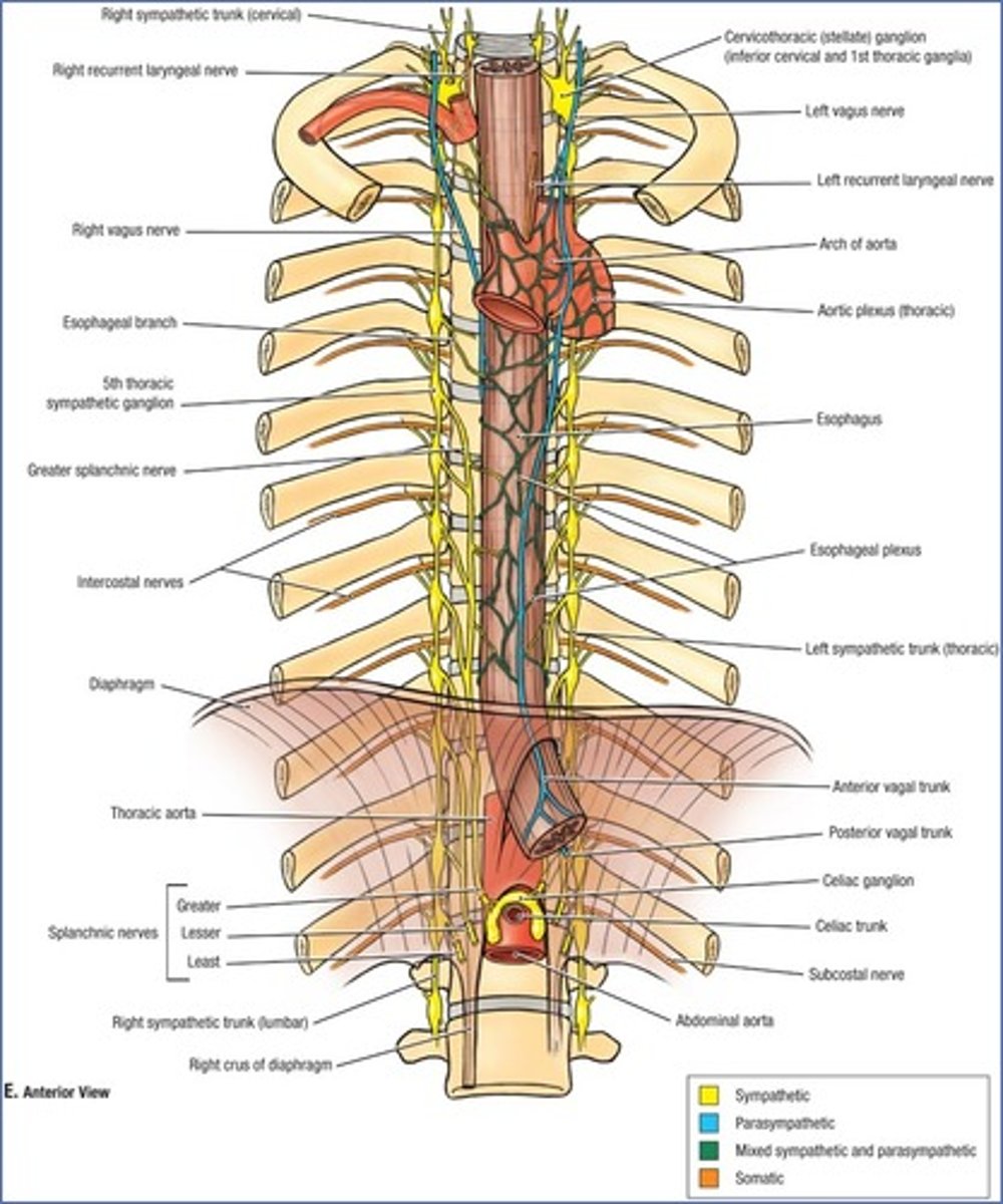
what are the parasympathetic nerves of the celiac ganglia and plexus?
vagus nerve
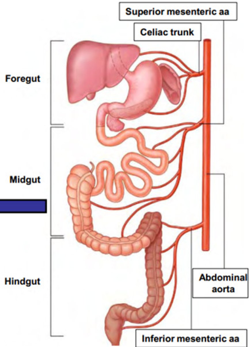
what does the celiac ganglia and plexus innervate?
liver, gallbladder, stomach, spleen, pancreas, and proximal part of duodenum
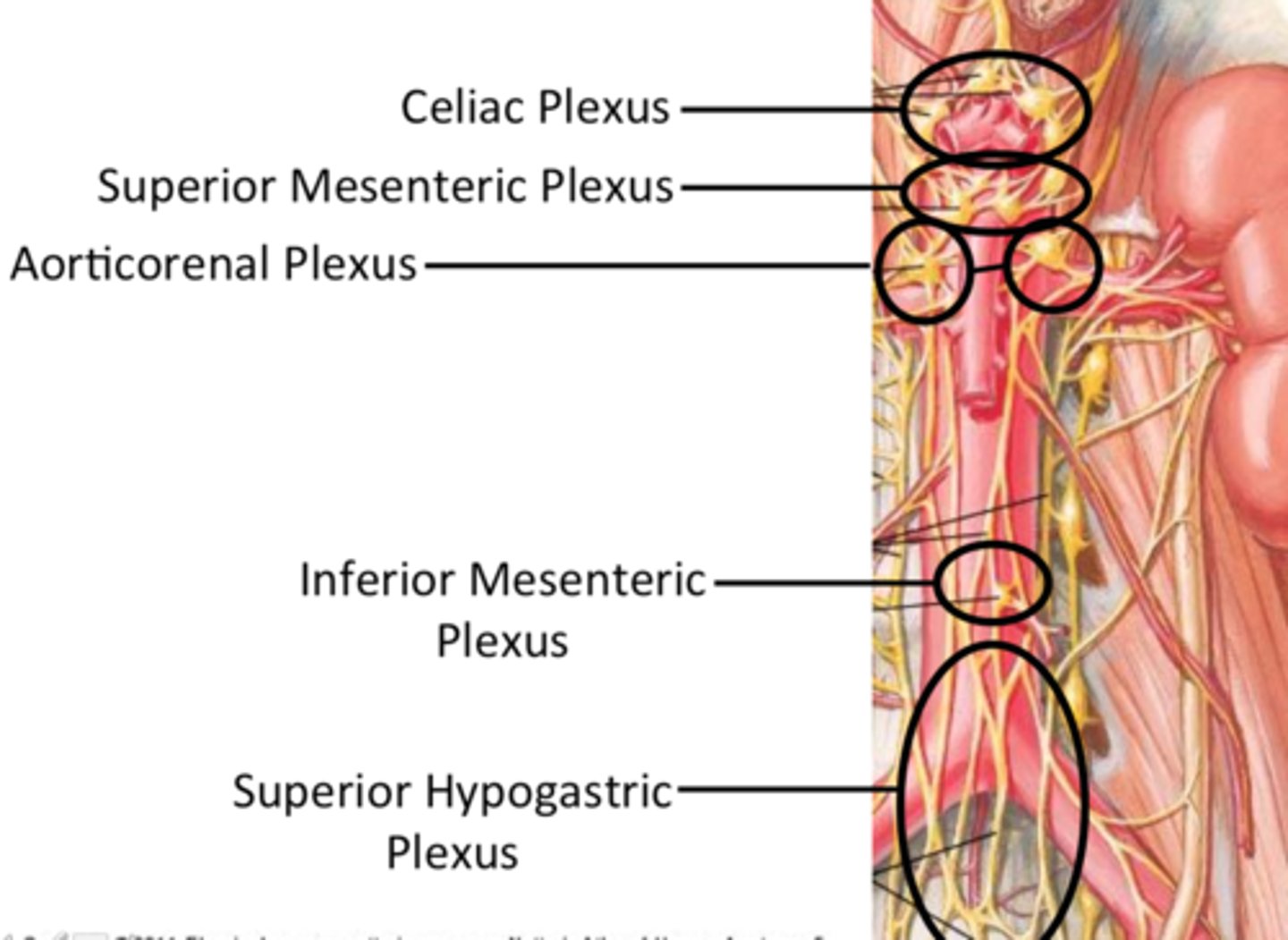
where is the location of the superior mesenteric ganglia and plexus?
around origin of SMA
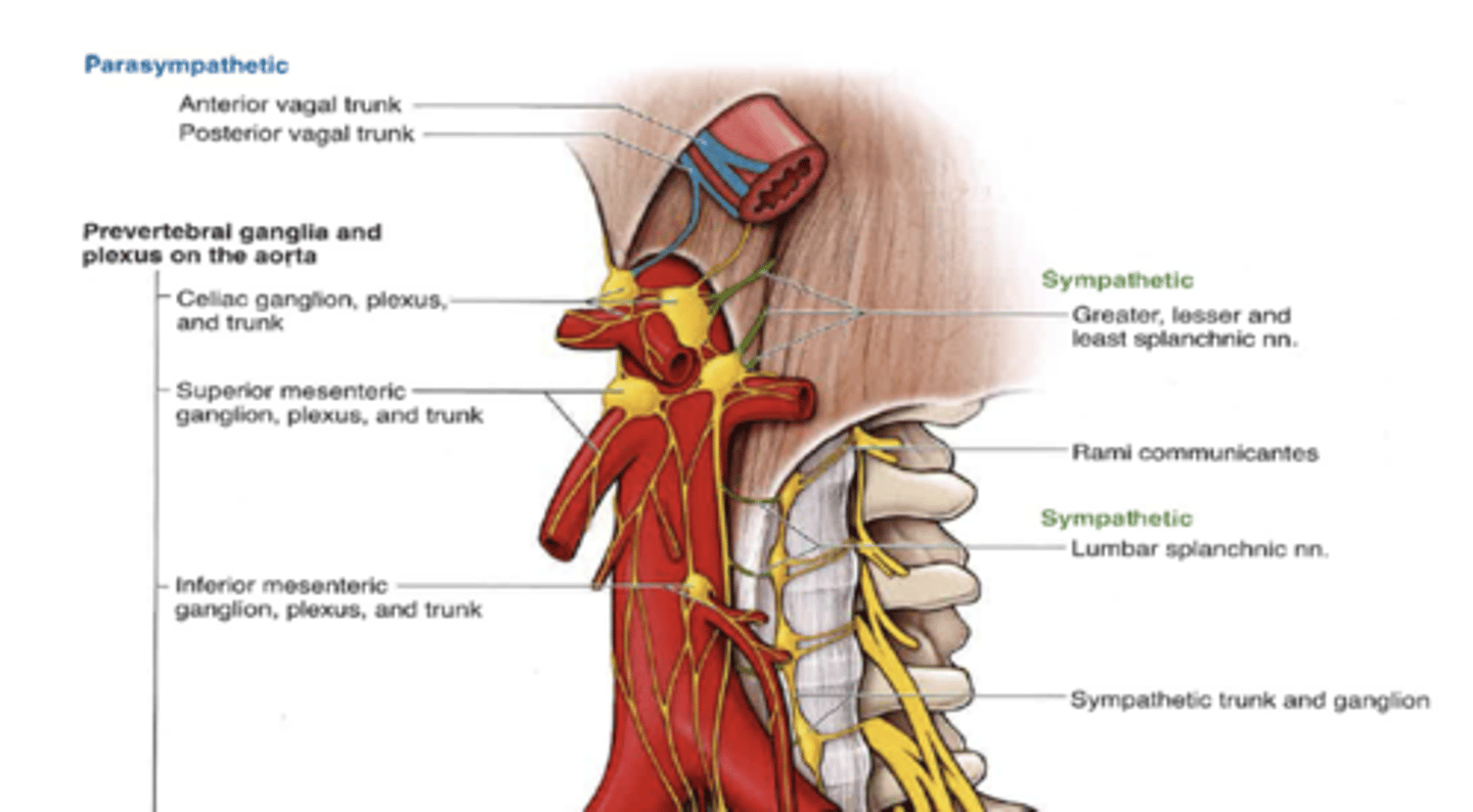
what is the superior mesenteric ganglia and plexus sympathetically innervated by?
lesser & least splanchnic nerves
what is the superior mesenteric ganglia and plexus parasympathetically innervated by?
vagus nerve
what does the superior mesenteric ganglia innervate?
head of pancreas, distal duodenum, jejunum, ileum, cecum, ascending and transverse colon (MIDGUT)
where is the location of the inferior mesenteric ganglia and plexus?
around origin of IMA
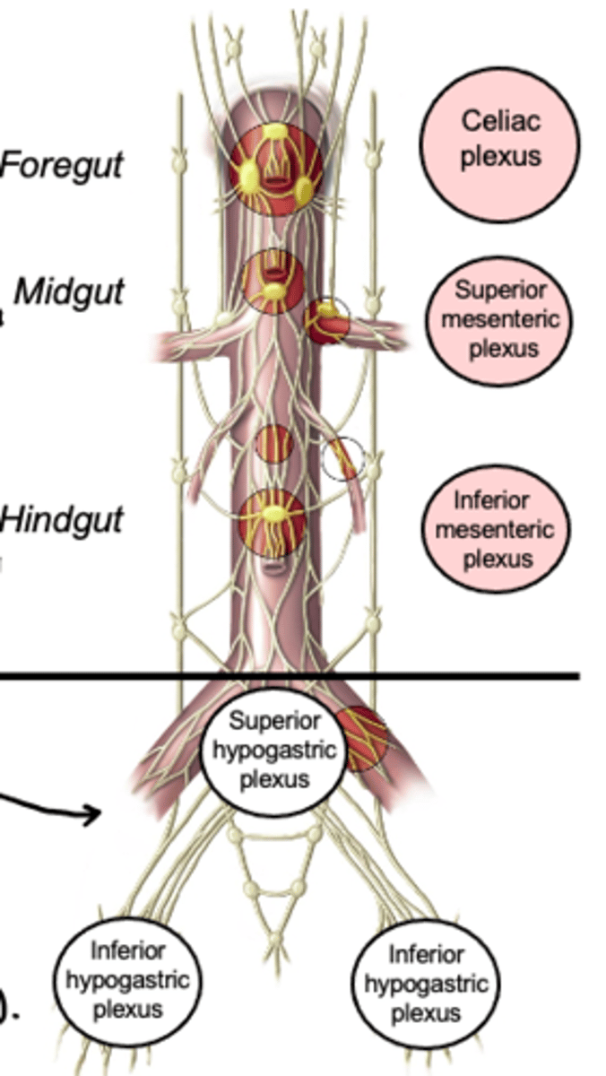
what is the Inferior mesenteric ganglia and plexus sympathetically innervated by?
lumbar splanchnic (L1 - L3)
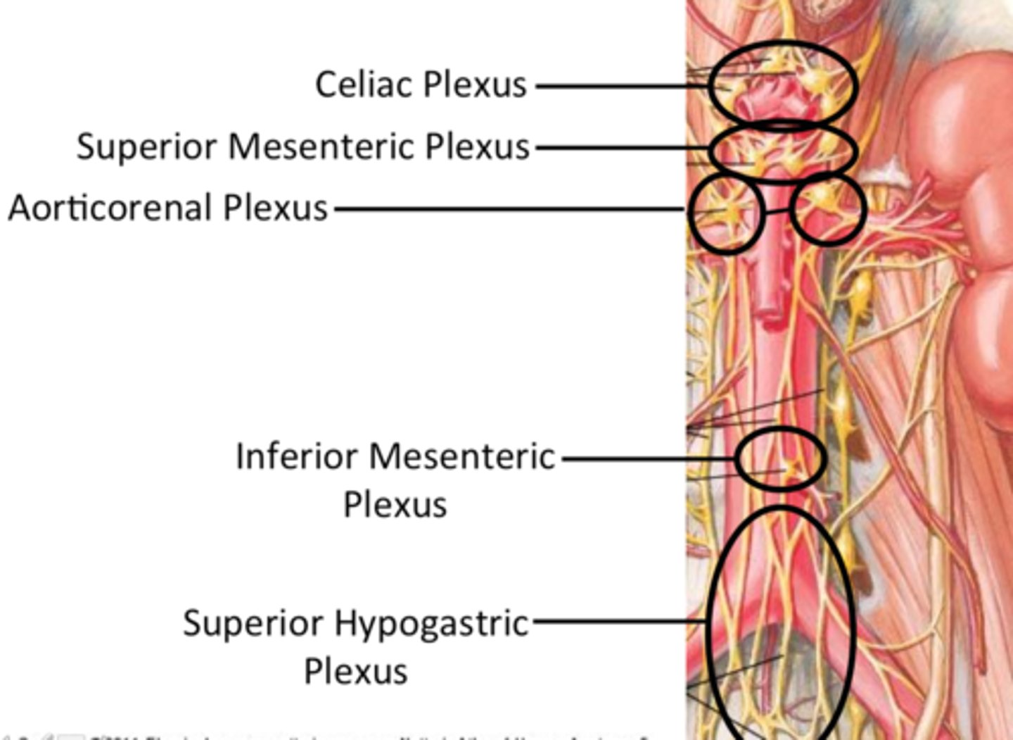
what is the inferior mesenteric ganglia and plexus parasympathetically and sympathetically (mixed) innervated by?
superior mesenteric & superior hypogastric plexuses
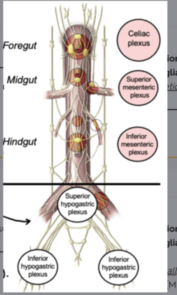
what does the Inferior mesenteric ganglia and plexus innervate?
descending colon, sigmoid colon, and upper rectum (hindgut)
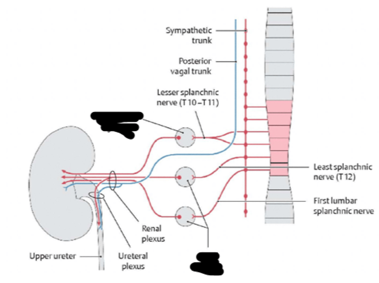
what is the location of the aorticorenal ganglion and renal plexus?
around origin of renal artery
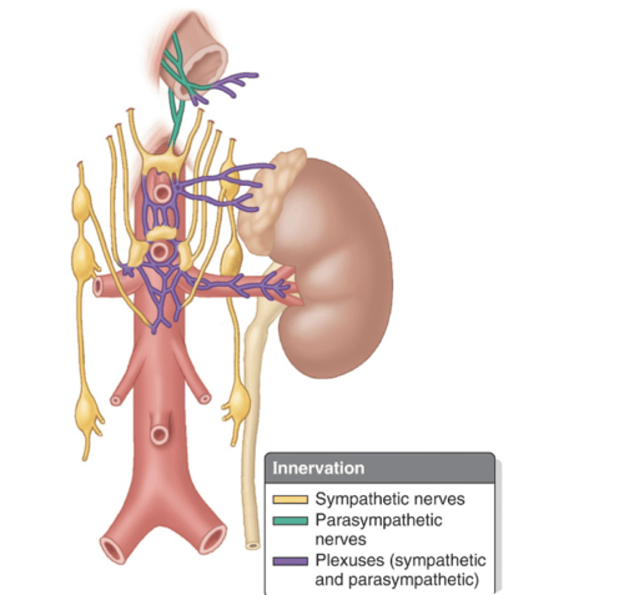
what is the aorticorenal ganglion and renal plexus sympathetically innervated by?
least splanchnic nerve (aorticorenal ganglion)
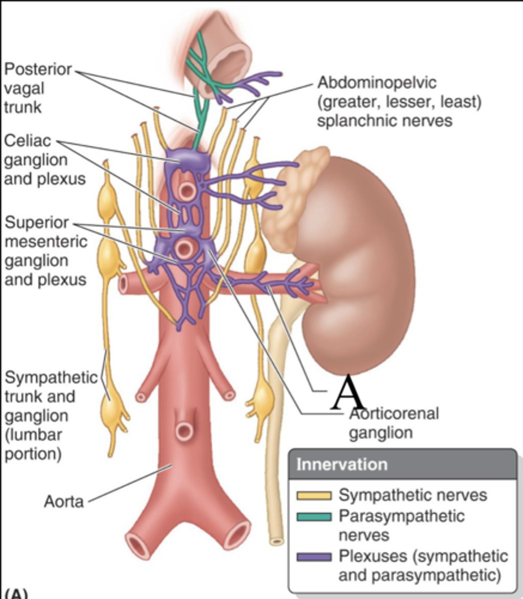
what is the aorticorenal ganglion and renal plexus parasympathetically innervated by?
vagus nerve
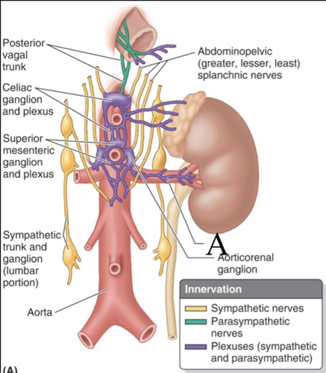
what is the aorticorenal ganglion and renal plexus renal plexus (mixed) innervated by?
celiac plexus, the aorticorenal ganglion, and the superior mesenteric plexus
what is the aorticorenal ganglion and renal plexus innervate?
adrenal glands, kidneys, and proximal ureters
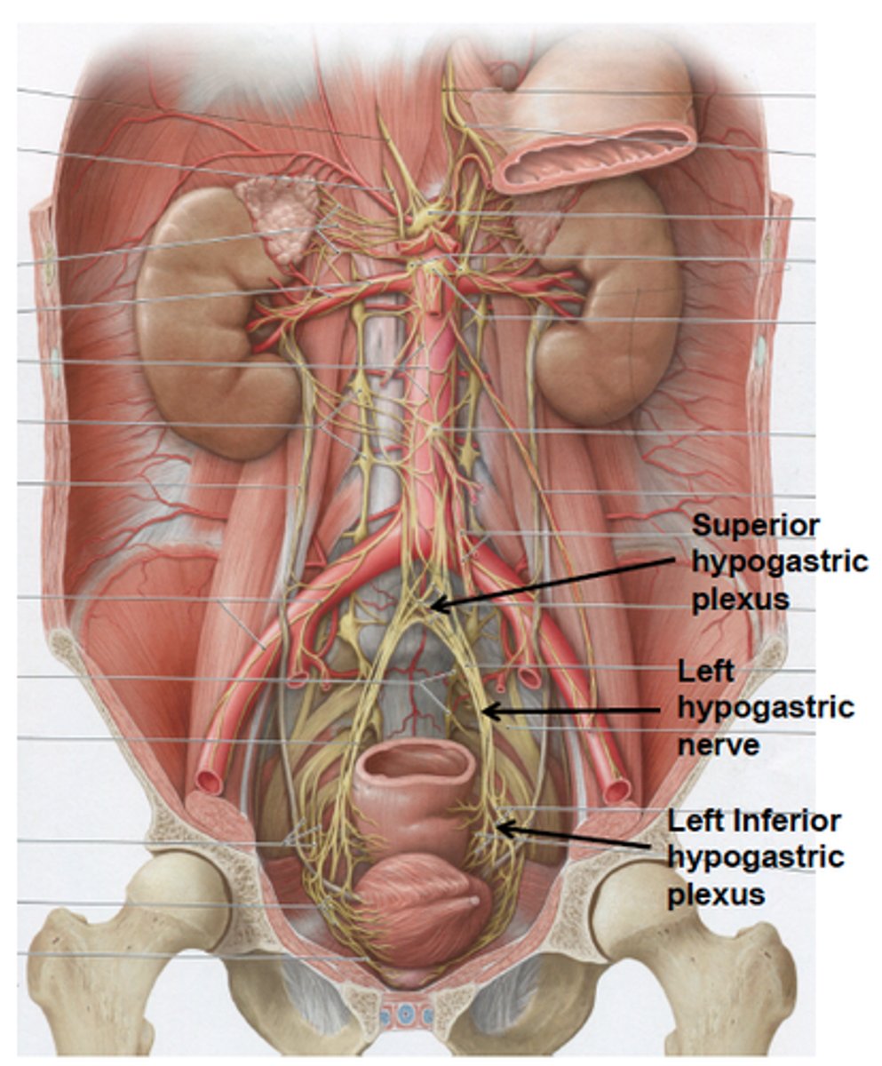
what is the location of the superior hypogastric plexus? what is it joined by?
inferior to bifurcation of aorta. it is joined to the inferior hypogastric plexus by the hypogastric nerves
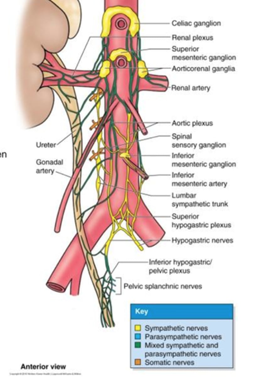
what is the superior hypogastric plexus sympathetically innervated by?
lumbar splanchnic (L1 - L3)
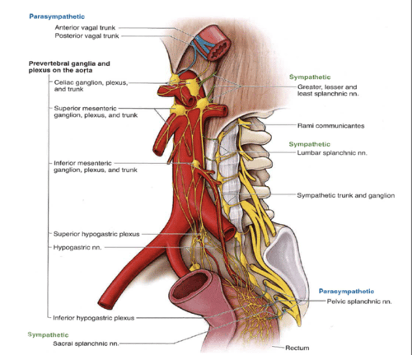
what is the superior hypogastric plexus parasympathetically sympathetically (mixed) innervated by?
inferior mesenteric plexus, inferior hypogastric plexus (mostly PSP from pelvic splanchnic nn.)
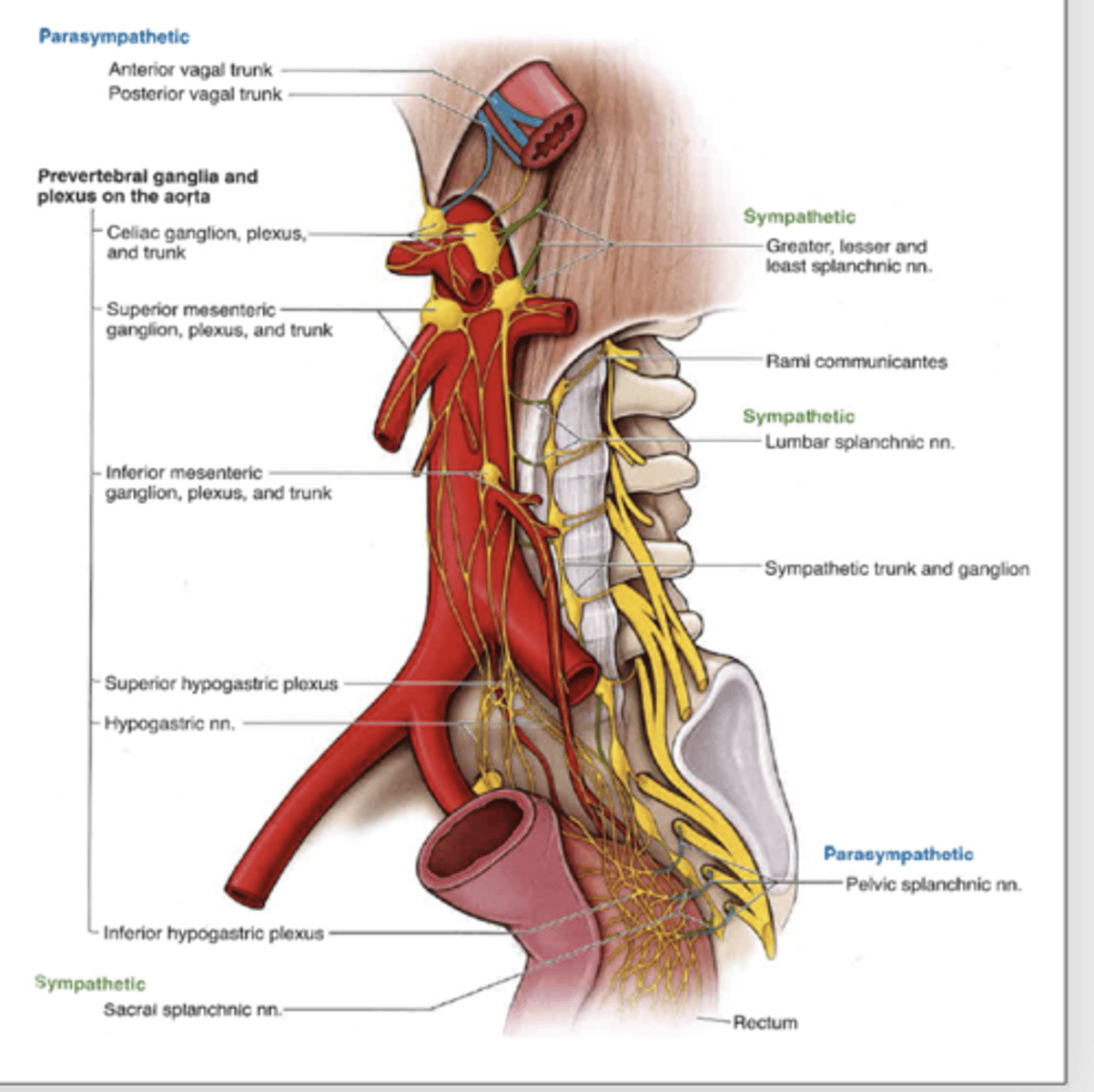
what does the superior hypogastric plexus innervate?
ureteric and testicular plexuses
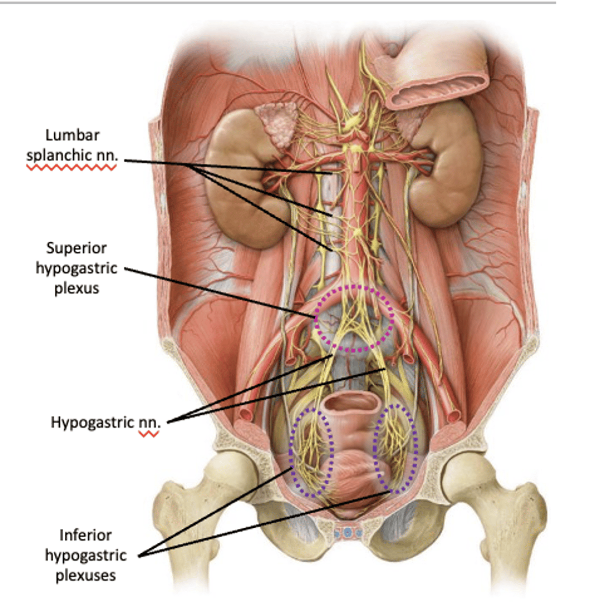
where is the location of the inferior hypogastric plexus?
on both sides of the rectum, cervix of uterus, and urinary bladder
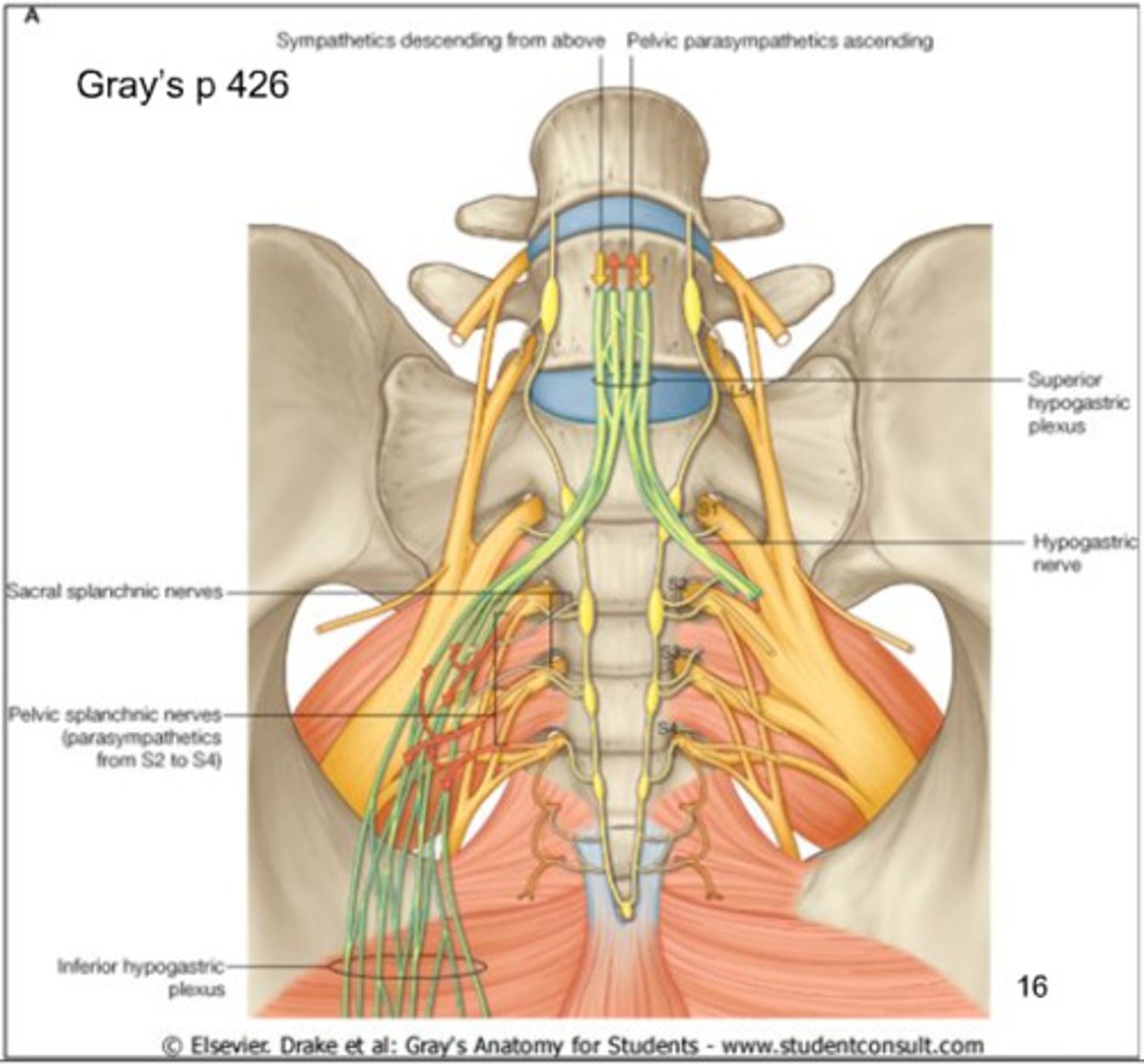
what is the Inferior hypogastric plexus sympathetically innervated by?
sacral splanchnic nerve
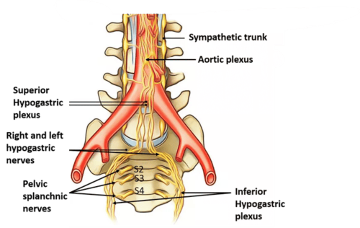
what is the inferior hypogastric plexus parasympathetically innervated by?
pelvic splanchnic nerve
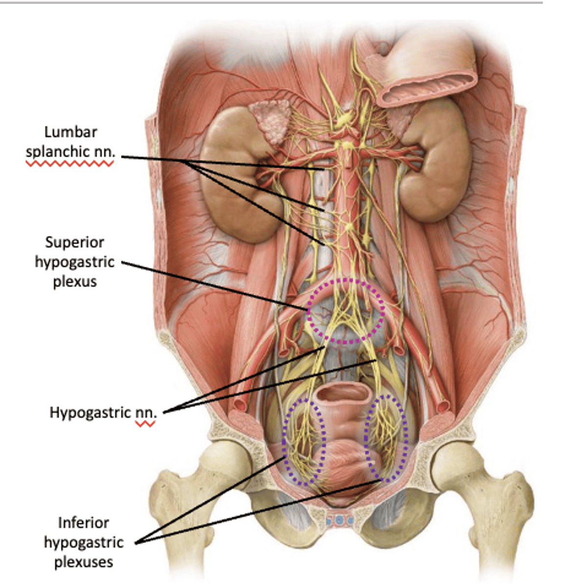
what does the inferior hypogastric plexus innervate?
rectal and vesical plexuses
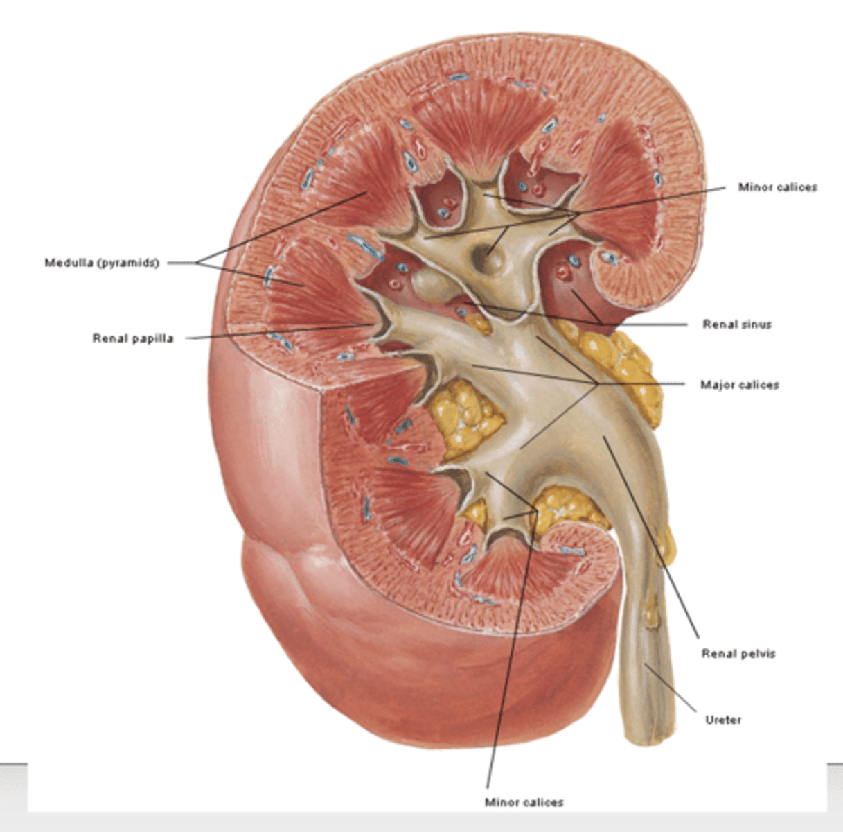
what kind of organ is the kidneys?
retroperitoneal on the posterior abdominal wall
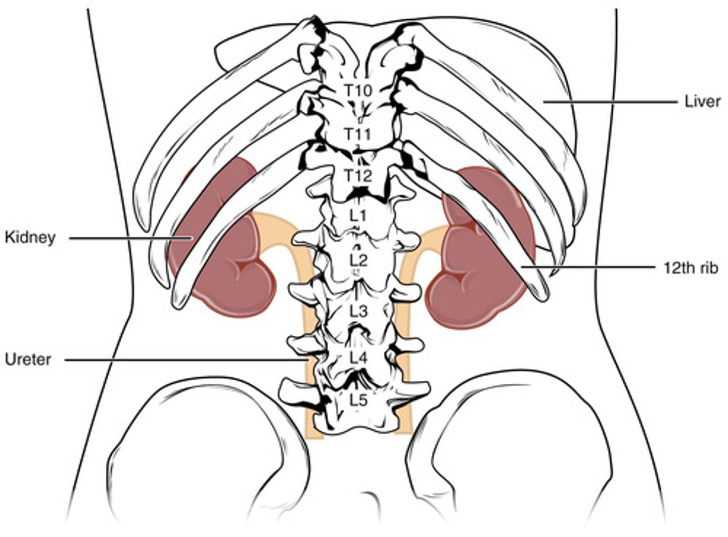
where is the kidney located? at what level vertebrae are they?
one on each side of the vertebral column at the level of T12 – L3 vertebrae
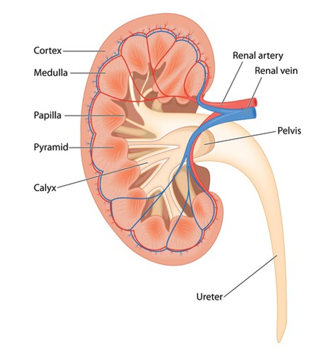
what is the function of the kidneys?
filters blood, and maintains fluid, salt, and pH balance
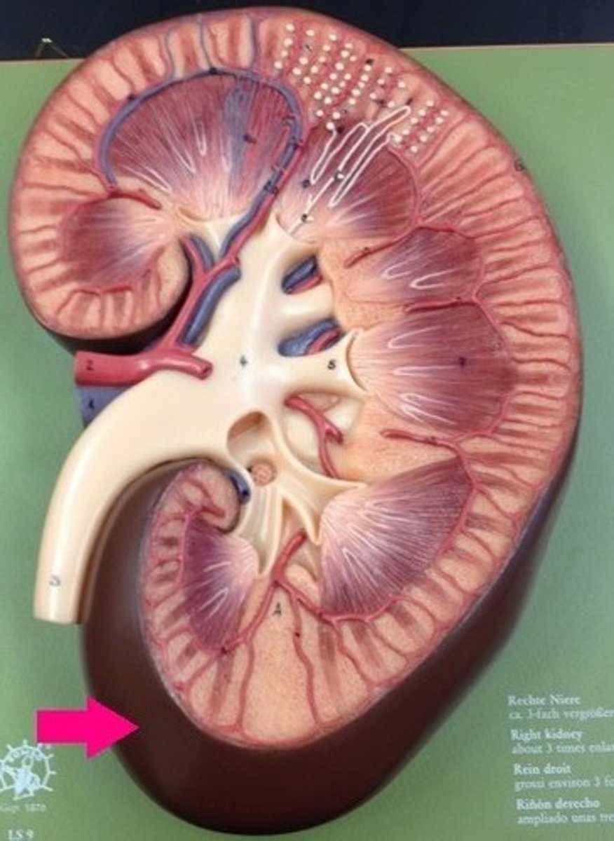
what is the renal capsule of the kidney?
tough, fibrous layer
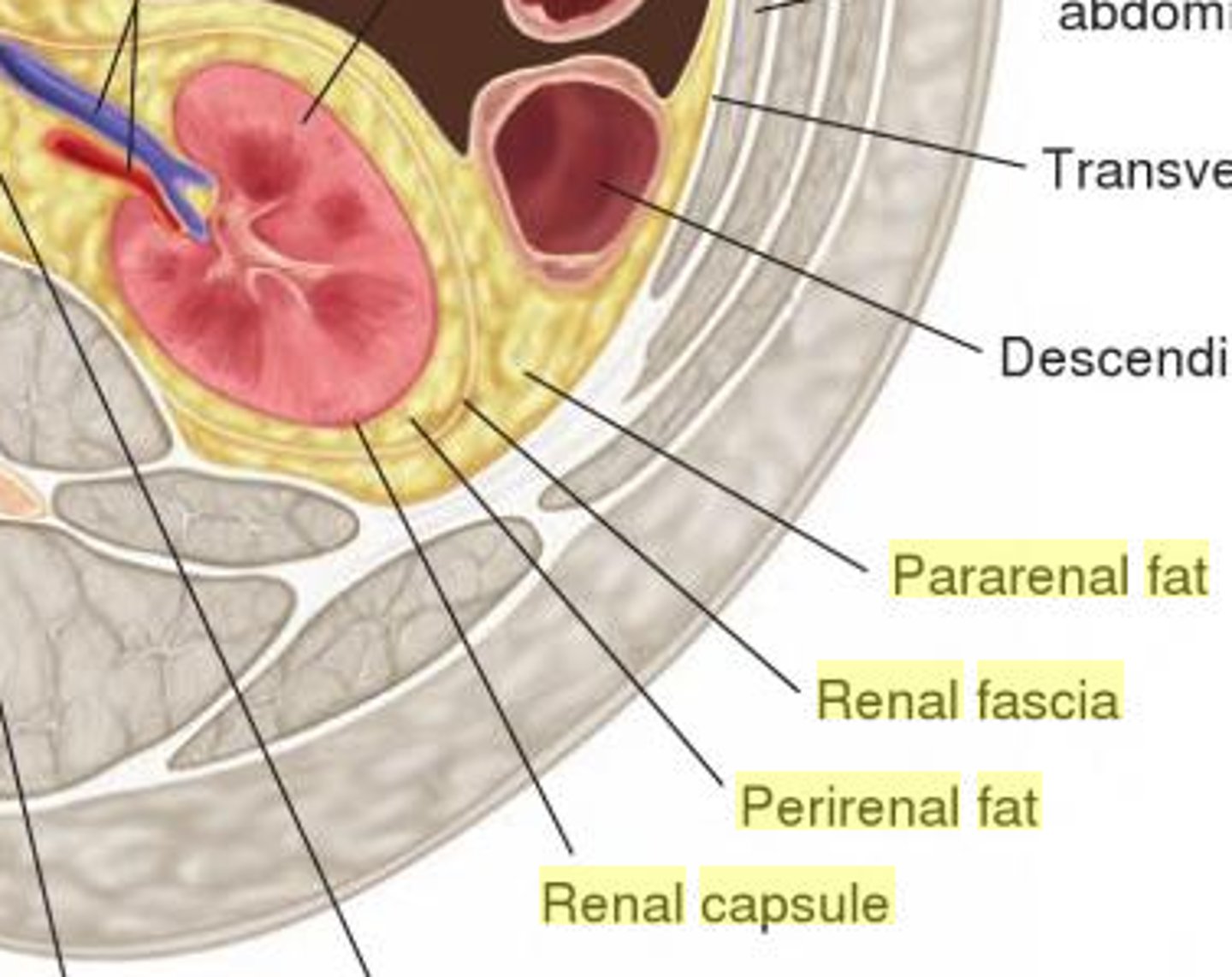
what does the perinephric fat of the kidney surround?
surrounds kidneys and their vessels
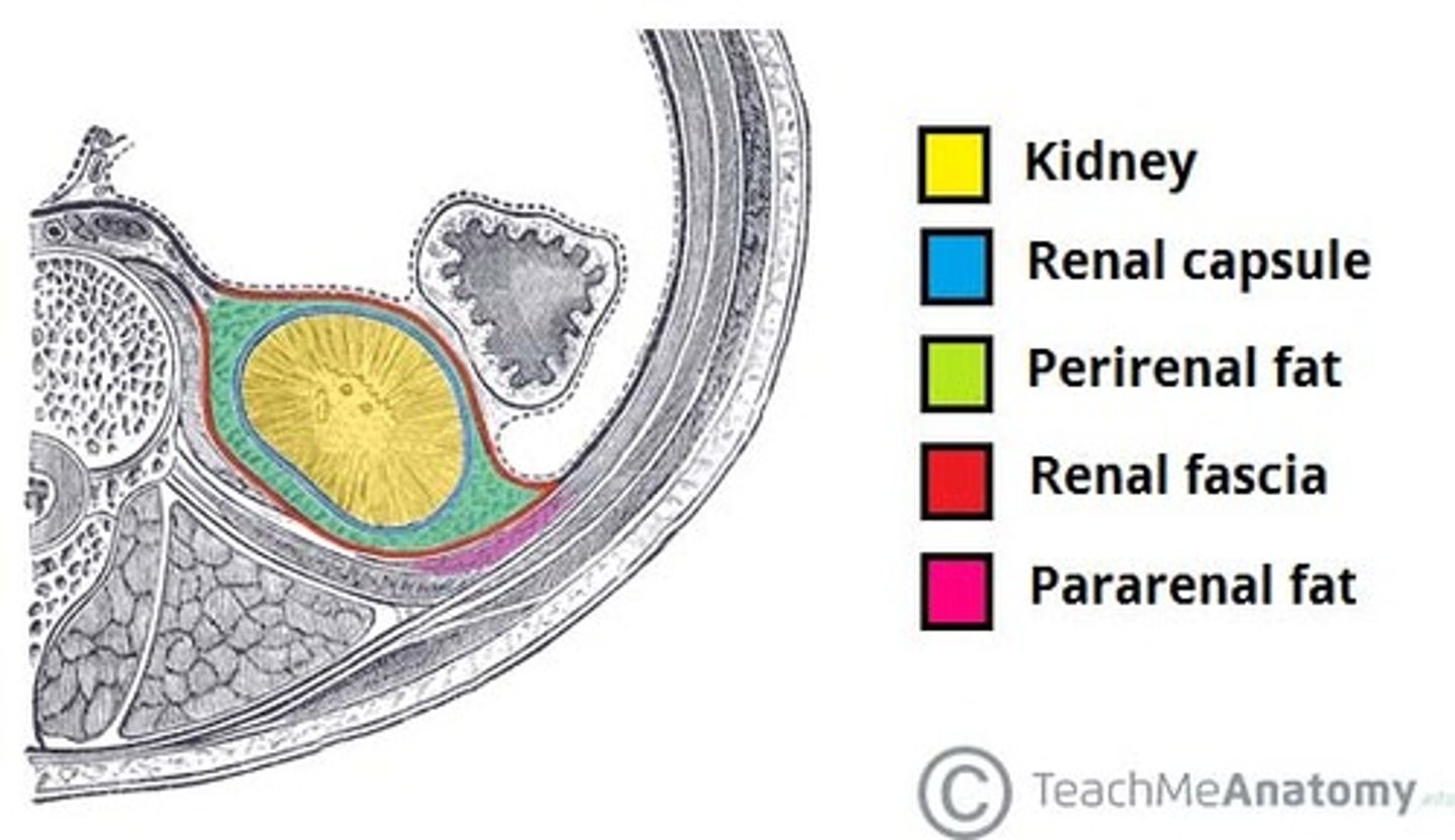
what is the renal fascia of the kidney? what is it around?
membranous layer around kidneys, adrenals, and perinephric fat
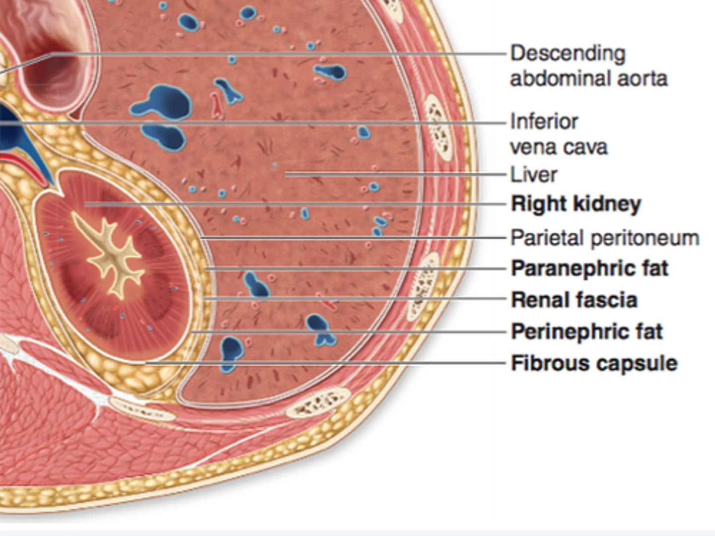
what is paranephric fat of the kidney? what is it external to? what does it attach?
extraperitoneal fat, external to renal fascia that attaches kidneys to posterior wall
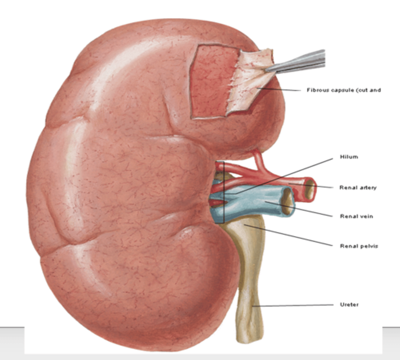
what is the renal hilum of the kidney?
vertical cleft where ureter, renal vessels, and nerves enter and leave the renal sinus
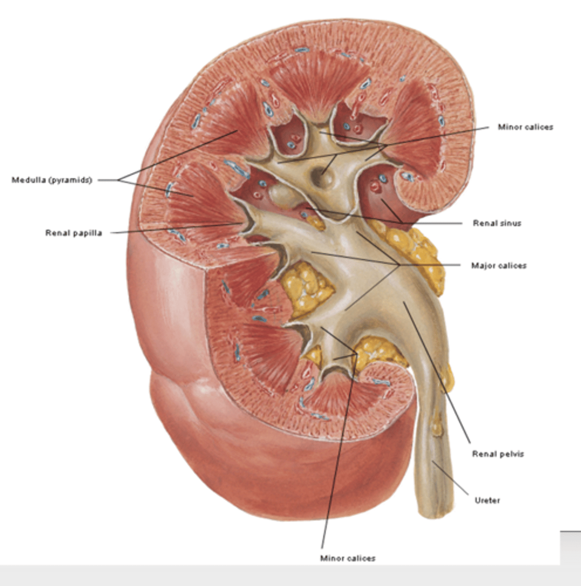
what is the renal sinus of the kidneys?
hollow center that receives renal vessels, nerves, and ureters
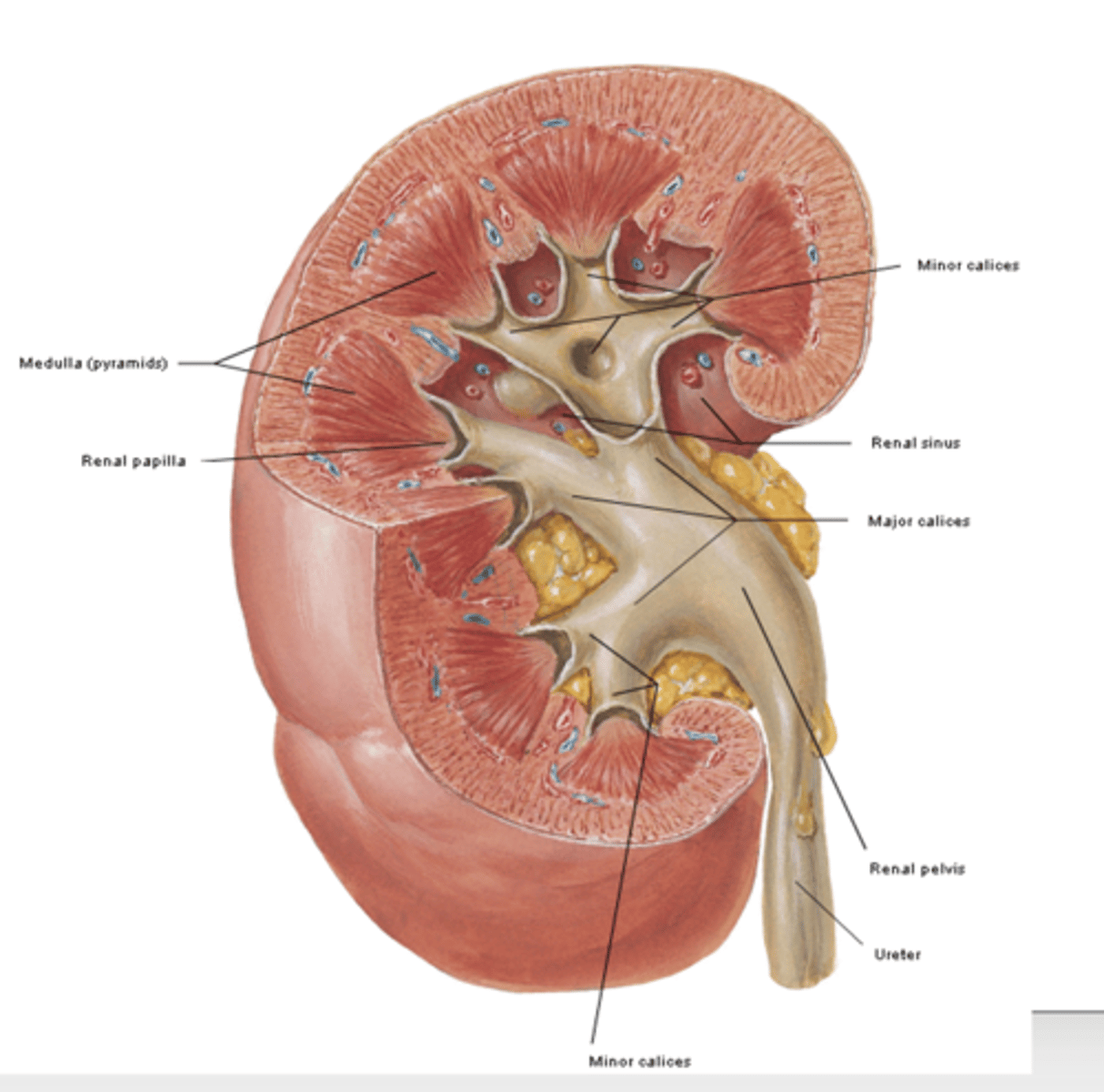
what is the renal pelvis of the kidneys? what does it receive?
flattened, funnel-shaped expansion of the superior end of the ureter. receives the major calices
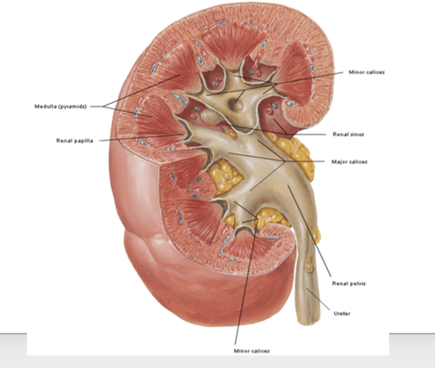
what are the major and minor calices of the kidneys? what do they form and go into?
minor calices (usually 3) > form the major calices > ureters
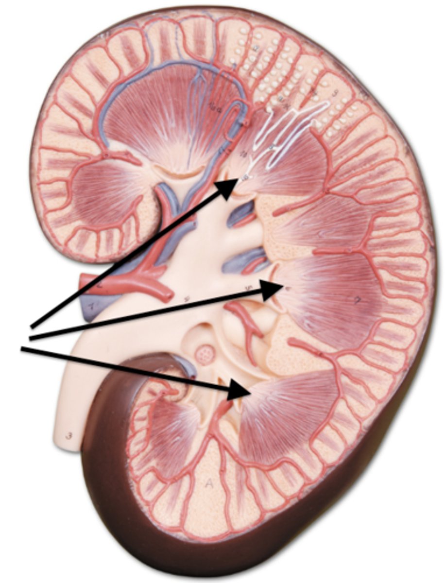
what is the renal papilla of the kidneys? what do they empty into?
where medullary pyramids empty into the minor calices
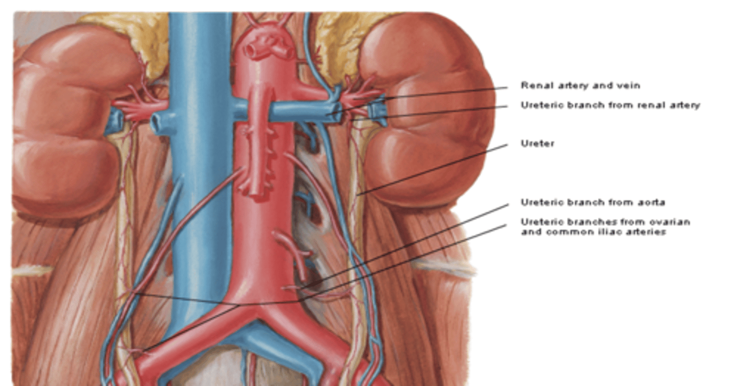
what is the vasculature of the kidneys? what are they innervated by?
vasculature
•renal arteries and veins
innervation
•renal plexus
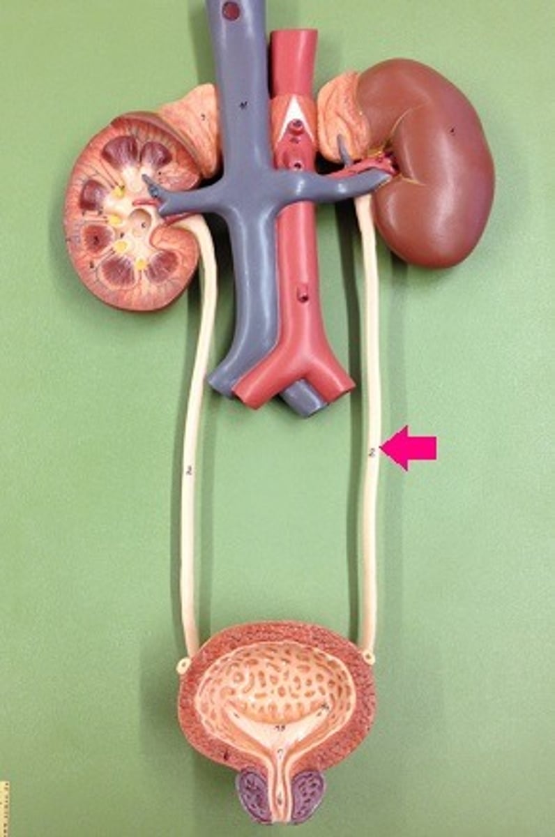
what are the ureters?
muscular ducts (25 - 30 cm long)
what does the ureters run inferiority from? what do they run anterior to? what do they pass over? What do they finally join?
that run inferiorly from the apices of the renal pelves, anterior to transverse processes of lumbar vertebrae, and passing over the common iliac vessels at the pelvic brim to join the urinary bladder
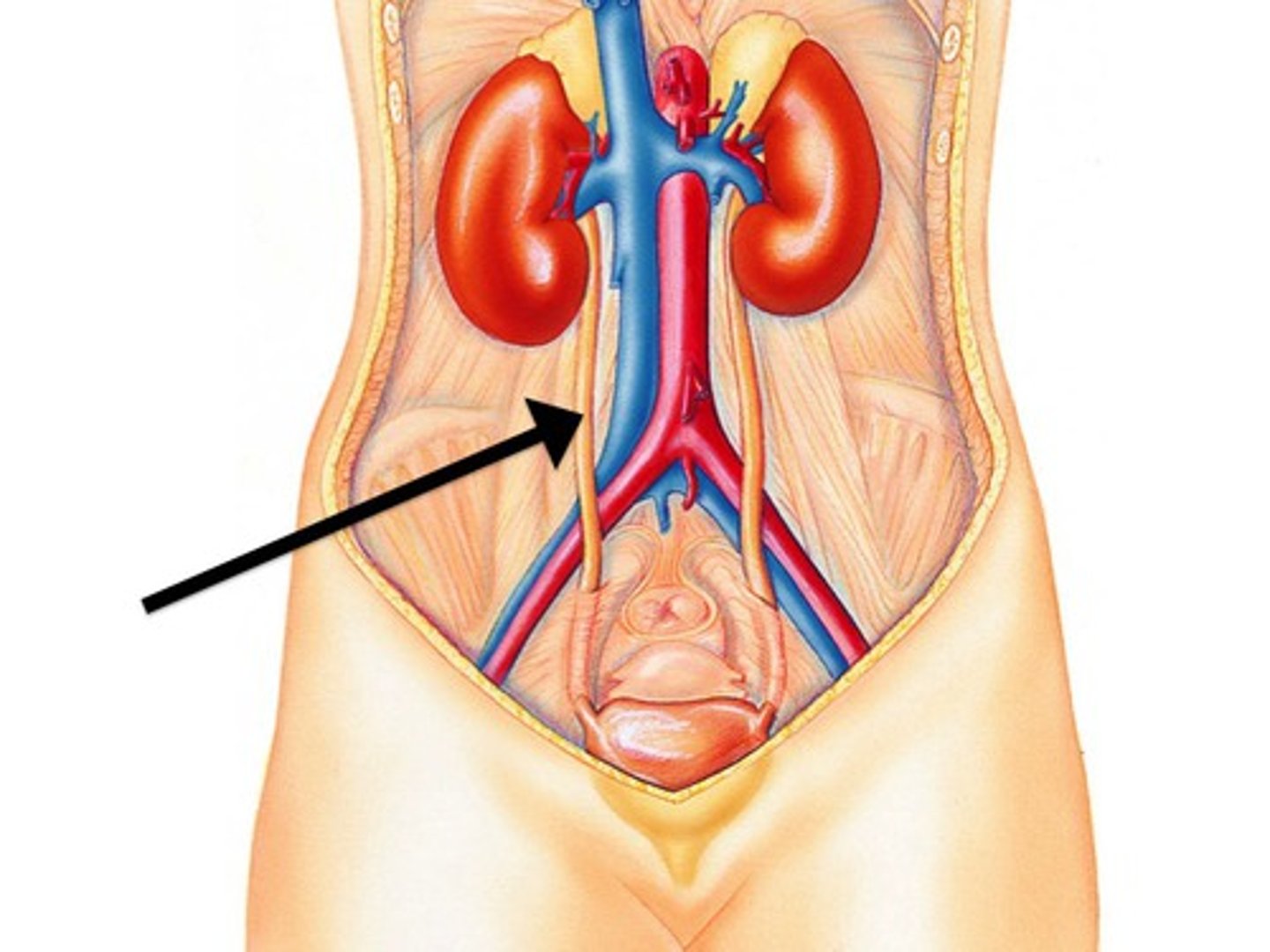
what is the function of the ureters?
carries urine from kidneys to urinary bladder
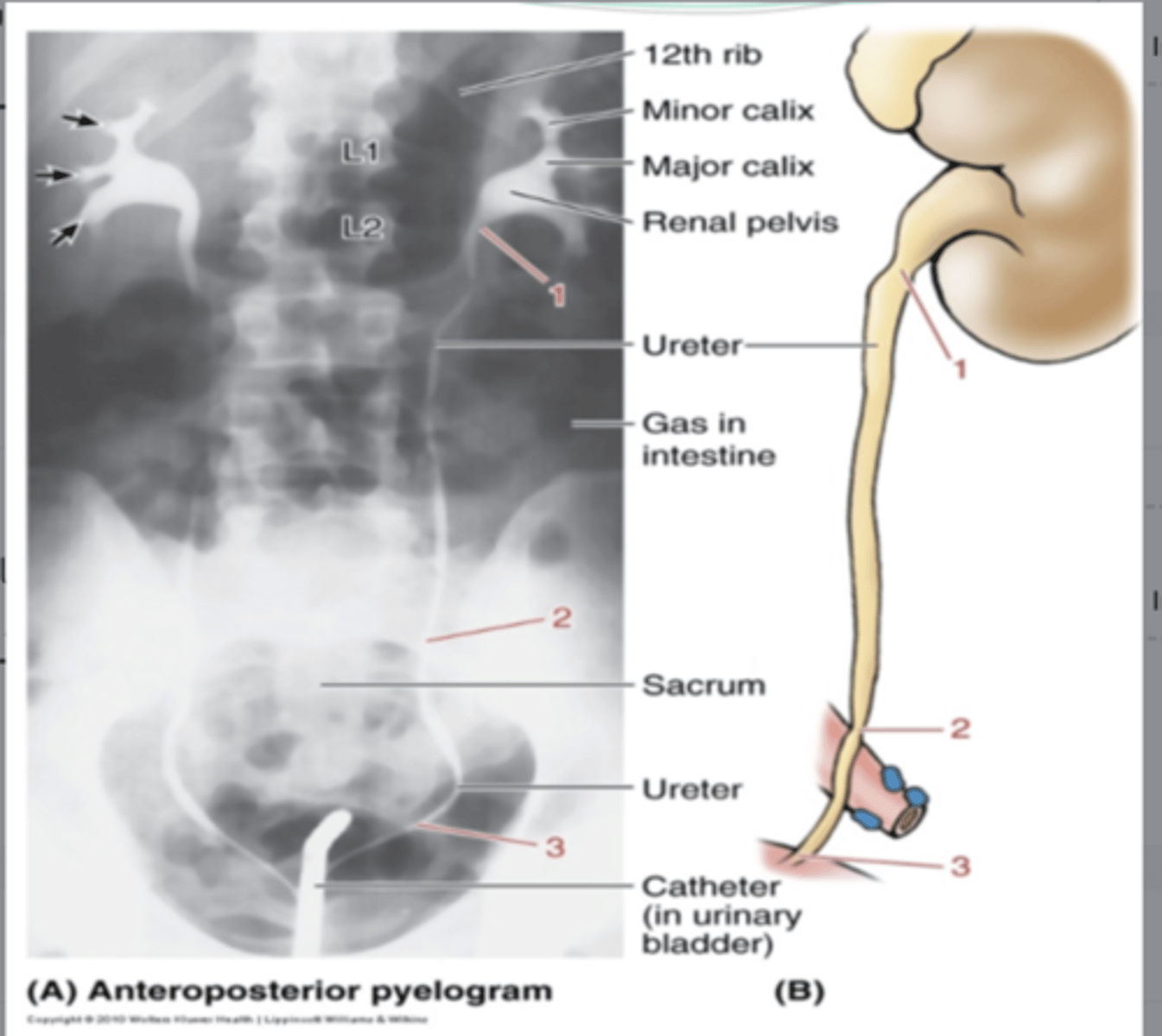
what are constrictions of the ureters?
potential sites of obstruction by ureteric stones
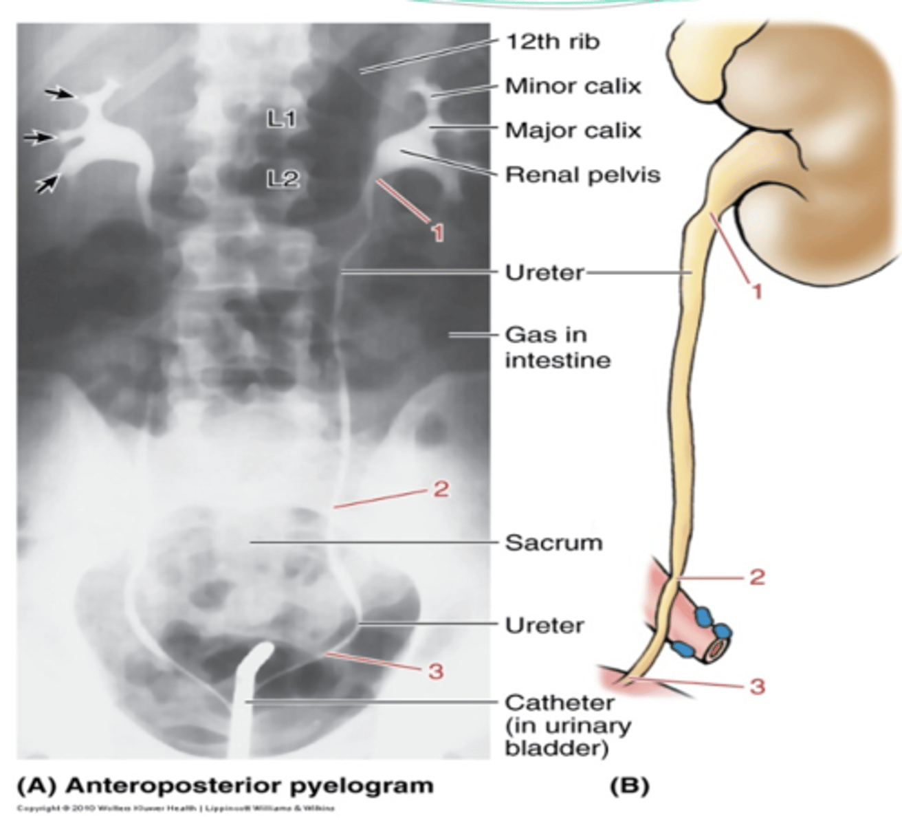
what are the three constrictions of the ureters?
1. junction of ureters and renal pelves
2. at the crossing of the pelvic brim
3. during passage through the walls of the urinary bladder
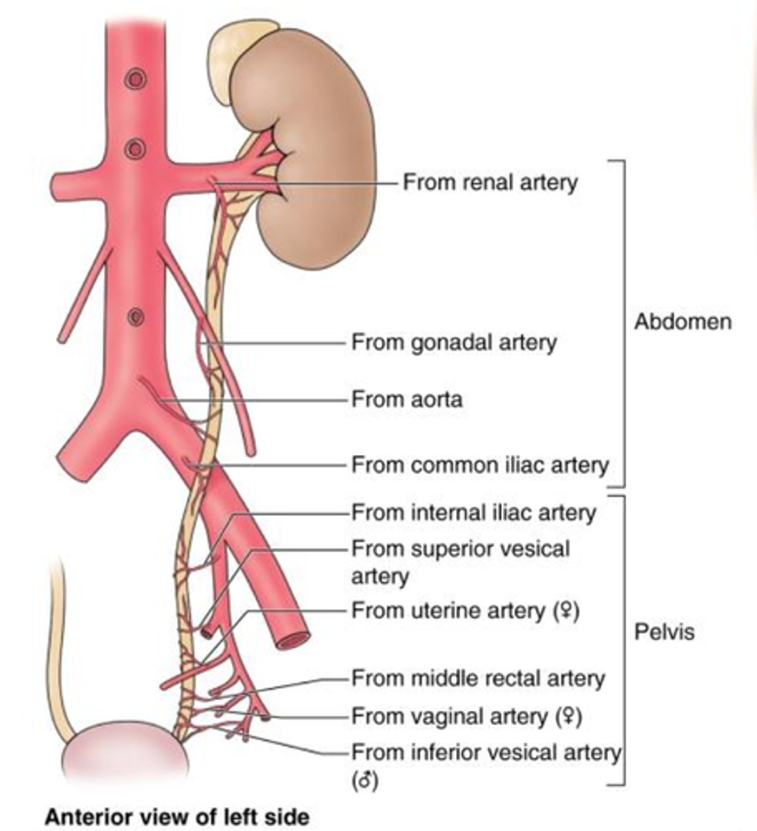
what is the vasculature of the ureters? what are they innervated by?
•vasculature
-branches of aorta, renal, gonadal, and common iliac artery and vein
•innervation
-renal, abdominal aortic, and superior hypogastric plexuses