Lesson 20: Basic Organs and Structures in the Abdominal and Pelvic Cavity
1/73
There's no tags or description
Looks like no tags are added yet.
Name | Mastery | Learn | Test | Matching | Spaced | Call with Kai |
|---|
No analytics yet
Send a link to your students to track their progress
74 Terms
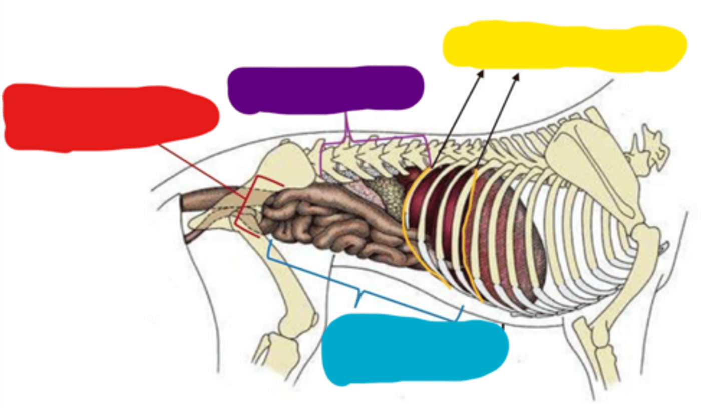
Caudal border (pelvic inlet from left and right hip bones)
Abdominal cavity boundaries:
Red

Dorsal border (thoracic and lumbar vertebrae)
Abdominal cavity boundaries:
Purple

Cranial border (last ribs, costal arch, and diaphragm)
Abdominal cavity boundaries:
Yellow

Ventral border (lateral and ventral abdominal muscles)
Abdominal cavity boundaries:
Blue
Liver
Left abdominal cavity:
7 and 8

Stomach
Left abdominal cavity:
9
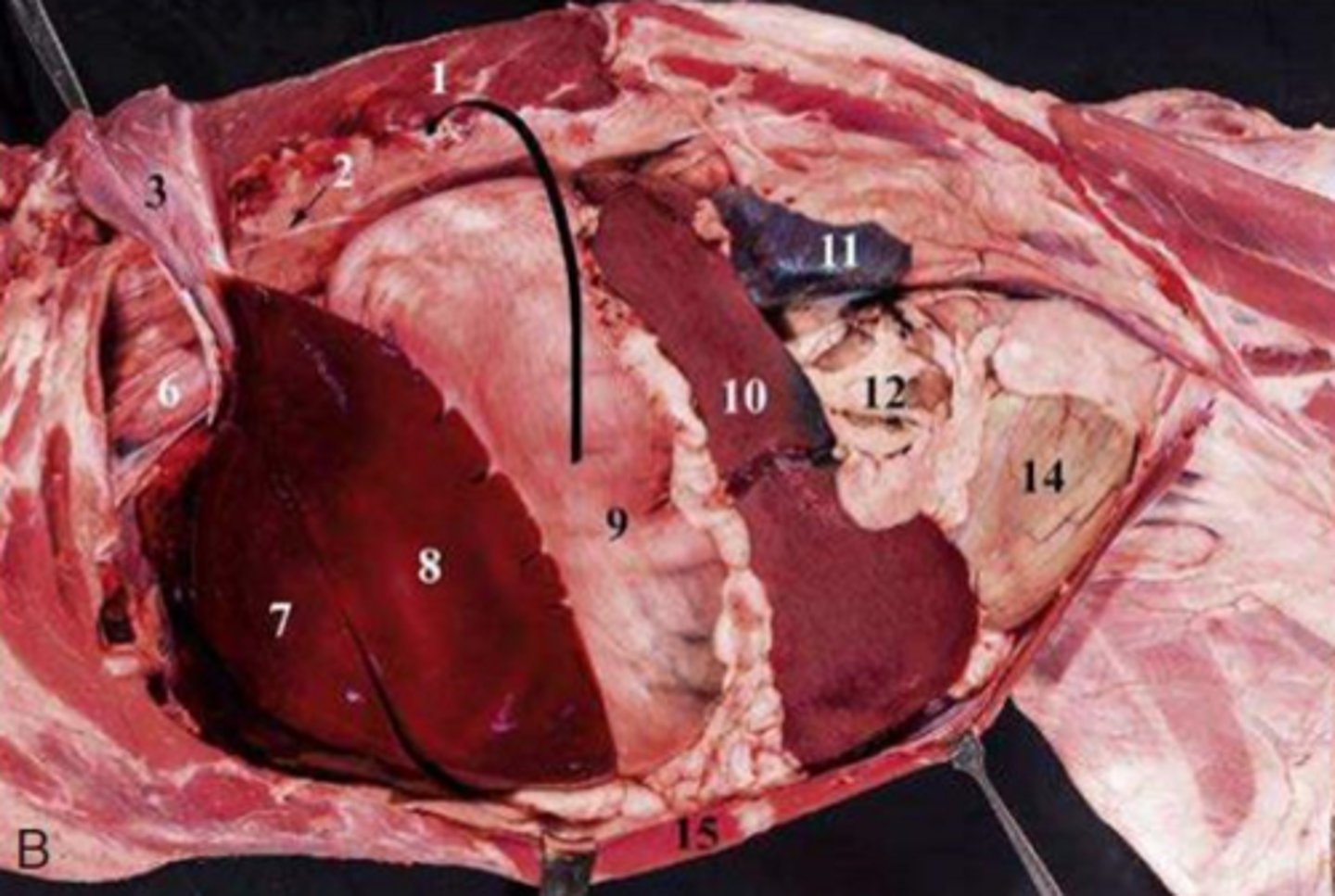
Spleen
Left abdominal cavity:
10

Left kidney
Left abdominal cavity:
11

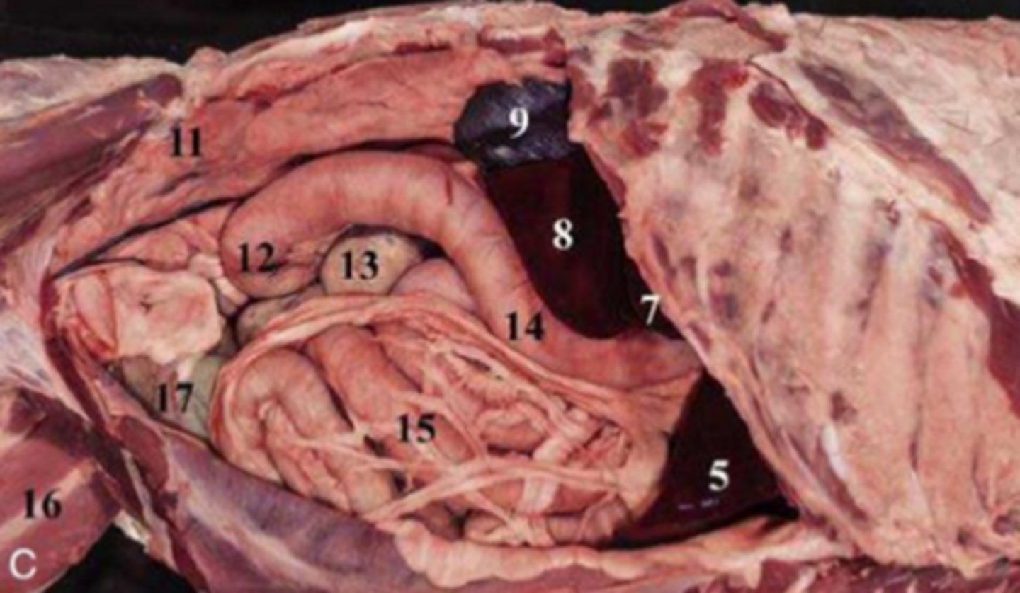
Jejunal loops
Left abdominal cavity:
12

Bladder
Left abdominal cavity:
14

Liver
Right abdominal cavity:
8, 7, and 5
Right kidney
Right abdominal cavity:
9

Cecum
Right abdominal cavity:
13

Descending duodenum
Right abdominal cavity:
14

Caval foramen
Diaphragm:
1

Aortic hiatus (aorta, azygous vein, thoracic duct)
Diaphragm:
2
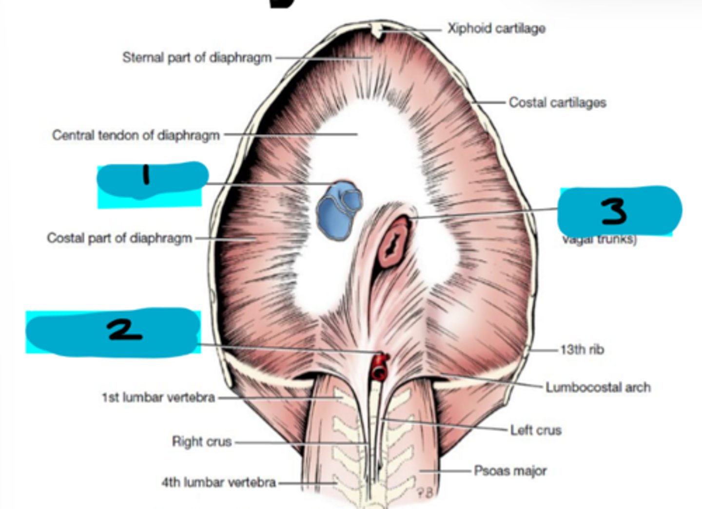
Esophageal hiatus
Diaphragm:
3

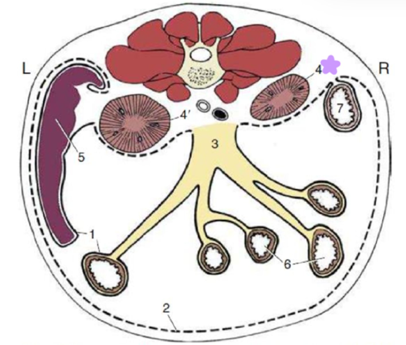
Visceral peritoneum
Peritoneal lining:
1
Lines organs

Parietal peritoneum
Peritoneal lining:
2
Lines walls

Retroperitoneal right and left kidneys
Peritoneal lining:
4 and 4'
Located behind the peritoneum
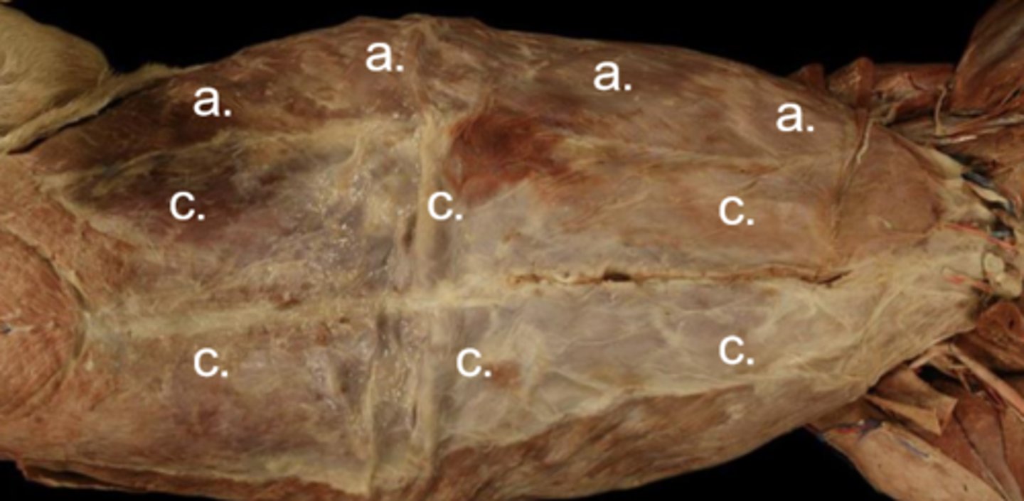
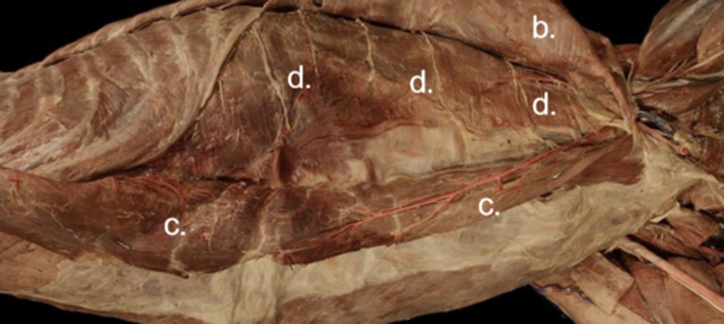
External abdominal m.
Abdominal cavity muscles:
Origin: costal arch, thoracolumbar fascia
Insertion: linea alba, prepubic tendon
External abdominal m.
Abdominal cavity muscles:
a
Origin: costal arch, thoracolumbar fascia
Insertion: linea alba, prepubic tendon
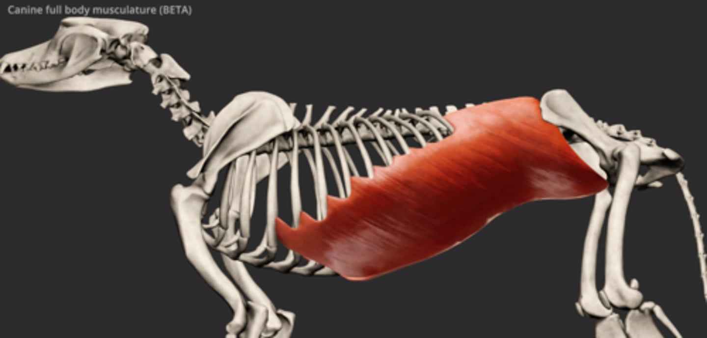
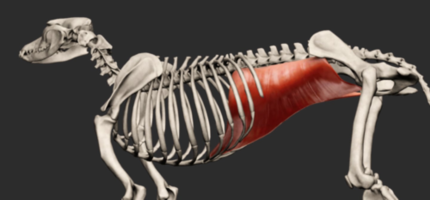
Internal abdominal m.
Abdominal cavity muscles:
Origin: tuber coxae, thoracolumbar fascia
Insertion: costal arch, linea alba, prepubic tendon
Internal abdominal m.
Abdominal cavity muscles:
b
Origin: tuber coxae, thoracolumbar fascia
Insertion: costal arch, linea alba, prepubic tendon
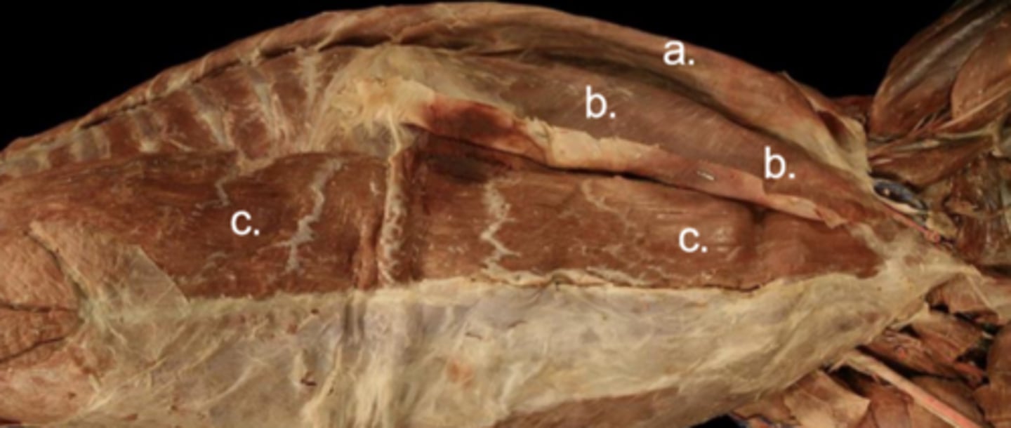
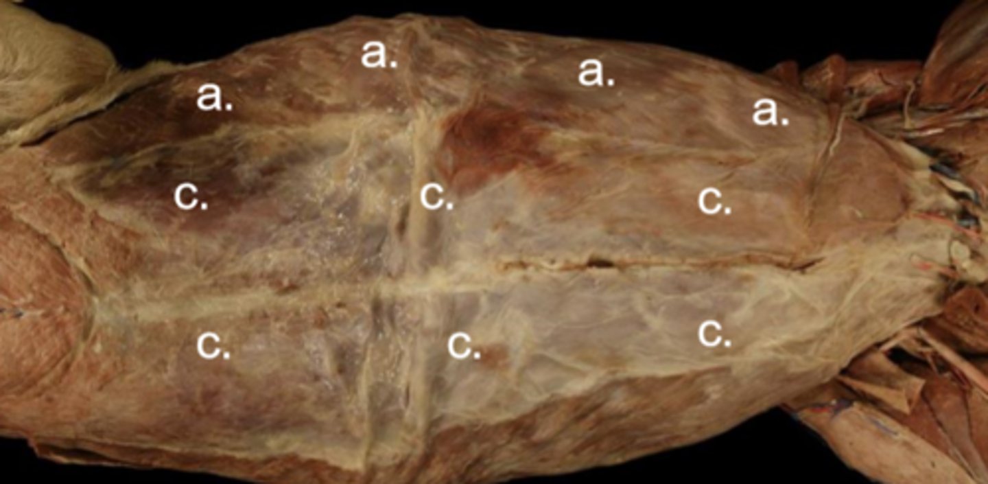
Rectus abdominis m.
Abdominal cavity muscles:
Origin: xiphoid process
Insertion: as prepubic tendon on the pecten of the pubis
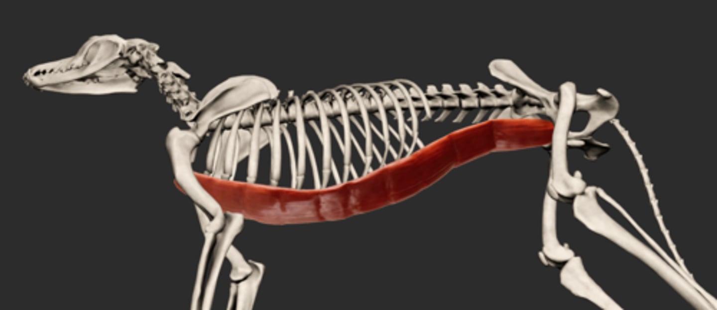
Rectus abdominis m.
Abdominal cavity muscles:
c
Origin: xiphoid process
Insertion: as prepubic tendon on the pecten of the pubis
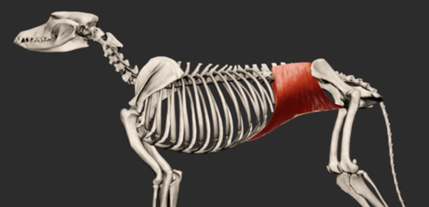
Transversus abdominis m.
Abdominal cavity muscles
Origin: transverse process of lumbar vertebrae, last ribs, thoracolumbar fascia (deep)
Insertion: linea alba, prepubic tendon
Transversus abdominis m.
Abdominal cavity muscles:
d
Origin: transverse process of lumbar vertebrae, last ribs, thoracolumbar fascia (deep)
Insertion: linea alba, prepubic tendon
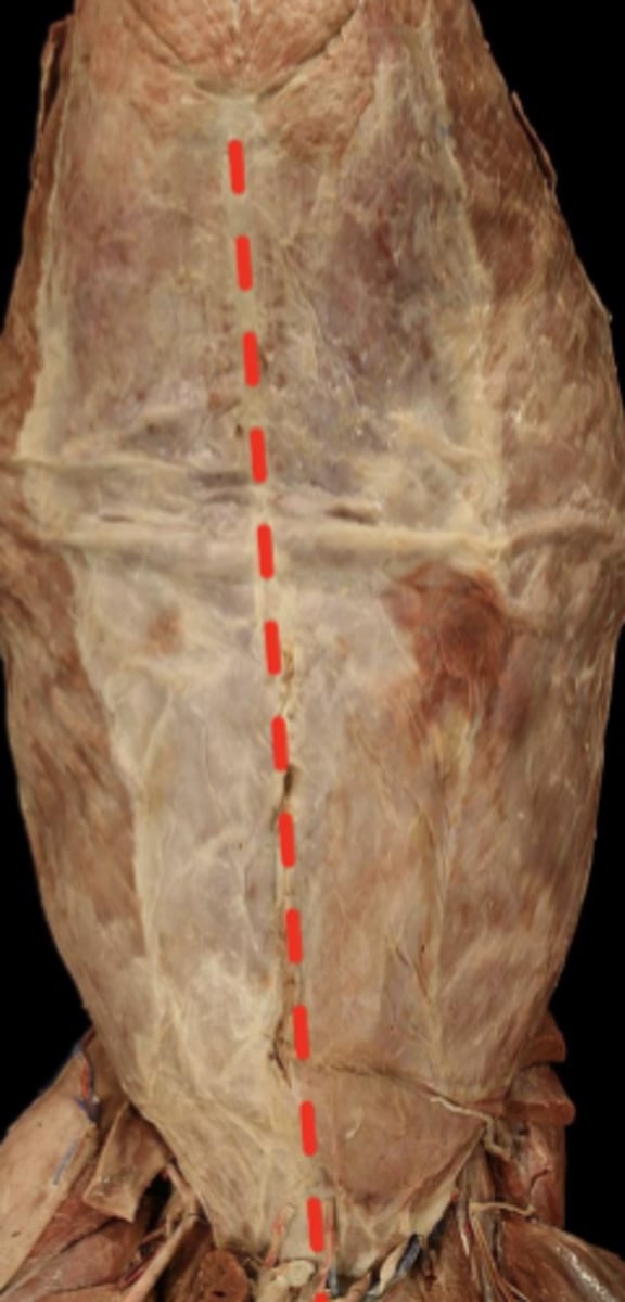
Linea alba (white line)
Rectus sheath
The tendonous wrapping around the rectus abdominis muscle
Heavy abdominal contents need reinforcement

External rectus sheath
Rectus sheath:
1
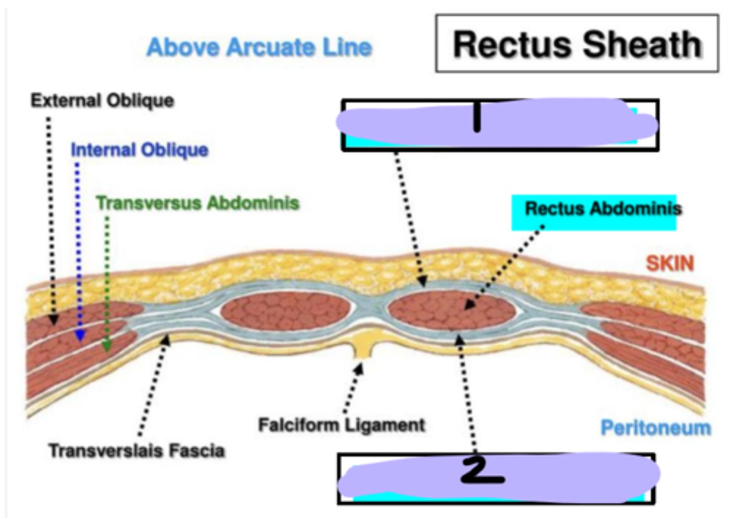
Internal rectus sheath
Rectus sheath:
2

Caudo-dorsal quadrant
Quadrants of abdominal wall:
1
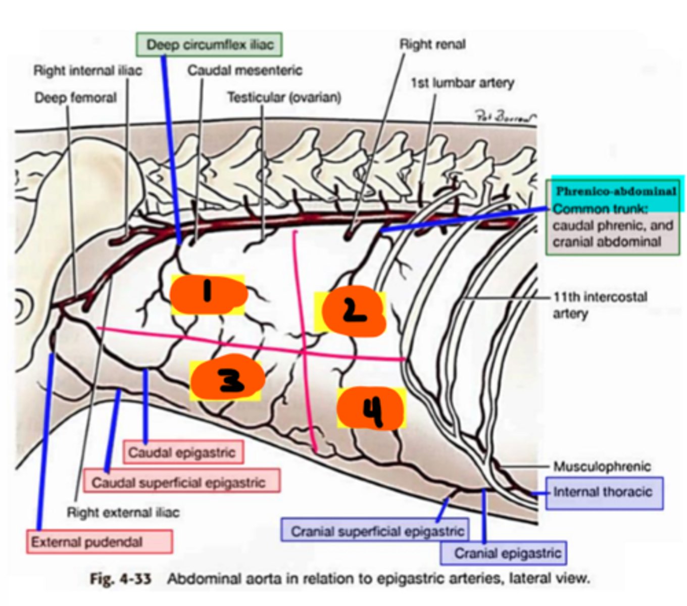
Cranio-ventral quadrant
Quadrants of abdominal wall:
4

Caudo-ventral quadrant
Quadrants of abdominal wall:
3

Cranio-dorsal quadrant
Quadrants of abdominal wall:
2

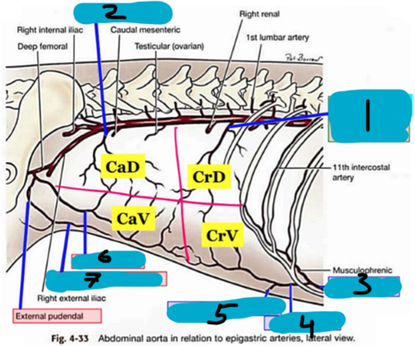
Phrenico-abdominal artery (common trunk)
Abdominal wall blood vessels:
1
Branches the cranial abdominal and caudal phrenic arteries

Phrenico-abdominal artery (common trunk)
Abdominal wall blood vessels:
1

Deep circumflex iliac (caudal abdominal artery)
Abdominal wall blood vessels:
2

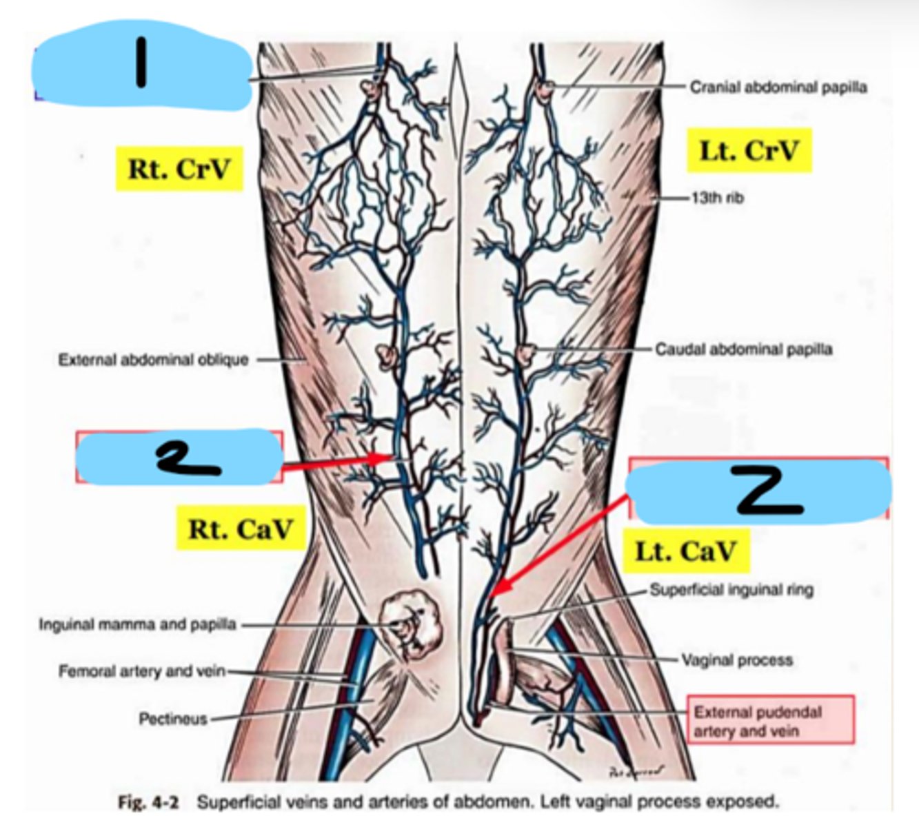
Caudal superficial epigastric artery
Abdominal wall blood vessels:
7

Caudal epigastric artery
Abdominal wall blood vessels:
6

Cranial epigastric artery
Abdominal wall blood vessels:
5

Cranial superficial epigastric artery
Abdominal wall blood vessels:
5

Cranial superficial epigastric artery
Abdominal wall blood vessels:
1
Cranial epigastric artery
Abdominal wall blood vessels:
4

Caudal epigastric artery
Abdominal wall blood vessels:
3
Internal thoracic artery
Abdominal wall blood vessels:
3
Caudal superficial epigastric artery
Abdominal wall blood vessels:
2

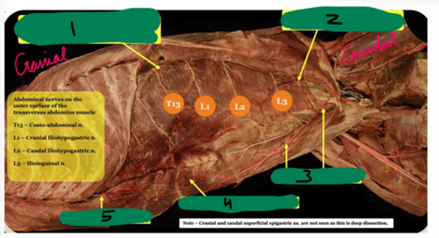
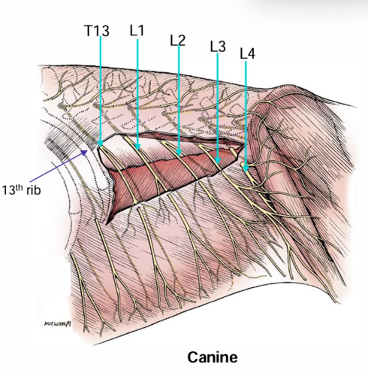
Costo-abdominal n.
DOG Abdominal wall nerves (outer surface):
T13

Costo-abdominal n.
DOG Abdominal wall nerves (outer surface):
T13
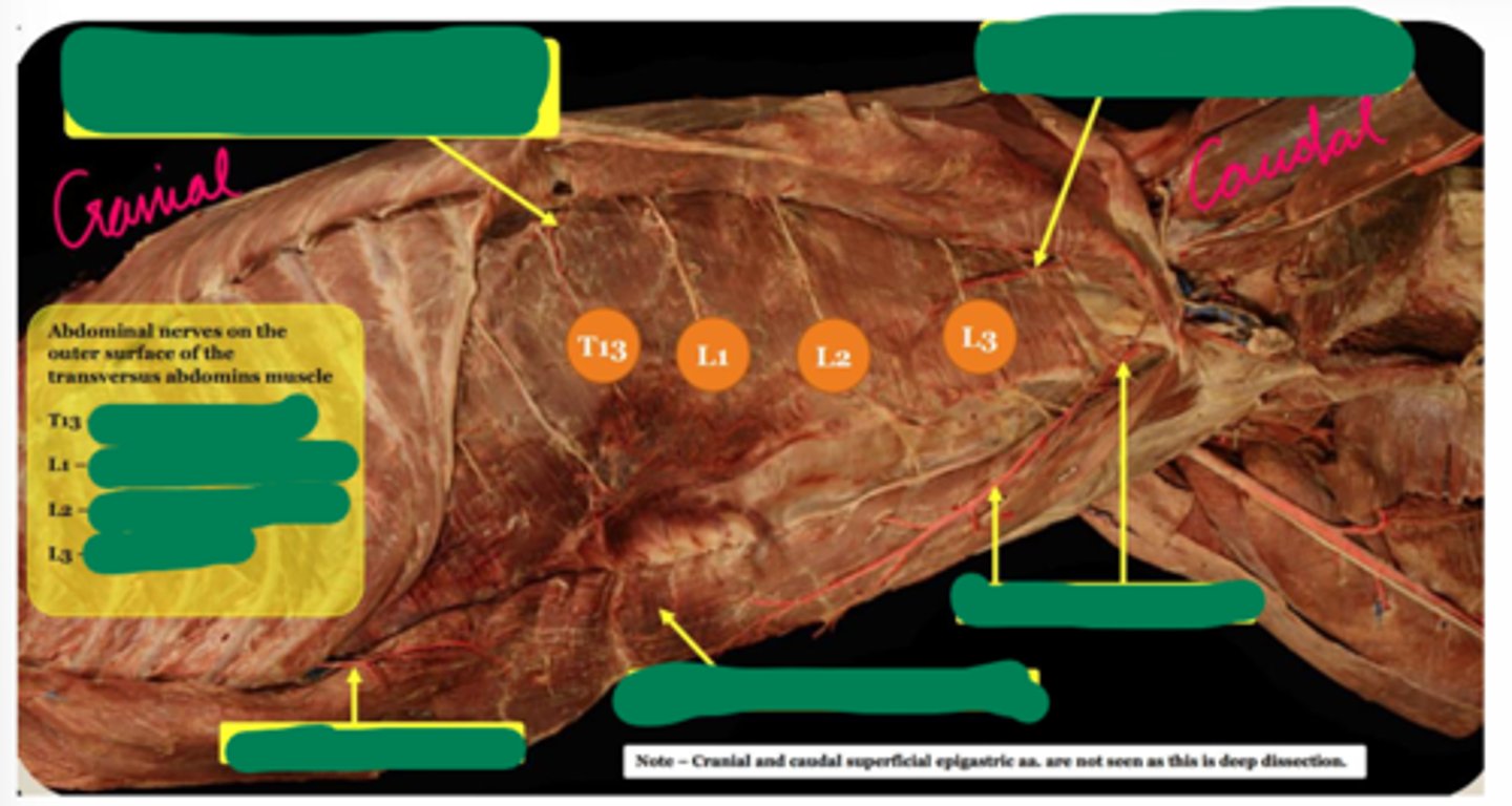
Cranial iliohypogastric n.
DOG Abdominal wall nerves (outer surface):
L1

Cranial iliohypogastric n.
DOG Abdominal wall nerves (outer surface):
L1

Caudal iliohypogastric n.
DOG Abdominal wall nerves (outer surface):
L2

Caudal iliohypogastric n.
DOG Abdominal wall nerves (outer surface):
L2

Ilioinguinal n.
DOG Abdominal wall nerves (outer surface):
L3

Ilioinguinal n.
DOG Abdominal wall nerves (outer surface):
L3

Lateral cutaneous femoral n.
DOG Abdominal wall nerves (outer surface):
L4
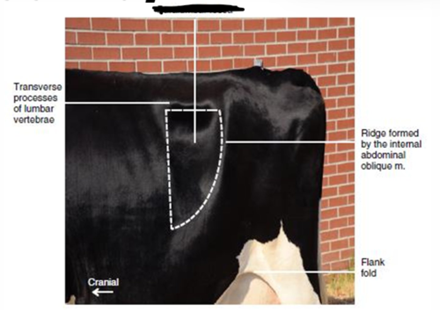
Paralumbar fossa
Bovine important surgical and clinical area
Boundaries-
Cranially: last rib
Dorsally: transverse processes of lumbar vertebrae
Caudally: craniodorsal edge of the internal abdominal oblique m.
Costoabdominal n.
Bovine nerves of paralumbar fossa:
T13

Iliohypogastric n.
Bovine nerves of paralumbar fossa:
L1

Ilioinguinal n.
Bovine nerves of paralumbar fossa:
L2
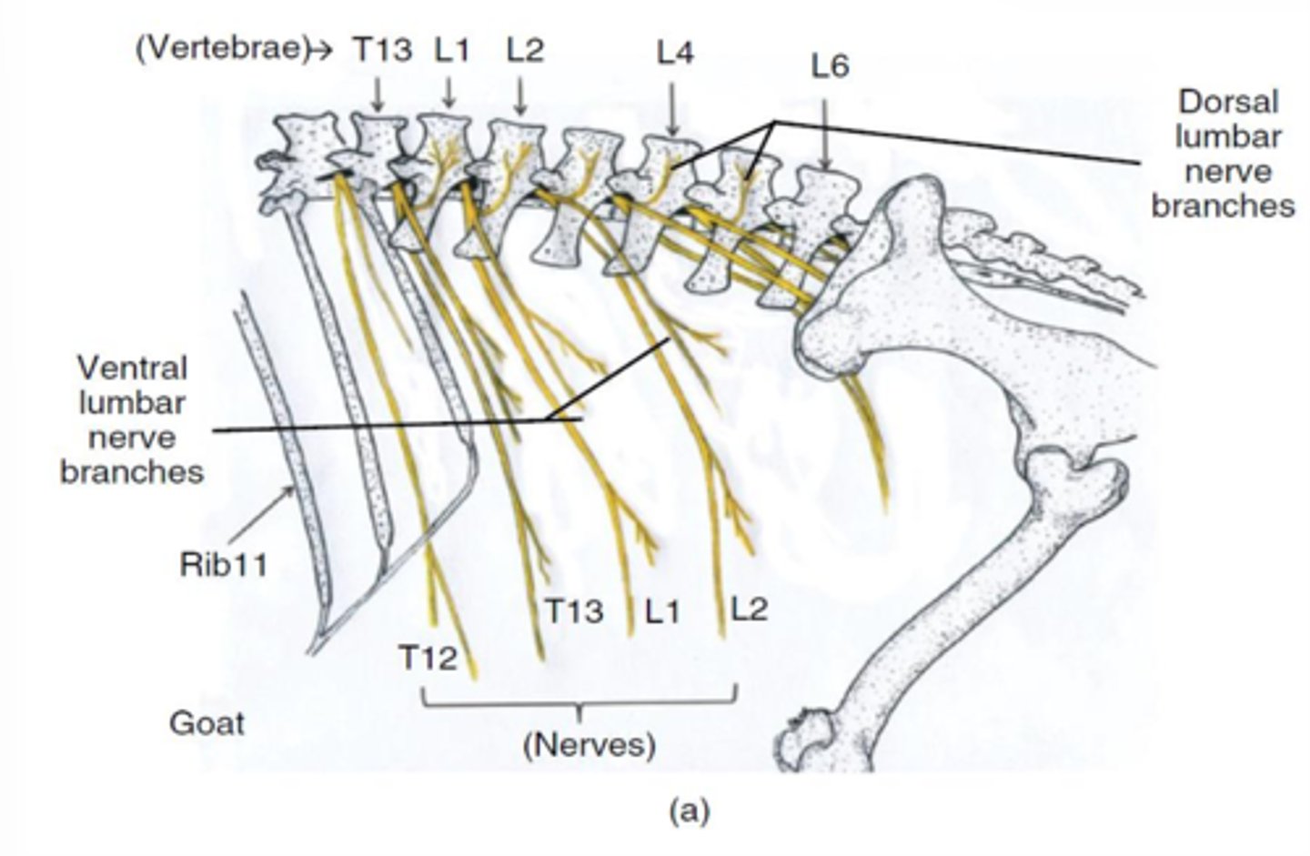
Genitofemoral n.
Bovine nerves of paralumbar fossa:
L3

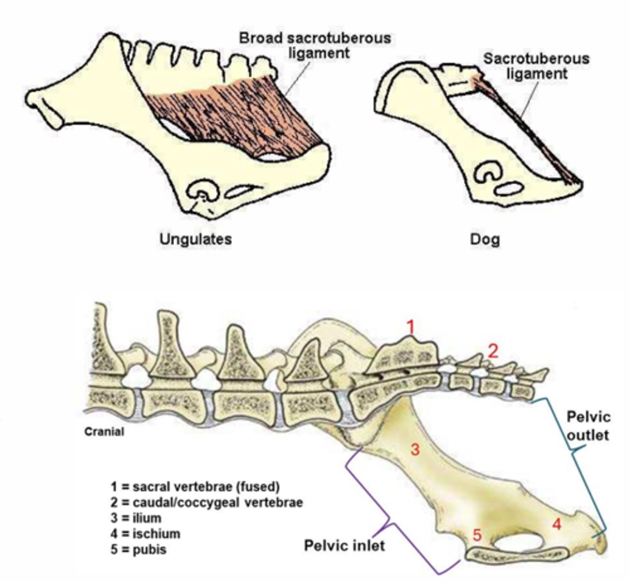
Dorsal border
Pelvic cavity borders:
sacrum, first few caudal vertebrae

Lateral border
Pelvic cavity borders:
pelvic bones (ilium, ischium) and broad sacrotuberous/sacrosciatic ligament

Ventral border
Pelvic cavity borders:
pelvic symphysis, floor of pelvic canal, extends between pelvic inlet and outlet

Caudal border
Pelvic cavity borders:
closed off by muscles that create a pelvic diaphragm and a urogenital diaphragm
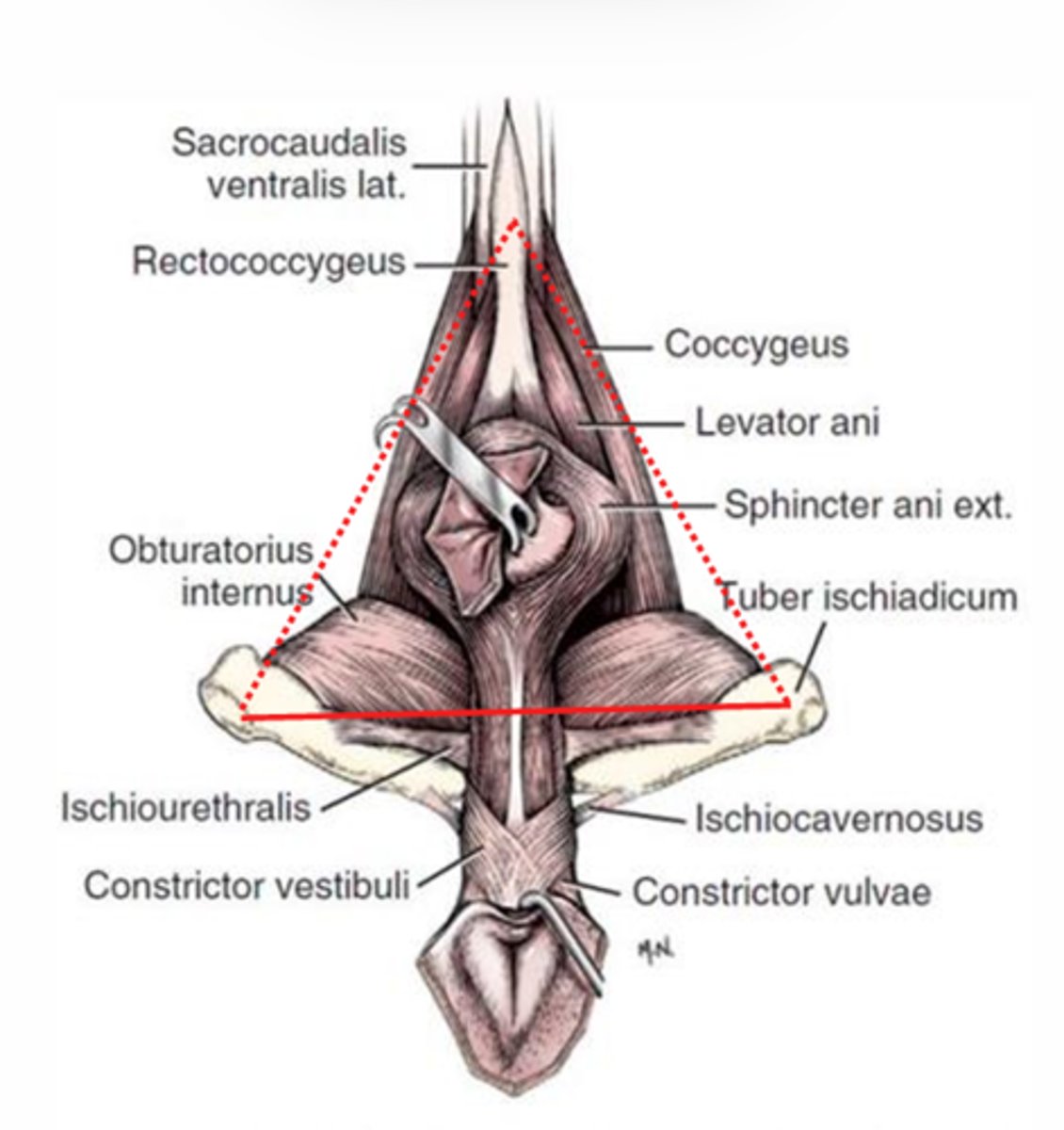
Anal triangle
Pelvic cavity boundaries:
Top triangle
Dorsal: caudal vertebrae
Lateral: deeper sacrosciatic ligament
Ventral: line between ischiatic tuberosities/deeper structure will be ischial arch
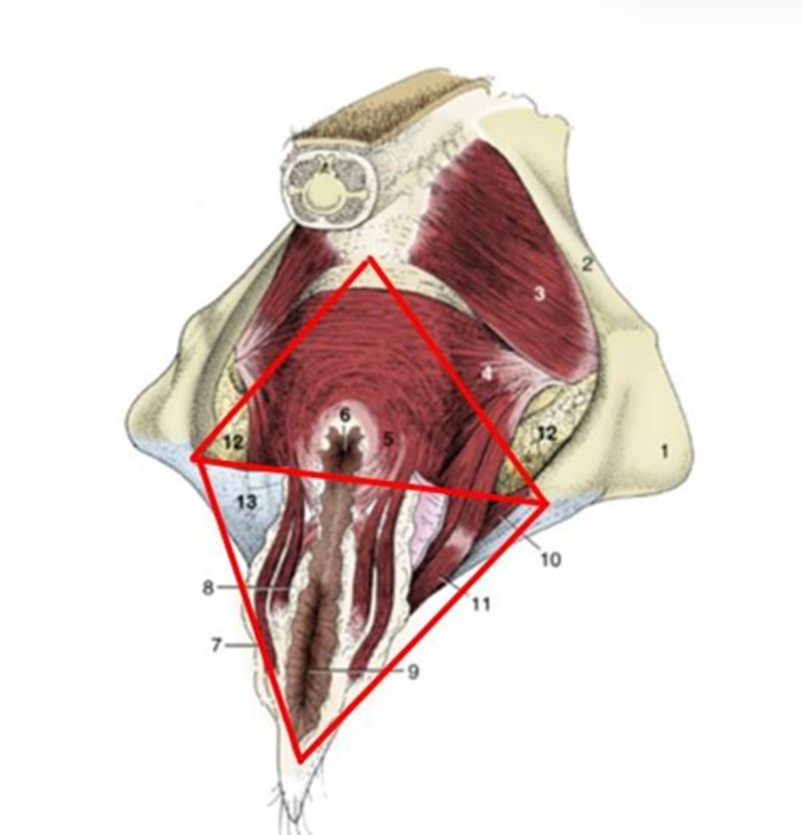
Pelvic diaphragm
Closes off the anal triangle
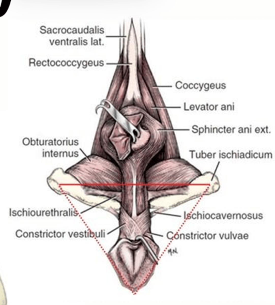
Urogenital triangle
Pelvic cavity boundaries:
Bottom triangle
Dorsal: line between ischiatic tuberosities
Ventral: ventral commissure of the vulva

Urogenital diaphragm
Closes off the urogenital triangle
Includes the urogenital muscles
Pararectal fossa
Compartments of the pelvic cavity:
1
the region/space dorsal to the rectum and ventral the sacral/caudal vertebrae

Rectogenital pouch
Compartments of the pelvic cavity:
2
the region/space between the rectum dorsally and the reproductive organs ventrally

Vesicogenital pouch
Compartments of the pelvic cavity:
3
the region/space between the reproductive organs dorsally and the bladder ventrally
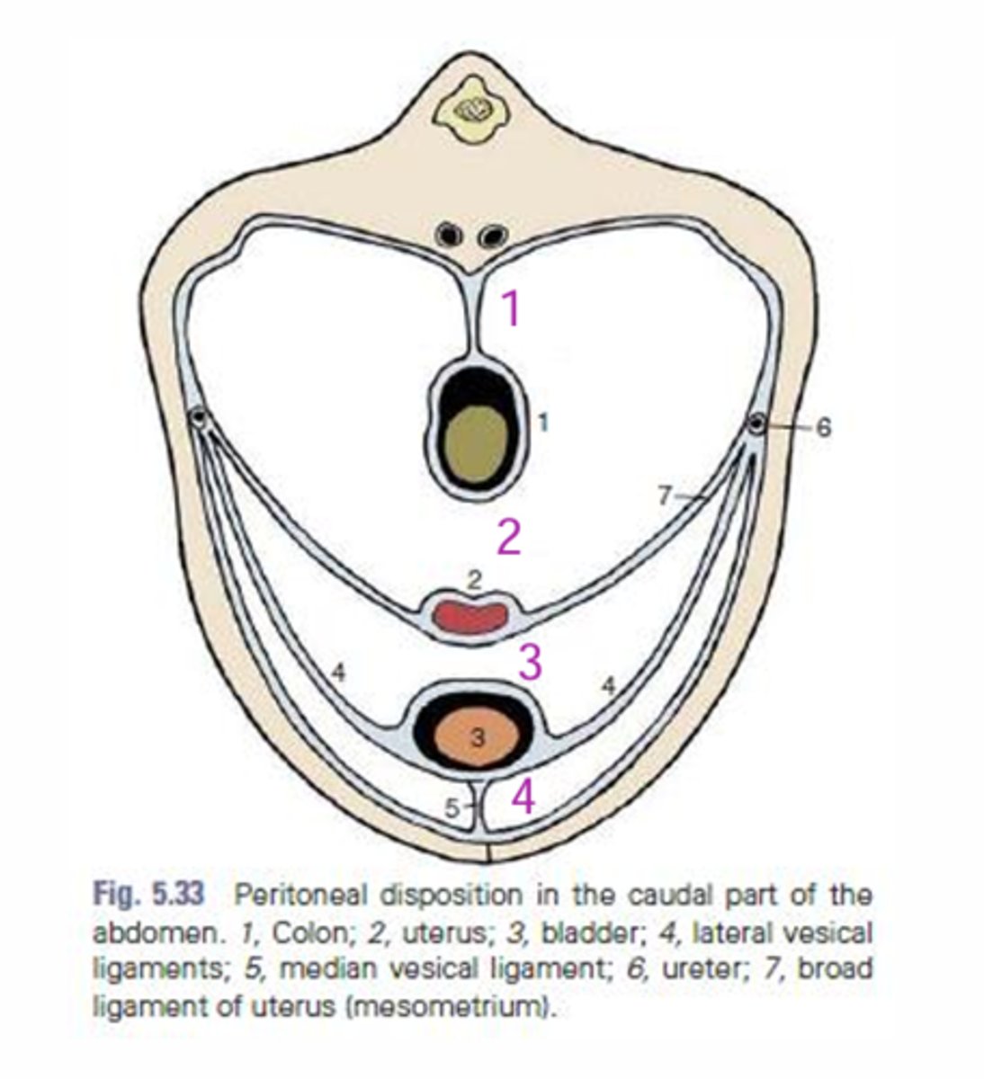
Pubovesical pouch
Compartments of the pelvic cavity:
4
the region/space between the bladder dorsally and the pubis or ventral abdominal floor
