Higher Brain Centers Anatomy
 Call Kai
 Learn
Learn Practice Test
 Spaced Repetition
 Match
Match Flashcards
 Knowt Play
1/48

There's no tags or description
Looks like no tags are added yet.
Name | Mastery | Learn | Test | Matching | Spaced | Call with Kai | Chat |
|---|
No analytics yet
Send a link to your students to track their progress
49 Terms
Cerebellum Location
Dorsal to the brainstem; separated from the brainstem by the 4th ventricle

Cerebellum function
Movement; motor coordination, error correction, motor learning, timing of movements
Diencephalon Components
Thalamus and Hypothalamus
Thalamus Structure
Two egg shaped structures (one in each hemisphere)
Thalamus location
Located near the midline at the base of the brain; one on either side of the 3rd ventricle
Thalamus function
contains thalamic nuclei
Projects to and feedback projections from the cortex
Sends sensory information to primary sensory cortices
Hypothalamus location
Ventral to the thalamus, near the bottom of the 3rd ventricle; attached to the pituitary gland along its ventral surface
Hypothalamus function
Contains small nuclei; regulates hormone release from the pituitary gland; major visceral motor center that regulates the autonomic NS (“head ganglion”)
Basal Ganglia Location
Inferior and medial to the cerebral cortex, lateral to the diencephelon
Basal ganglia make-up
Composed of interconnected groups of neurons called “ganglia”
Basal ganglia shape
Caudate nucleus (part of the basal ganglia) curves in a backwards “C” shape
Basal ganglia function
Motor planning/gating
Cerebral cortex divisions
Frontal, parietal, temporal, occipital, and insular (all are part of the telencephelon, named after the bones of the skull that fused during development (except for the insular cortex))
Sulci definition
Shallow folds in the cerebral cortex
Fissures definition
sulci folds that are deeper
Longitudinal fissure
Divides the cerebral cortex into two halves, creating 2 hemispheres
Lateral fissure
Separates the temporal lobe from the front and parietal lobe
Where is the insula?
Since the cortex of the lateral fissure is so deep, it’s held within its depths
Gyri/Gyrus
Rounded regions between the Sulci
Sutures
separate the bone (plates) of the skull
Name the sutures of the skull
Coronal, Sagittal, Lamboid, Squamous
Coronal Suture
Separates the frontal and parietal bones (“crown”)
Sagittal Suture
Lies along the midline and separates the two parietal bones
Lamboid Suture
Separates the parietal and occipital bones (named after Greek Lambda )
Squamous Suture
Separates the temporal bone from the parietal bone (temporalis = temple)
Types of cortical areas
Primary and association
Describe primary sensory cortices
Receive sensory input from the thalamus and association cortices (olfactory does not receive this information); involved in early processing of sensory input
Types of primary sensory cortices
Primary visual cortex, primary somatosensory cortex, primary auditory cortex, primary olfactory cortex, primary gustatory (taste) cortex
Describe primary motor cortex
Sends motor commands to the lower areas of the motor system
Describe association areas
Send and receive information only from other cortical areas; involved in higher order sensory, motor, and cognitive functions
Occipital lobe location
Back of the brain, below the parietal lobe and above the cerebellum
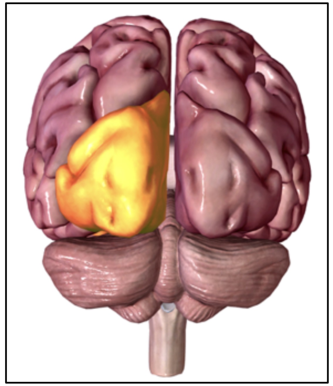
Primary cortex of the occipital lobe
Primary visual cortex
Function of the occipital lobe
Primary cortex is Organized as a “map” of the retina; association cortices involved in visual processing at earlier stages than parietal lobe
Primary cortex of the parietal lobe
Primary somatosensory cortex
Parietal cortex location
Located in the post-central gyrus, caudal to the central sulcus

Function of the parietal lobe
Organized topographically (somatosensory homunculus); somatosensory input to each hemisphere come from the contra lateral side of the body
Describe the association cortex of the parietal lobe
Posterior parietal cortex includes multiple association cortices, including those involved in high-level visual processing, and perception of spacial organization
Primary cortex of the frontal lobe
primary motor cortex
Primary motor cortex location
in the precentral gyrus, rostral to the central sulcus
Motor cortex organization
organized topographically; “motor homunculus”; the amount of cortical tissue for a given region of the body is proportional to the number of motor neurons used to control that region
Motor cortex function
controls voluntary movements of the contralateral body
Association areas of the frontal lobe
broca’s area (language); multiple divisions of the prefrontal cortex are involved in functions such as long-term planning, working memory, and judgement
Describe the corpus callosum
axons that cross the midline to connect the two cerebral hemispheres
Temporal Lobe Location
ventral side of the brain, below parietal lobe
Primary cortex of the temporal lobe
primary auditory cortex (organized according to the pitch of sounds)
Association Cortices in the temporal lobe
Wernicke’s area (critical for language); areas involved in recognizing objects and people, areas involved in memory
Insular lobe location
Located inside the lateral fissure
Primary cortex for the insula
gustatory cortex, involved in taste
Insular lobe function
taste and receiving visceral sensory inputs