BIO 11.1 - Exer 3A
1/31
There's no tags or description
Looks like no tags are added yet.
Name | Mastery | Learn | Test | Matching | Spaced | Call with Kai |
|---|
No analytics yet
Send a link to your students to track their progress
32 Terms
Anton van Leeuwenhoek (1632-1723)
The earliest records of microscopic observations were done by ________________________
Magnification
It is the magnitude of amplification of the image of the specimen by the microscope
Theoretical magnification = magnification of ocular lens * magnification of objective lens
Actual magnification = apparent size of specimen/actual size of specimen
Magnification can be measured in two ways, these are and their formulas?
Linear magnification
It is the magnitude of amplification of the image of the specimen by the objective lens only.
Resolving power (R)
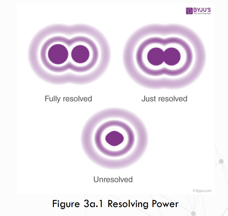
It is the ability of the microscope to distinguish two adjacent points as distinct and separate.
Limit of resolution (R)
It is the smallest distance between two adjacent points a microscope can distinguish.
smaller; higher
The ____________ the value of the limit of resolution, the ___________the resolving power.
Numerical aperture (NA)
NA of LPO = 0.25
NA of HPO = 0.65
An index of the light-gathering power of the objective lenses
It is inversely proportional to the limit of resolution (r).
Refractive index (RI)
Air and oil have refractive indices equal to 1.0 and 1.56, respectively.
It is the ability of a medium to deflect light.
A high ____________ value means less deflection, hence, more light enters the objective.
Working distance
LPO - 5.00 mm
HPO - 0.46 mm
OIO - 0.13 mm
It is the distance between the specimen and the objective lens when the specimen is in focus.
Field of vision
The diameter of the field of vision of each objective is:
LPO - 1.50 mm
HPO - 0.35 mm
OIO - 0.18 mm
It is the area visible under the microscope.
Depth of focus
It is a property of the microscope that indicates how deep the space the microscope can bring into clear view at any one time.
Depth of focus
Range of distances for which the object is imaged with an acceptable sharpness on the image plane

What is the relationship between all the microscope terms?
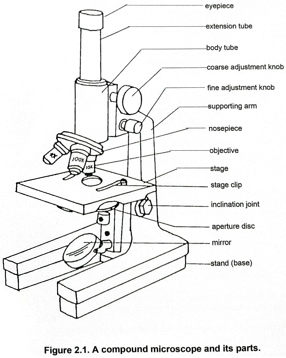
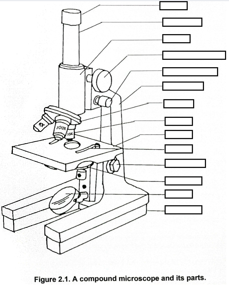
Eyepiece
It is a small, removable tube containing lenses with the magnifying power etched on its surface. The observer sees the amplified image of the minute specimen through the __________.
Extension tube
Also containing lenses, encases the bottom portion of the eyepiece. Through this tube, the image of the specimen is projected over a distance.
Body tube
A wide tube usually without lenes is found below the extension tube. It provides a short distance for the image to pass through.
revolving nosepiece
Attached beneath the body tube is the ___________ which serves as the base for one or more objectives. It can be rotated to position the appropriate objective to be used.
objectives
In most compound microscopes, the shortest objective (10x, 5 mm) is the low power objective (LPO), next (40x, 0.46 mm) is the high power objective (HPO) and the longest, if present (100x, 0.13 mm) is the oil immersion objective (OIO).
The __________ are small narrow tubes containing compound lenses for magnification. These _________ have marks identifying their magnification power and the working distance.
scanner objective (4x)
A _____________ is found in some microscopes in place of the OIO. The _________ objective which is shorter than the LPO allows a wider area of the specimen to be viewed.
arm
The curved portion connecting the body tube to the base of the microscope is the _______. This is where the microscope is held for carrying or tilting. It supports the body tube and the adjustment knobs.
coarse adjustment knob; fine adjustment knob
The adjustments knobs are the two pairs of knobs found on both sides of the arm.
The larger pair of knobs, the _____________, adjusts or moves the body tube together with the objectives, up and down easily. (Or in other microscopes, the stage is the one that moves up and down). It is used to bring into the focus the specimen to be observed.
The smaller pair of knobs, the _____________, adjusts slowly and is used to sharpen the focus.
inclination joint
Found at the base of the arm of some microscopes is the ______________, which allows the upper portion of the microscope to be tilted.
stage; stage clips
The glass slide which contains the specimen to be observed is placed on the ________. It is a square platform which contains ___________ for holding the slide in place, and a hole where light passes through.
aperture disc
A movable ______________ connected to and beneath the stage contains a series of holes with different sizes for the regulation of the incoming light.
substage condenser
A third system of lens, found in some microscopes, is the _______________ used to concentrate the incoming light.
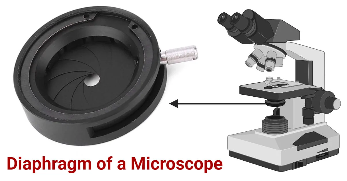
iris diaphragm
Below the condenser is the _____________ with a movable lever, also for regulating incoming light.
pillar
Finally, the _________ refers to the region connecting the inclination joint with the stand at the base of the microscope. Together they support and hold the microscope in a steady position.
Stereomicroscope (Dissecting Microscope)
This microscope can produce three-dimensional (3-D) images of objects that can be magnified from 10x to 60x depending on the model.
The observer views the object as a 3-D image due to the different perspectives perceived by the right and left eyes.
Student Microscope
The common monocular compound microscope with ocular comes with different objectives and a plano-concave mirror for transmitted-light illumination.
Some models include a third lens system, the substage condenser.
This is used for general brightfield microscopy but can be upgraded for other advanced techniques.
Research Microscopes
These microscopes are versatile and multi- featured for various applications.
They usually have a binocular or a trinocular head, built-in illumination and added accessories for various microscopic works and techniques.