Eye embryology
 Call Kai
 Learn
Learn Practice Test
 Spaced Repetition
 Match
Match Flashcards
 Knowt Play
1/24

There's no tags or description
Looks like no tags are added yet.
Name | Mastery | Learn | Test | Matching | Spaced | Call with Kai | Chat |
|---|
No analytics yet
Send a link to your students to track their progress
25 Terms
Developement of eye
Early Embryology & Genetics
Initiation: begins at 22 days of gestation as an outpocketing/extension of the diencephalon (prosencephalon).
Key Gene: PAX6 .
Gene Defects: Mutations in PAX6 or related pathways (such as RAX, PAX2) lead to severe congenital malformations like anophthalmia or microphthalmia.
Choroidal Fissure: A ventral groove/gap in the optic stalk through which embryonic vasculature passes; it normally closes by 6–7 weeks of gestation.
Precess
Early -
As the neural tube closes, from procencephalon the lateral outpouchings(diencephalon) expand outward to form the optic vesicles on both sides.
optic vesicle expands further against the surface ectoderm, to form the narrow optic stalk
physical contact in surface ectoderm and vesicles induces the surface ectoderm to thicken, forming the early lens placode.
Lens formation
Lens placode invaginates → lens pit.
The pit then pinches off from surface ectoderm → lens vesicle. [Lens develope from surface ectoderm]
The lens vesicle is initially a hollow spherical structure with:
Anterior wall
Posterior wall
Cavity of lens vesicle
Cells of the posterior wall of the lens vesicle become elongated by converting cuboidal cells in columnar cells
These elongated cells grow toward the anterior wall.
They become primary lens fibers.
cells from the equatorial region continue to form secondary lens fibers throughout life.
No posterior wall
The anterior wall remains as a single layer of cuboidal cells → becomes the lens epithelium.
Eyeball close from → superotemporal to inferior nasal direction
Structure of lens
Lens has:
Anterior capsule
Anterior lens epithelium
Cortex
Nucleus
Posterior capsule
Lens epithelium is present only anteriorly.
There is NO posterior lens epithelium.
At the periphery, the lens fibers meet at the equator.
Center of anterior part - anterior pole
Center of posterior part - posterior pole
The central older part is the nucleus.
The peripheral newer fibers form the cortex.
types of nuclei of lens
Embryonic nucleus
Formed earliest.(0-3 monthsof gestation)
Corresponds to the earliest embryonic lens fibers. (Oldest)
Fetal nucleus
Formed during fetal life.(3-8 months of gestation)
Surrounds the embryonic nucleus.
Infantile nucleus
Fibers formed after birth during early childhood. (8 months to puberty)
Adult nucleus
Continues to form after childhood.(Puberty to adults)
Newer fibers are added around the older nucleus.(Youngest)
Lens sutures
Sutures are microscopic junctions/gaps between lens fibers.
They are formed because the ends of lens fibers meet in characteristic patterns.
From front surface
Has an upright Y-shaped suture.
From back surface
Has an inverted Y-shaped suture.
Strongest basement membrane of body
Lens capsule
During developent which vessels supply lens?
Hyaloid artery - 3 branches
Anterior pupillary vessels
Capsulopupillary vessels
Posterior pupillary vessels
After developement , lns become avascular
Developed lens nutrition
The lens is avascular.
Therefore, it does not have its own blood vessels.
It receives nutrients by diffusion.
Main sources:
Anterior lens
→ mainly from aqueous humor
Posterior lens
→ mainly from vitreous humor
[Option choice → AH+VH > AH > VH]
Which connective tissue present all around eye balll,lens in orbit?
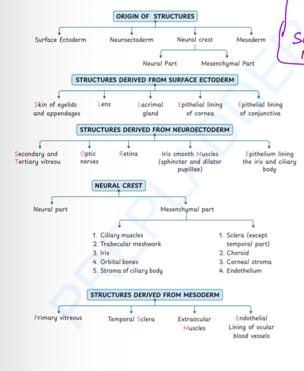
Mesenchyme. It will further differentiate in neural crest and mesoderm.
Neural Crest vs Mesoderm
Neural crest | Mesoderm |
Ciliary muscle | Primary vitreous |
Trabecular meshwork | Temporal sclera |
Orbital bones | Extraocular muscles |
Stroma of ciliary body , supercilliary lamina | Endothelium lining ocular blood vessels |
Sclera (except temporal part) | |
Choroid | |
Corneal stroma | |
Corneal endothelium |
Condensed part of viterous called. As
Hyaloid membrane
Remains of hyaloid artery affter regression
Cloquet canal
PHPV (Persistent hyperplastic primary viterous)
During normal eye development, the primary vitreous and hyaloid vascular system normally regress.
If this Entire artery regression/resorption does not occur → PHPV develops.
Your notes mention:
Non-resorption of hyaloid artery/system
Association with unilateral microphthalmos.
It can cause:
Vitreous haemorrhage
Poor visual prognosis.(because retinal detachment and cataract)
Persistent partial hyaloid vessels
Muscae volitantes
A small fragment of the persistent hyaloid vessel can remain.
It may appear as floaters in the visual field.
Hence:
Persistent hyaloid vessel fragment → Muscae volitantes (floaters).
Mittendorf’s dot
The anterior part of the hyaloid vessel persists.
It remains attached to the posterior lens capsule.
Seen as a small white dot on the posterior lens capsule.
Bergmeister’s papilla
The posterior part of the hyaloid vessel persists.
It remains attached near the optic disc on retina.
Appears as a small tuft/papilla over the optic disc.
Retina formation
Optic stalk convert in optic nerve
Optic cup is made up of neuroectoderm,so. Retina also neuroectodermal
The developing optic cup has two layers:
Outer layer
Becomes the retinal pigment epithelium (RPE).
Inner layer -
Becomes the neuro-sensory retina.
Made up of 9 layers
Formation of Ciliary body
Innermost(Inner limiting membrane) and outermost layer(Retinal pigmented epithelium) of retina further invaginate to form ciliary body
Layers of ciliary body (5) →
Inner limiting membrane
Non-pigmented epithelium (middle 8 layer of retina form 1 layer)
Retinal pigmented epithelium
Ciliary body stroma → derived from neural crest/mesenchyme.
Supracilliary lamina → derived from neural crest/mesenchyme.
Ciliary muscles →
use for accomodation
Secreted from stroma of ciliary body (Neural crest origin)
Formation of iris
Only non pigmented and pigmented layers moves forward to form iris but Nonpigmented convert in pigmented and pigmented convert in non pigmented
Hence, inner layer of iris is pigmented and outer is nonpigmented
Above that mesenchyme will form stroma and anterior limiting layer
All layers of iris →
Pigmented layer
Non pigmented layer
Stroma
Anterior limiting layer
Iris muscles →
Secreted by anterior non pigmented epithelium
2 types →
Spinchter pupillae
Dilator pupillae
Formation of viterous humor
Primary vitreous
Present till hyaloid artery remains.
mesodermal if hyaloid artery present.
Secondary vitreous
Forms the major adult vitreous(after hyaloid artery regress).
neuroectoderm origin as secreted by optic cup
Tertiary vitreous
Forms the zonular fibres.
This hyaloid zonules are condensed viterous which surrounded around lens forms suspensory ligaments of lens
3 parts →
Anterior zonular limb
Equatorial zonular limb
Posterior zonular limb
Canals and spaces in eye
1. Between pre-equatorial and post-equatorial zonule
→ Hannover’s canal
2. Between hyaloid zonules and posterior/equatorial limb
→ Petit’s canal
3. Area where lens is situated, formed by condensed vitreous
→ Patellar fossa
4. Space between anterior hyaloid membrane and patellar fossa
→ Berger’s space
Developement of cornea
In front of lens ,2 waves of neural crest cells form to make corneal layer
1st wave
Neural crest cells migrate into the developing cornea.
They form the corneal endothelium and Dua’’s dasement membrane
2nd wave
Another population of neural crest cells migrates into the cornea.
They form the corneal stroma and Bowmans membrane
Outer that Corneal epithelium present.
All layers in order →
orneal structure | Origin |
Epithelium(Outer) | Surface ectoderm |
Bowman’s membrane | Anterior stroma / neural crest |
Stroma | Neural crest |
Descemet’s membrane | Corneal endothelium / neural crest |
Endothelium (Inner) | Neural crest |
Formatiuon of eyelids,conjunctival sac, lacriomal gland
Eyelid formation -
Upper and lower eyelid folds develop around the eye from surface ectoderm.
They grow toward each other.
Eventually, the eyelids fuse temporarily
Lacrimal gland formation -
The lacrimal gland develops from an epithelial bud of surface ectoderm at juction of fold.
It develops from the superolateral conjunctival epithelium.
The epithelial bud grows into the surrounding mesenchyme.
It branches repeatedly → forms the lacrimal gland and its ducts.
Conjuctival sac formation →
The eyelids initially fuse.
The space between the eyelids and eyeball develops into the conjunctival sac.
When the eyelids separate, the conjunctival sac becomes continuous with the external environment.
Ankyloblephron
Ankyloblepharon = abnormal adhesion/fusion of the eyelid margins.
Normally:
Eyelids fuse temporarily during development.
They later separate.
If separation fails or is incomplete:
→ Ankyloblepharon
Symblephron
Adhesion between the palpebral conjunctiva and bulbar conjunctiva.
Coloboma
Coloboma = defect caused by failure of closure of the optic fissure
It can involve:
Iris
Lens
Eyelid
Optic nerve
Choroid, etc.
Important examples
Iris coloboma
Typical inferonasal/keyhole-shaped defect.
Due to failure of closure involving the optic fissure.
Lens coloboma
Deficiency of lens tissue, commonly associated with abnormal zonular development.
Choroid coloboma
Can produce a visual field defect.
Eyelid coloboma
Defect in the eyelid tissue.
Myelinated nerve fibers
Myelination starts around the lateral geniculate body during 7 months intrauterine life.
It then progresses toward the eye.
After birth, it normally reaches the lamina cribrosa.
Normally, myelination does not cross the lamina cribrosa.
If myelination crosses the lamina cribrosa
The myelinated fibres become visible on the fundus.
They appear as:
White
Feathery
Flame-like patches around the optic disc.
Clinical significance
They can produce:
Pseudopapilledema
Pseudopapillitis
Enlargement of the blind spot
Summary