clin test 3 eyes, skin and bandages
 Call Kai
 Learn
Learn Practice Test
 Spaced Repetition
 Match
Match Flashcards
 Knowt Play
1/315

There's no tags or description
Looks like no tags are added yet.
Name | Mastery | Learn | Test | Matching | Spaced | Call with Kai | Chat |
|---|
No analytics yet
Send a link to your students to track their progress
316 Terms
Otodectic Mange ( ear mites)
Otodectes cynotis ( ear mites)
Occurs in dogs and cats
More common in cats
Occurs in the external ear canal ( looks like coffee grounds)
What are the clinical signs of Ear mites
Often bilateral ( both)
Thick black discharge
Intense pruritus ( itchy) causes scratching, head shaking and droopy ears
how to diagnosis otodectes cynotis
Done using a ear cytology
get the gunk from the ear out using a cotton tipped applicator place on slide
Use mineral oil on the slide and then exam the slide for ear mites and eggs
What is the treatment for otodectes cynotis
Topical ear treatments like
Milbemite ( milbemycin)
Acarexx ( ivermectin)
Topical prevention
Revolution
Advantage multi
Sometimes need multiple treatments die to the presence of the eggs
Cuteerebra larvae ( botfly larvae)
The larvae of Cuterebra, commonly known as botfly larvae, infest various mammals including dogs, cats, ferrets, and rabbits. They dwell beneath the skin of the host, creating a noticeable breathing hole and may migrate to the central nervous system or respiratory system, requiring removal with forceps for treatment.
What do skin scrapes show
Mites like Sarcoptes and Demodex.
Skin cytology diagnosis what?
yeast and bacteria
Ear swabs diagnosis what
Bacteria and yeast
Scotch tape prep diagnosis what
Cheyletiella ( walking dandruff mite), yeast,and bacteria
What color should anal gland material be
Should be a light brown color and thin
Where are anal glands located
At 4 and 8 o’clock at the rectum superficial
What should be done before a bath
Brush prior to bathing
Clean ears
Place artificial tears or paralyze ointment to protect corneas
Place cotton balls in the ears
How often should medicated baths be done for
2-3 times a week
In healthy skin patients you don’t want to bathe more than every 4-6 weeks
What should not be done when bathing a patient
Never leave patient in the tub
Never allow water to collect in tub
Never leave blower on animals for too long as it can lead to hyperthermia
Used clippers to remove matted areas if unable to remove with Matt combs
Do not use scissors
Should a flea and tick topical be applied before or after bathing
It is important to avoid baths 1-2 days prior to or after applying the topical flea and tick prevention.
Miconoazole
Shampoo that kills yeast
Benzoyl peroxide
Shampoo that flushes the hair follicles
Ketoconazloe
kills yeast
chlorhexidine
Antibacterial and at higher concentrations yeast killing agent effective for infections.
Aloe and oatmeal
Shampoo that is good for slightly upset skin
When is a skin scrapping performed
When the patient has lots of skin lesions
How to preform a skin scrapping
Squeeze skin beforehand
Use a dull blade
Dip it in mineral oil
Hold skin taught
Scrape toward your self
keep the blade at an angle the top should not touch the skin
Scrape till you see capillary bleeding
Trichogram
Used hemostat to pull hairs surrounding lesions
Place hairs in drops of mineral oil
View under 10x annd 40x
Does work for demodex in some cases
What is the correct order for dip quick staining
Fixative ( light Blue methanol)
Eosin ( red Eosin)
Counter stain ( purple polychrome stain/thiazide)
Step one of dip quick staining
Fixative ( light Blue methanol)
step two of f dip quick staining
Eosin ( red Eosin)
what is step three of dip quick staining
Counter stain ( purple polychrome stain/thiazide)
Culture and sensitivity steps
Wear sterilize gloves
Swab lesion of concern
Place swab in media tube
Send to lab
Used for chronic cases or cases that fail to respond to antibiotics
Punch Biopsy
Administer lidocaine as a local block
Used punch biopsy to remove a small section of the skin
Place section on tongue depressor
Suture area closed
Submit to the lab
Cataracts
Opacity of lens that causes reduced vision
most common disease of the lens
Caused by genetics, Diabetes or trauma to the lens
What causes cataracts
Diabetes
Genetics
Trauma to the lens
What are the signs of cataracts
Progressive loss of vision
Opaque pupillary opening
How to diagnosis cataracts
requires using an ophthalmoscope to look through the pupil. If you cannot see the back of the eye, specifically the tapetum and retinal vessels, then cataracts are present.
Nuclear sclerosis
Changes to the lens of the eye in dogs that does not affect vision
Looks like cataracts
how are cataracts different from nuclear sclerosis
Cataracts cause vision loss while nuclear sclerosis does not significantly affect vision.
when looking at the eye with an ophthalmoscope you can see through the lens with cataracts you can’t
Glaucoma
Caused by increased intramural pressure (IOP)
Normal IOP values 15-25mmHg
Often caused by genetics
Can cause an animal to go blind very quickly
Requires life long treatment
Glaucoma symptoms
Painful red, squinting, bulging eye
Can result in enucleation of one or both eyes
If the pressure is elevated over 24 hours, permanent vision loss may occur
Normal IOP values
15-25mmHG
What pressure value diagnosis glaucoma
> 30mmHG
Glaucoma definition
Increased intraocular pressure due to excess production or decreased drainage of aqueous humor leading to potential optic nerve damage and vision loss.
What is the aqueous humor
provides nourishment to lens and cornea
Normally the amount of fluid produced equals the amount of fluid leaving the eye
Is glaucoma an emergency
Yes it is an emergency
Underlying diseases that cause secondary glaucoma
Neoplasia, trauma, uveitis, cataracts
How to diagnosis Glaucoma
test the intraocular pressure of the eye a high pressure ( >30) indicates glaucoma.
What needs to be used with a tonopen
A topical anesthetic like proparacaine
Proparacaine
Topical anesthetics
Two drops can last up to 25 minutes in dogs
1 drop would be 15 minutes in dogs
One drop lasts 5 minutes in cats
Schiotz tonometer
applanation technique to measure eye pressure
The old way of getting an eye pressure
Tonopen uses what technique
applanation technique
A small metal plate or tip touches the central part of the cornea directly and sits there briefly to measure the pressure. This method requires a local anesthetic like proparacaine.
Tonovet uses what method
The rebound technology.
No topical anesthetic needed
A tiny ball on a stick quickly touches the cornea and bounces off to measure the pressure. This method is so fast that it does not require a local anesthetic.
What can cause false elevated IOP
Excessive restraint
Patient stress
Excessive pressure on the globe
Squinting of patient
Corneal disease
Uveitis
Inflammation of the eye that supplies blood to the eye ( uveal tract)
Low intraocular pressure
Many causes can be which can be trauma, viruses, immune mediated and many systemic diseases
this is painful
the color of the eye may be different from each other
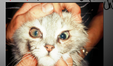
what is this?
Uveitis
what causes Uveitis
Uveitis can be caused by trauma, viral infections, autoimmune reactions, and systemic diseases that affect the uveal tract.
What type of pressure does Uveitis have
low ocular pressure
ocular neoplasia
Ocular neoplasia refers to the presence of tumors in or around the eye, which can be malignant or benign and may affect vision and surrounding tissues.
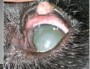
what is this?
ocular neoplasia
Proptosis
When the globe of the eye moves forward out if its orbit
Globe is luxated or displaced
Causes lots of swelling and is painful
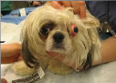
what is this?
Proptosis
What is proptosis causes by
HBC and big dog little dog fight
Brachycephalic dogs are prone to this
Trauma
Conformation
Retrobulbar abscess or neoplasia
What are the clinical signs of proptosed
Protrusion of the globe ( coming out of the socket real gross)
Eyelids unable to close may be trapped behind globe
Treatment of proptosed
Lubricate immediately
Reduce the globe into the socket ( put that eye back in) ASAP to reduce trauma to optic nerve, if the eye is viable
Enucleation if optic nerve severed
Systemic and topical antibiotics
entropion
Eyelids that roll in against the cornea
Caused by: Congenital, Trauma causing scarring and distortion of life
Severe blepharospasm ( eyelid open close/ blink all the time) from pain
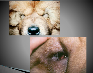
What is this?
entropion
What are the signs of entropion
Epiphora
Chemosis
Conjunctivitis
Pain
Corneal ulceration
Photophobia
Ectropion
Eyelids rolling away from the eye
Caused by congenital defects or an over- correction of entropion
Seen in Great Danes and Bassett hounds
What are the signs of ectropion
Conjunctivitis
Epiphora
Keratitis ( corneal inflammation/ scarring)
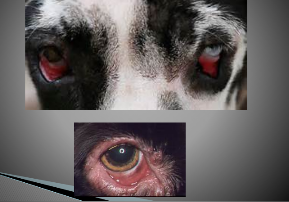
what is this
ectropion
What is the treatment of ectropion
Surgery to shorten the eyelid
Not needed unless the condition is very bad
Chronic superficial keratitis ( pannus)
Superficial corneal vascularization/scar tissue
it is Progressive, bilateral, can result in blindness
what is Chronic superficial keratitis ( pannus) caused by
Is an immune-mediated disease ( infiltration of cornea with lymphocytes, plasma cells)
Ultraviolet light/high altitudes increases incidence
What are the signs of chronic superficial keratitis ( pannus)
Opaque lesions that begin at limbus and extend into cornea
Milky, pink or tan lenses
What dog breed commonly gets chronic superficial keratitis
German Shepards
What is the treatment for chronic superficial keratitis ( pannus)
Corticosteroids often lifelong
Cyclosporine often lifelong
Antibiotic eye ointment
Information clients need to know about Pannnus
There is no cure
If treatment is stopped, disease will return and progress
High altitudes and sun will predispose animals to it
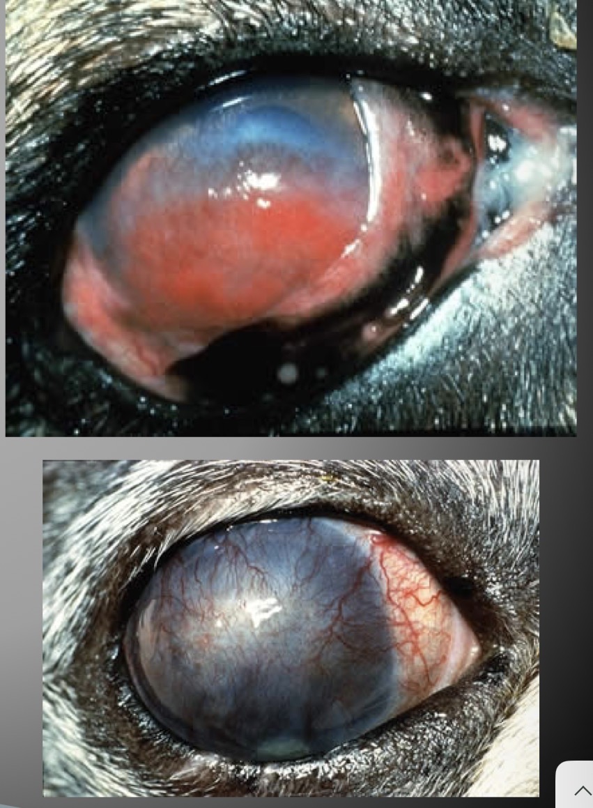
What is this
Chronic superficial keratitis
Progressive Retinal atrophy
A group of hereditary disorders causing loss of rods, cones and/or blood supply to the retina which leads to progressive vision loss.
Signs of progressive retinal atrophy
Slow onset of blindness
Loss of night vision ( rods)
Loss of day vision ( cones)
How to diagnosis progressive retinal atrophy
Ophthalmic exam
the retina will be Gray, and have a granular appearance
Narrowing of blood vessels will be seen and the optic nerve will be atrophy
Is there a treatment for progressive retinal atrophy
No there is no treatment
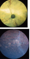
what is this?
progressive retinal atrophy
Information that should be given to the client about Progressive Retinal Atrophy
It is an inherited disease
Avoid buying affects dogs
Have ocular exam by board certified ophthalmologist
Blind animals will adapt well, but may have trouble with strange surrounding
Cats needs a well balanced diet
Taurine deficiency can lead to PRA
Detached Retina
Usually related to trauma
Retina will look folded down form the back of the eye
Animal will appear blind in affected eye
No treatment
Does Detached retina have a treatment
No there is no treatment
What test measures tear production
Schirmer tear test
What test is used to diagnose glaucoma or uveitis
IOP
What test is used to diagnose corneal ulcers
Fluorescein Stain
What is the scientific term for dry eye
Keratoconjunctivitis Sicca (KCS)
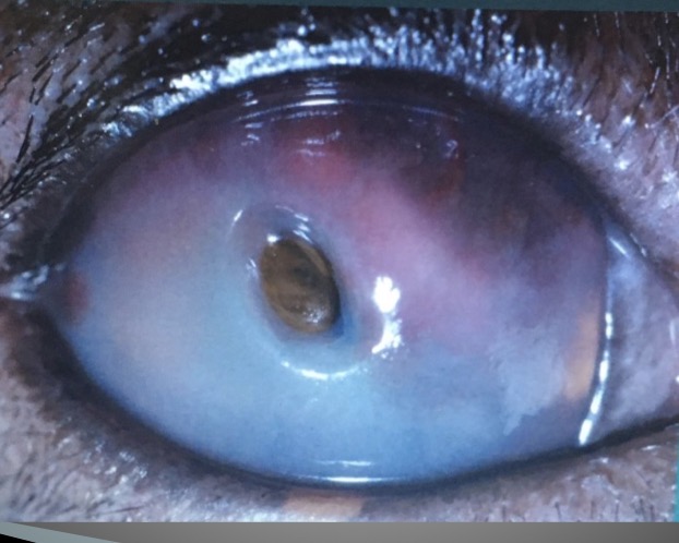
What is this
Descemetoceles
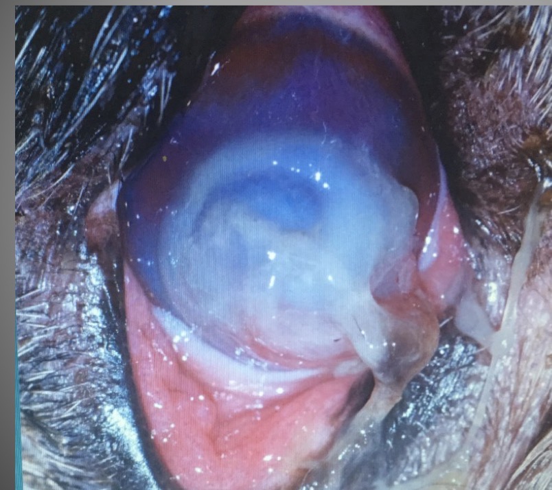
What is this
Melting ulcer
Blepharospasm
Eye that is painful but they keep blinking repeatedly
Strabismus or Nystagmus
Cross eyes
May happen in many different directions
What does an ocular physical exam include
Palpebral reflex
Menace response
Check pupil symmetry
Pupillary light reflexes
Maze
Schirmer tear test
Fluorescein stain
Tonometry
Schirmer tear test ( STT)
Always do this first
Measures tear production
Less than 10mm/Min is a KCS diagnostic
10-15 mm/min is a gray area
Normal > 18-23 mm/min
what is a normal STT
18-23 mm/min
Fluorescein stain test
Detects defects in the cornea
After flushing the eye and any stain is retained is diagnostic as a corneal ulcer
tonometry ( Tonopen)
Measures intraocular pressure ( IOP)
Normal ranges are 15-25 mmHG
What are the normal ranges of foR IOP
15-25 mmHG
How does the Fluorescein stain work
Corneal Structure: The outermost layer of the cornea, the epithelial layer, is composed of lipids. Beneath this is the stroma, which is water-soluble, and then Descemet's membrane, which is lipid.
Intact Cornea: In an intact, healthy cornea with no defects, when the water-soluble fluorescein stain is applied, the lipid epithelial layer will repel it, causing the stain to simply wash off.
Corneal Defects (Erosions/Ulcers): If there is an erosion (a superficial defect in the epithelial layer) or an ulcer (a deeper defect), the lipid epithelial layer is compromised or gone. This allows the water-soluble fluorescein to penetrate and bind to the exposed, water-soluble stroma underneath.
When fluorescein stain is applied to the cornea, the lipid epithelial layer repels the stain if intact, causing it to wash off. In the presence of corneal defects, such as erosions or ulcers, the stain penetrates the compromised epithelial layer and binds to the stroma, making defects visible.
Rose Bengal stain
Used to determine the mucin composition of the tear film ( defect in aqueous part)
Used in feline herpes virus corneal ulcers ( more superficial erosions/ulcers)
Used to help diagnose KCS TOO ( Used the same way as the other stain)
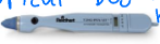
Tonopen
Requires topical anesthetic to measure intraocular pressure using a small, handheld device that gently applanates on the cornea.