Spatial resolution
1/61
There's no tags or description
Looks like no tags are added yet.
Name | Mastery | Learn | Test | Matching | Spaced | Call with Kai |
|---|
No analytics yet
Send a link to your students to track their progress
62 Terms
Spatial resolution aka
sharpness, definition, recorded detail, or detail
Spatial resolution
degree of sharpness or accuracy of the anatomical structure lines in an image
Film-screen unit of resolution
line pairs per mm (Ip/mm) or cycles per mm
Resolution test tools
set of lines that have some distances from one another
the closer the lines are to one another, the better the spatial resolution
the point where the viewer can see the close line pairs apart is the Ip/mm reading (limited to range of 5)
systems cannot provide this lvl of resolution
the greater the Ip/mm, the higher the resolution
when Ip/mm increase
resolution increase
reciprocal of line pairs per mm
number of line pairs (usually 1) / number of lines
spatial resolution determined by
matrix size, pixel size, and grayscale bit depth (aka spatial frequency)
higher the spatial resolution/frequency
pairs of lines are very close together
when two objects can be seen smaller and closer together
spatial resolution high
when image lacks sharp definitions of fine details
poor resolution, caused by penumbra
realistically regarding information from images
some information in images are always lost
factors like patient movement increase unsharpness
assessing spatial resolution
expressed in DR images as x-axis, y-axis, and grayscale bit depth (z-axis)
z-axis (aka grayscale bit depth)
the different shades of gray in an image represents the added depth to an image
comes from combining the gray levels of each pixel
digital imaging characteristics
recorded as a matrix (combo of rows and columns of small squares called pixels)
each pixel has a number representing the amount of brightness
pixel location on the matrix represents area within patient or volume of tissue
field of view (FOV)
dimensions of anatomic area
greater the matrix size
greater amount of pixels, better resolution
relationship between pixel size, FOV and matrix size (equation)
pixel size = FOV/matrix size
bit depth
each pixel has bit depth
uses system expressed as 2^n (n= # of bits)
large bit depth, greater number of shades of gray, better resolution
point spread function (PSF)
measures penumbra
quantify digital system spatial resolution
uses a single point from an extended line
smaller PSF, less blur, better resolution
Line spread function (LSF)
uses a narrow slit in a sheet of lead
Edge spread function (ESF)
uses sharp edge instead of a line or point
line spread function (LSF), edge spread function (ESF) and PSF
express the boundaries/edges of the image
penumbra or blur
PSF in other modalities
X-ray: tiny hole in lead sheet then producing x-ray image of the hole
NM: point source of radioactivity
CT: a thin metal wire
US: a monofilament
MRI: water filled hole in a phantom
PSF graph
narrower the peak of the graph, better spatial resolution
spatial frequency
high freq signals, high spatial resolution, better image of smaller objects
modulation transfer function (MTF)
measures the accuracy of an image compared to the original object from a scale of 0-1
fidelity (or trueness) of an image
record available spatial freq
the overall values of PSF, LSF, and ESF
All system components must be working or else fidelity of patient is less accurate and MTF decrease
High MTF at high spatial resolution are desired
MTF decrease
when spatial freq increase but spatial resolution is high
system noise
noise coming from digital equipment
Ambient noise
noise coming from radiation in the background
quantum noise
lack of quantity of photons causing quantum mottle
improper kvp and mas
signal to noise ratio (SNR)
the amount of radiation exposure to the detector (signal) compared to the noise
high SNR means signal stronger than noise
low SNR means noise and signal about the same (not good)
higher the SNR, better image will be, better spatial resolution
contrast to noise ratio (CNR)
how big the differences bw contrast resolution and noise are
high CNR, stronger contrast than noise, better resolution
low CNR, contrast weak/ too much noise, low resolution/blurry image
Digital sampling (nyquist criterion)
bc digital systems collect signals at discrete points, spatial resolution freq signals must be sampled twice per cycle
think of it as snapshots for a moving image (more snapshots taken of the image, more detailing)
more sampling, less details missing
uses the spacing between detector elements (or pixels) to figure out how often to take samples so that info is captured correctly
Aliasing
not enough sampling causing misinterpretation of the data
also occurs when the spacing between the detector elements (pixels) are too far apart
Factors affecting spatial resolution
motion
OID
focal spot size
intensifying screen
phosphor size
concentration
SID
Geometry of recorded detail
focal spot size and distance (SID and OID)
bc xray coming out of small area (focal spot) photons will diverge from source
(think cone shape and the tip of the cone is the x-ray tube)
distance affects on spatial resolution
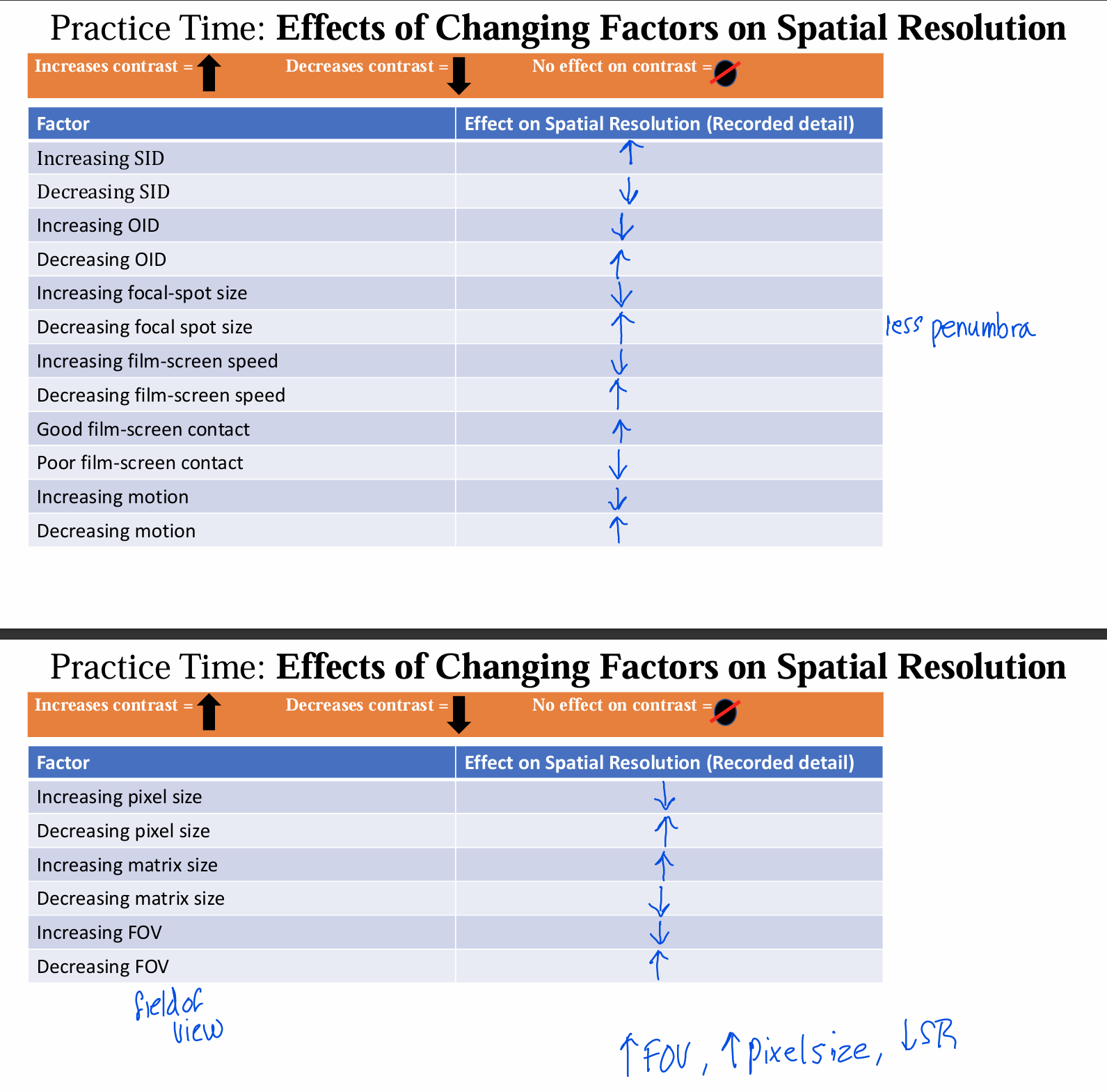
SID increase, spatial resolution increase
OID increase, spatial resolution decrease
focal spot size affects on spatial resolution
controlled by line focus principle
controls penumbra
focal spot size increase, penumbra increase, spatial resolution decrease
width of penumbra (formula)
penumbra= (focal spot size x OID)/ SOD
penumbra
unsharpness around the edges of an image
umbra is the sharp area of a shadow
penumbra surrounds umbra
attenuation absorption unsharpness
increases penumbra due to beam divergence as its attenuated by an object
too much space between the beam divergence and the object sides cause penumbra increase
IR of recorded detail
based off film screen systems or digital systems
digital systems: DR
uses silicon or selenium detectors
silicon limited by fill factor (quantity of photons that gets registered by detector)
high fill factor, high resolution
selenium detectors are thicker and can’t be small like silicon
both are limited by the size of the detector element
image processing also limits: matrix size, pixel size, and grayscale bit depth (xyz)
digital systems: CR
limitations similar to intensifying screens
phosphor size, layer thickness, and concentration, and image reader device (IRD)
film screen systems
classified by speed
when film screen speed increases, resolution decreases
intensifying screens cause poor resolution, to make it up, 20-100x mAs required
resolving power depend on phosphor size, phosphor layer thickness, and phosphor concentration
radiographic film construction
made up of the base and the emulsion
the base
polyester sheet
between two emulsions
dimensional stability: keeps size and shape through all processing conditions (temperature, pH, etc.)
blue tint added to base to enhance contrast, make image more pleasing, and absorb 15% of the light
the emulsion
gelatin substance surrounding the base
crystals of silver bromine in gelatin
gelatin characteristics: colloid (capable of suspending the crystals and coating each to separate them) amphoteric (can be used with acids or alkalis)
speed of film
relationship between exposure a film receives and amount of density called film speed
when film speed increase, resolution decrease
when density increase, sensitivity increase, resolution decrease
more exposure latitude allows more wiggle room
intensifying screens
made up of base and active layer
phosphor emits light when crystal hit with xray photons
light from intensifying screen exposes the film and make an image
problem with intensifying screen: light diffusion
decreases resolution bc of the distance between the film and phosphor, causing the light to spread out on the film surface
when light diffusion increase, resolution decrease
when crystal size increase, light diffusion increase, resolution decrease
problem with intensifying screen: screen speed
higher screen speed, more light emitted, resolution decrease
fast screens have larger phosphor crystals or have more phosphor layers, which produce more light and cause less resolution
problem with intensifying screen: poor screen contact
when distance between the film and screen increase, light diffusion increase, and resolution decrease
caused by dropping cassettes or bending them
when phosphor size increase
resolution decrease, patient dose decrease, film density increase
when layer thickness of phosphor increase
resolution decrease, patient dose decrease, film density increase
when phosphor concentration increases
resolution increases (bc crystals are getting packed closer together and decreases light diffusion), patient dose decreases, film density increases
quantum mottle and intensifying screens
when mAs is not increased when using high speed intensifying screens, not enough photons will reach the film causing quantum mottle
motion affecting spatial resolution
voluntary, involuntary, equipment
voluntary motion
patient has direct control of movement
communication is key
sometimes requires immobilization
involuntary motion
patient not in control of motion
decreasing exposure time or increasing kVp with mAs decrease (15% rule)
equipment motion
vibrations in the machine or x ray tube suspension system
report problem and don’t use machine till fixed
effects of changing factors on spatial resolution chart
