Animal Phys Exam 4
 Call Kai
 Learn
Learn Practice Test
 Spaced Repetition
 Match
Match Flashcards
 Knowt Play
1/226

There's no tags or description
Looks like no tags are added yet.
Name | Mastery | Learn | Test | Matching | Spaced | Call with Kai | Chat |
|---|
No analytics yet
Send a link to your students to track their progress
227 Terms
Cardiac Cycle
The process by which the heart cycles between contraction (systole) and relaxation (diastole).
Ventricular Filling (Phase 1)
Late diastole; both sets of chambers are relaxed and ventricles fill passively.
Isovolumetric Contraction (Phase 2)
The first phase of ventricular contraction; it pushes AV valves closed but does not create enough pressure to open the semilunar valves.
End-Diastolic Volume (EDV)
The maximum blood volume in the ventricles, reached during isovolumetric contraction.
Ventricular Ejection (Phase 3)
As ventricular pressure rises and exceeds pressure in the arteries, the semilunar valves open and blood is ejected.
Isovolumetric Relaxation (Phase 4)
As ventricles relax, pressure in the ventricles falls. Blood flows back into the cusps of the semilunar valves, snapping them closed.
End-Systolic Volume (ESV)
The minimum blood volume in the ventricles, reached during isovolumetric relaxation.
P Wave
Part of the ECG corresponding to the start of atrial systole.
QRS Complex
Part of the ECG associated with ventricular depolarization.
T Wave
Part of the ECG corresponding to the later stages of ventricular systole and the start of diastole.
Dicrotic Notch
A small plateau or dip in the aortic pressure curve, caused by the closure of the aortic valve.
Heart Sounds (S1)
Associated with the closing of the AV valves (start of ventricular systole).
Heart Sounds (S2)
Associated with the closing of the semilunar valves (start of ventricular diastole).
Cardiac Output (CO)
A measure of cardiac performance; defined as the volume of blood pumped by one ventricle in a given period of time. Formula: CO = Heart Rate * Stroke Volume. Average is 5 L/min.
Stroke Volume (SV)
The volume of blood pumped per contraction. Formula: SV = EDV - ESV. Average is 70 mL (for a 70-kg man at rest).
Contractility
The intrinsic ability of a cardiac muscle fiber to contract at any given fiber length.
Length-Tension Relationships
Determined by the volume of blood at the beginning of contraction; the degree of stretch is called preload.
Preload
The degree of stretch on the cardiac muscle fibers before they contract; determined by the volume of blood at the beginning of contraction.
Frank-Starling Law of the Heart
Stroke volume is proportional to EDV; the heart pumps all the blood that is returned to it.
Venous Return
The amount of blood returning to the heart, which determines EDV. It is affected by: (1) skeletal muscle pump, (2) respiratory pump, and (3) sympathetic innervation of veins.
Afterload
The combined load of EDV and arterial resistance during ventricular contraction.
Ejection Fraction
The percentage of EDV ejected with one contraction. Formula: Stroke Volume / EDV.
Inotropic Agent
Any chemical that affects contractility, producing an inotropic effect.
Positive Inotropes
Chemicals that increase contractility (e.g., epinephrine, norepinephrine, and digitalis). They work by increasing Ca2+ storage with phospholamban.
Negative Inotropes
Chemicals that decrease contractility.
Norepinephrine/Epinephrine (Effect on Heart Rate)
Increases heart rate by binding to adrenergic beta-receptors and activating the adenylate cyclase (AC) signal transduction pathway. This opens cation (funny) and T-type Ca2+ channels, increasing the rate of depolarization of the pacemaker potential.
Acetylcholine (Effect on Heart Rate)
Decreases heart rate by binding to muscarinic receptors. This activates a signal transduction pathway that closes Ca2+ channels and opens K+ channels. This prevents Ca2+ from entering and allows K+ to exit, causing a net hyperpolarization, which increases the time needed for the pacemaker potential to depolarize the cell to threshold.
Parasympathetic Control
Decreases heart rate: K+ permeability increases resulting in hyperpolarization, and Ca2+ permeability decreases which slows the rate of depolarization.
Sympathetic Control
Increases heart rate: Beta-1 adrenergic receptors on the autorhythmic cells are activated, increasing Na+ and Ca2+ permeability.
Phospholamban
A regulatory protein that alters sarcoplasmic reticulum (SR) Ca2+-ATPase activity.
Catecholamine Pathway (Cardiac Contraction)
Epinephrine/Norepinephrine bind to beta-1 receptors -> activate cAMP second messenger system -> phosphorylates (1) Voltage-gated Ca2+ channels (increases open time, increasing Ca2+ entry from ECF) and (2) Phospholamban (increases Ca2+-ATPase on SR, increasing Ca2+ stores in SR and removing Ca2+ from cytosol faster). This results in a more forceful contraction and shorter duration of contraction.
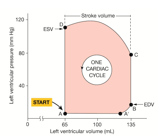
Stroke Volume Cardiac Cycle Graph
Ventricular Filling: A-B
Ventricular Ejection: C-D
Ventricular Isovolumic Contraction: B-C
Ventricular Isovolumic Relaxation: D-A
AV Closing: B
AV Opening: A
SL Closing: D
SL Opening: C
ESV: D-A
EDV: B-C
Atrial Contraction: A-B
Systolic Blood Pressure - 120
Diastolic Blood Pressure - 80
First Heart Sound(lub):
Second Heart Sound(dup):
Stroke Volume: C-D or EDV - ESV
Hemodynamics
The principles that govern blood flow in the cardiovascular system.
Blood Flow (Q)
The volume of blood that passes a given point per unit of time, determined by the pressure gradient and resistance to blood flow. Formula: Q = (P1 - P2) / R.
Velocity of Flow (v)
Measures how fast blood flows past a point, calculated as flow rate divided by cross-sectional area: v = Q / A.
Flow Rate vs. Velocity Relationship
Flow rate refers to volume over time, while velocity describes distance over time, increasing in narrower vessels.
Direction of Blood Flow
Blood flows from areas of higher pressure in the heart into lower pressure vessels, forming a closed loop.
Fluid Motion Pressure Loss
Pressure in a moving fluid decreases with distance due to friction in the vessels.
Hydrostatic Pressure
The pressure exerted by a fluid at rest, uniformly in all directions.
Resistance (R)
Opposition to blood flow, calculated using the formula R = 8ηL / (πr⁴).
Flow-Resistance Proportionality
Flow is inversely proportional to resistance (Flow ∝ 1/R).
Flow-Pressure Proportionality
Flow is directly proportional to the pressure gradient divided by resistance (Flow ∝ ΔP / R).
Poiseuille’s Law
Resistance is proportional to vessel length and fluid viscosity, and inversely proportional to the radius to the fourth power (R ∝ Lη / r⁴).
Vessel Length and Resistance Relationship
Resistance increases with the length of the blood vessel.
Fluid Viscosity and Resistance Relationship
Resistance increases with the thickness (viscosity) of the fluid.
Vessel Radius and Resistance Relationship
Resistance decreases as the vessel radius increases (R ∝ 1/r⁴).
Blood Vessel Wall Composition
Walls consist of smooth muscle, elastic connective tissue, and fibrous connective tissue, varying among vessel types.
Lumen
The internal open space of a blood vessel through which blood flows.
Endothelium
The inner layer of blood vessels that regulates blood pressure, growth, and absorption.
Vascular Smooth Muscle
Muscle tissue in blood vessel walls responsible for regulating vessel diameter via contraction.
Muscle Tone
The state of partial contraction maintained by vascular smooth muscle.
The Three Tunics (Vessel Layers)
Tunica intima; 2. Tunica media; 3. Tunica externa.
Capillary Wall Structure
Microscopic vessels with only a single layer of flattened endothelial cells.
Three Main Groups of Arteries
Elastic arteries, muscular arteries, and arterioles, categorized by size and function.
Elastic Arteries
Large arteries like the aorta with high elastic tissue content, serving as pressure reserves.
Pressure Reserve Mechanism
Blood pressure is stored in the elastic arteries during ventricular contraction and released during relaxation.
Muscular Arteries
Distributing arteries with more smooth muscle, involved in vasoconstriction.
Arterioles
Smallest arteries leading into capillary beds, primarily regulating blood flow.
Resistance Arteries
Arterioles, named for their role in changing resistance to control blood flow.
Capillaries (General Characteristics)
Microscopic vessels with small diameters, ensuring single RBC passage.
Capillary Function
Main site for exchanging gases, nutrients, and wastes between blood and tissues.
Capillary Density
The concentration of capillaries in a tissue area, linked to tissue metabolic activity.
Three Types of Capillaries
Continuous; 2. Fenestrated; 3. Sinusoidal.
Capillary Bed
Network of capillaries between arterioles and venules.
Postcapillary Venule
Vessels capillaries drain into after exiting a capillary bed.
Capillary Bed Flow Control
Blood flow is regulated by the diameter of terminal arterioles.
Local Chemical & Vasomotor Regulation
Local conditions and vasomotor fibers regulate blood entry into capillary beds.
Vascular Shunt
A capillary arrangement that directly connects an arteriole and a venule.
Precapillary Sphincter
Smooth muscle cuff regulating blood flow into true capillaries based on local chemical conditions.
Venules
Small vessels formed from united capillaries, allowing fluid movement into tissues.
Veins (Pressure and Resistance)
Lower pressure vessels with large diameters enabling blood return to the heart.
Venous Valves
Prevent backflow of blood in veins, especially in limbs.
Systemic Circulation Pressure Waves
Pressure waves from ventricular contraction that diminish in amplitude with distance.
Pulse Pressure
The difference between systolic and diastolic blood pressure.
Mean Arterial Pressure (MAP) Equation
MAP = diastolic pressure + (1/3) (pulse pressure).
Basic Determinants of Mean Arterial Pressure
MAP is determined by blood volume, cardiac output, resistance, and blood distribution.
Blood Volume Determinants
Fluid intake and loss balance blood volume.
Effectiveness of the Heart as a Pump (Cardiac Output) Determinants
Heart rate and stroke volume dictate cardiac output.
Resistance of the System to Blood Flow Determinants
Arterial diameter affects systemic peripheral resistance.
Relative Distribution of Blood Determinants
Arterial and venous diameter changes shift blood volume distribution.
Sphygmomanometry
Method of measuring arterial blood pressure using a cuff and gauge.
Korotkoff Sounds
Sounds heard during sphygmomanometry, indicating changes in blood flow through a partially compressed artery.
Variable Blood Distribution
Blood distribution changes based on tissue metabolic needs.
Cerebral Blood Flow
The consistent supply of blood to the brain, unaffected by systemic demands.
Coronary Blood Flow Regulation
Coronary blood flow matches heart workload; low tissue oxygen triggers arterial dilation.
Arteriolar Resistance Influences
Arteriolar resistance is regulated by local and systemic control mechanisms.
Systemic Control: Sympathetic Reflexes
Neural control over blood vessels mediated by adrenergic receptors.
Systemic Control: Hormonal Regulation
Hormones like vasopressin and angiotensin II adjust vessel diameters.
Local Control: Myogenic Autoregulation
Vascular muscle regulates contraction based on pressure stretch.
Local Control: Paracrine Signals
Chemical signals that adjust vessel tone, such as nitric oxide.
Local Control: Metabolic Indicators
Chemical signals from active tissues that alter blood flow.
Cardiovascular Control Center (CVCC)
The central brainstem structure coordinating cardiovascular function.
Baroreceptor Reflex
Autonomic reflex controlling systemic blood pressure based on mechanoreceptor feedback.
Orthostatic Hypotension
A sudden drop in blood pressure from rapid postural changes.
Capillary Velocity Principles
At constant flow, capillary velocity is highest in arteries and slowest in capillaries.
Capillary Fluid Exchange Formula
Net Pressure across a capillary wall: Net Pressure = Hydrostatic Pressure - Colloid Osmotic Pressure.
Filtration vs. Absorption
Directional fluid movement depending on net pressure across capillary walls.
Net Systemic Fluid Flow Out
Slightly more filtration than absorption in capillaries results in 3 L/day fluid movement into tissues.
Lymphatic System Functions
Retrieves fluids and proteins, absorbs fats, and filters pathogens.
Lymphatic Architecture
An open-ended system with blind-ended lymph capillaries and semilunar valves.