Functional Anatomy 1 - Lecture 1b
1/77
There's no tags or description
Looks like no tags are added yet.
Name | Mastery | Learn | Test | Matching | Spaced | Call with Kai | Chat |
|---|
No analytics yet
Send a link to your students to track their progress
78 Terms
Anatomical Position
The standard position of the body used as a reference point in anatomy; the individual stands upright, facing forward, with arms at the sides and palms facing forward.
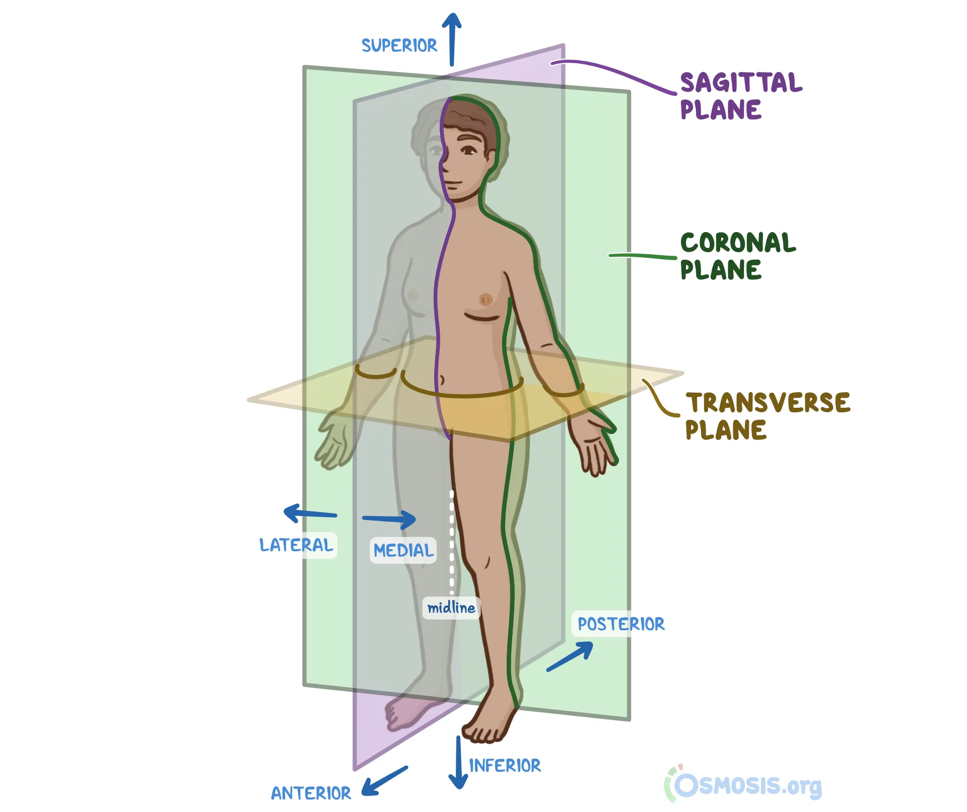
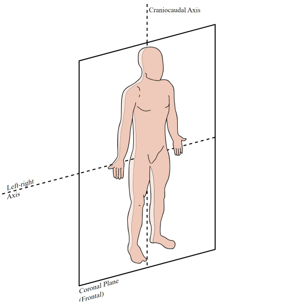
frontal (coronal) plane
An anatomical plane that divides the body into anterior and posterior sections
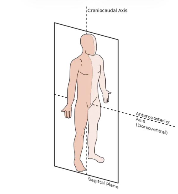
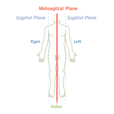
Sagittal Plane
Divides the body into left and right sections, can be anywhere on the body
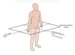
transverse (axial) plane
Divides the body superiorly and inferiorly
Median sagittal plane
sagittal plane always midline
Anteroposterior (AP) axis
axis of the frontal plane
Mediolateral (ML) axis
axis of the sagittal plane
Vertical axis
axis of the transverse plane
superficial
nearer to surface
intermediate
between a superficial and a deep structure
deep
farther from surface
medial
nearer to median plane
lateral
farther from median plane
posterior (dorsal)
nearer to back
superior (cranial)
nearer to head
anterior (ventral)
nearer to front
distal
farther from trunk or point of origin (ex: of a limb)
proximal
nearer to trunk or point of origin (ex: of a limb)
plantar
inferior foot surface (sole)
dorsal
superior foot surface (dorsum)
palmar
anterior hand (palm)
dorsal (hand)
posterior hand (dorsum)
inferior (caudal)
nearer to feet
bilateral
both sides of the body
unilateral
one side of the body
ipsilateral
towards same side of reference structure
contralateral
away from/opposite side of reference structure
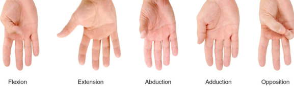
flexion/extension
movement in the sagittal plane rotating around a medial/lateral axis
abduction/adduction
movement in the frontal plane rotating around the AP axis
abduction
moving away from midline
adduction
moving toward midline
internal/external rotation
movement in the transverse plane rotating around a superior/inferior (longitudinal) axis
sagittal plane joint motions
a. flexion and extension of upper limb at shoulder joint and lower limb at hip joint
b. flexion and extension of forearm at elbow joint and of leg at knee joint
c. flexion and extension of vertebral column at intervertebral joints
d. flexion and extension of hand at wrist joint
e. dorsiflexion and plantarflexion of foot at ankle joint
frontal plane joint motions
abduction
adduction
lateral bending
transverse plane joint motions
rotation of the head, neck, lower limb
lateral and medial rotation
circumduction
a combination of flexion, extension, abduction, adduction
ipsilateral rotation
ex: muscle on right side rotating to right
contralateral rotation
ex: muscle on left rotating to the right
supination
forearm rotates palm upward
pronation
forearm turns palm of hand downwards
eversion of foot
turns sole outward, away from midline
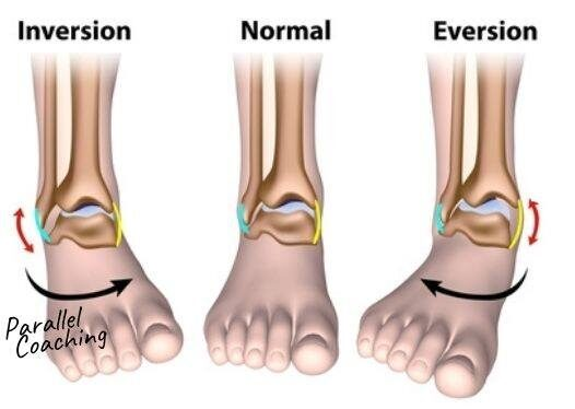
inversion of foot
turns sole of food inward, toward the midline

opposition
brings pad of thumb into direct contact with pads of other fingers on same hand, essential for fine motor skills
reposition
returns thumb back to neutral, resting anatomical position
thumb movements
Axial Skeleton
skull, vertebrae, ribs
Appendicular Skeleton
clavicle, scapula, pelvis, upper extremities, lower extremities
fibrous joints
bones united by fibrous tissue
variable amount of movement within different types
no joint capsule
direct connection between bones via fibrous tissue
examples: sutures, gomphosis (tooth), syndesmosis (radioulnar)
suture
the apposed bony surfaces are united by fibrous tissue, permitting no movement, fibrous joint
ex: coronal
gomphosis
teeth, no movement, fibrous joint
syndesmosis
partially moveable, bones connected by interosseous membrane, fibrous joint
cartilaginous joints
bones are united by hyaline cartilage or fibrocartilage
no joint cavity or joint capsule, direct connection via tissue
2 types: synchondrosis, symphyses
hyaline cartilage
firm, lots of collagen, same cartilate that makes articular cartilage
fibrocartilage
flexible yet tough
synchondrosis
primary, hyaline cartilage, not to be confused with synarthroses
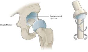
ex: head of femur
hyaline cartilage unites bone
growth plate - epiphyseal plate
permit growth of the bone, convert to solid bone after puberty
used to assess boney maturity
costochondral joints
between the anterior end of rib and costal cartilage
symphysis
secondary, fibrocartilage
ex: intervertebral disc
fibrocartilage unites bone
slightly movable joints that provide strength with flexibility
intervertebral discs or pubic symphysis
synovial joints
most common joint type in body
diarthroses - allow most motion
joint capsule
fibrous outer layer blends with periosteum, synovial membrane
joint cavity
synovial fluid secreted by synovial membrane
articular (hyaline) cartilage
covers articulating surfaces of the bone
reinforced with ligaments
diarthroses
allow most motion
synovial fluid
a viscous fluid
nourishes cartilage
lubricates joint surface
reinforcing ligaments
often are thickened parts of the fibrous capsule
connect bone to bone
some are intracapsular ligaments - located internal to the joint capsule
ACL, PCL
some are extracapsular ligaments - located outside the joint capsule
LCL, MCL
synovial joints are richly supplied with sensory nerves
in fibrous capsule and ligaments
proprioception
detect pain
monitor stretching of the capsule
synovial joints have a rich blood supply
synovial membrane and ligaments (not the cartilage)
extensive capillary beds produce basis of synovial fluid
some synovial joints contain an articular disc
temporomandibular joint, wrist, and knee joint
joints whose articulating bones have different shapes
help to increase joint congruency
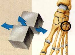
plane (gliding) joint
articular surfaces are flat planes
small gliding movements
intertarsal and intercarpal joints
scapulothoracic joint
movements are non-axial
gliding does not involve rotation around any axis
joints of hand, wrist, foot
hinge joint
cylindrical (convex) end of one bone fits into a trough (concave surface) on another bone
angular movement in one plane
elbow, ankle, knee, interphalangeal
movement is uniaxial (only flexion/extension)
strong lateral ligaments, bony structure
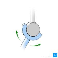
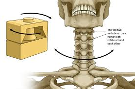
pivot joints
classified as uniaxial - rotating bone only turns around its long axis (only rotation)
examples
joint between atlas and axis (atlanto-axial joint)
proximal radioulnar joint
pronation and supination of forearm
condyloid joint
biaxial: movement occurs around 2 axes
wrist joint (radio/ulnar articulation with carpals)
MCP
MTP
one bone is concave, one is convex
allows
abduction/adduction (frontal plane)
flexion/extension (sagittal plane)
no rotation
similar to, but more restrictive than ball in socket joint
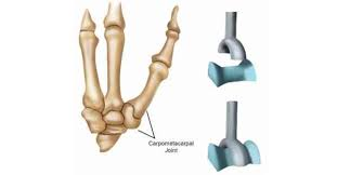
saddle joint
each articular surface has concave and convex surfaces
biaxial joints
allows flexion, extension, adduction, abduction, circumduction
no axial rotation
1st carpometacarpal joint
allows opposition of the thumb
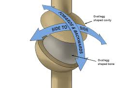
ball-and-socket joints
spherical head of one bone fits into round socket of another
classified as multiaxial
shoulder and hip
sacrifice stability for mobility
factors influencing stability of synovial joints
articular surfaces
shapes of articulating surfaces determine movements possible
ligaments
the more ligaments, usually stronger and more stable
what joint relies heavily on ligaments for support/function? knee and shoulder
muscle tone
helps stabilize joints by keeping tension on tendons
mobile
the more stable a joint, the less ____ it is
stable
the more mobile a joint, the less ____ it is
sheath
tendinous movement over joint
bursa
between bone and muscle, skin, tendons, or ligaments
fat pad
between joint/bone and tendons
articular disc, labrum
improve joint congruency, help shock absorption
osteokinematics
movements are described by the actual direction that the bones move and the axis about which they move
flexion of the forearm in the sagittal plane around the medial/lateral axis of the elbow
arthrokinematics
the movement of the joint may be different than the motion of the axial bones
relationship of the movement between the two joint surfaces
ex: roll, glide, slide