Kopp 2012 (SOX9)
 Call Kai
 Learn
Learn Practice Test
 Spaced Repetition
 Match
Match Flashcards
 Knowt Play
1/31

There's no tags or description
Looks like no tags are added yet.
Name | Mastery | Learn | Test | Matching | Spaced | Call with Kai | Chat |
|---|
No analytics yet
Send a link to your students to track their progress
32 Terms
What is the main finding of this paper?
1. Acinar cells turn into ductal cells with SOX9
2. When acinar cells acquire KRAS mutations, they turn into precursors of PDA
(not centroacinar or ductal cells)

Figure 1: main conclusion
SOX9+ state is associated with PDA initiation in early PanIN’s
What are IPMN’s?
Intraductal papillary mucinous neoplasms (very early stage)
What are MCN’s?
Mucinous Cystic Neoplasm
(early stage)
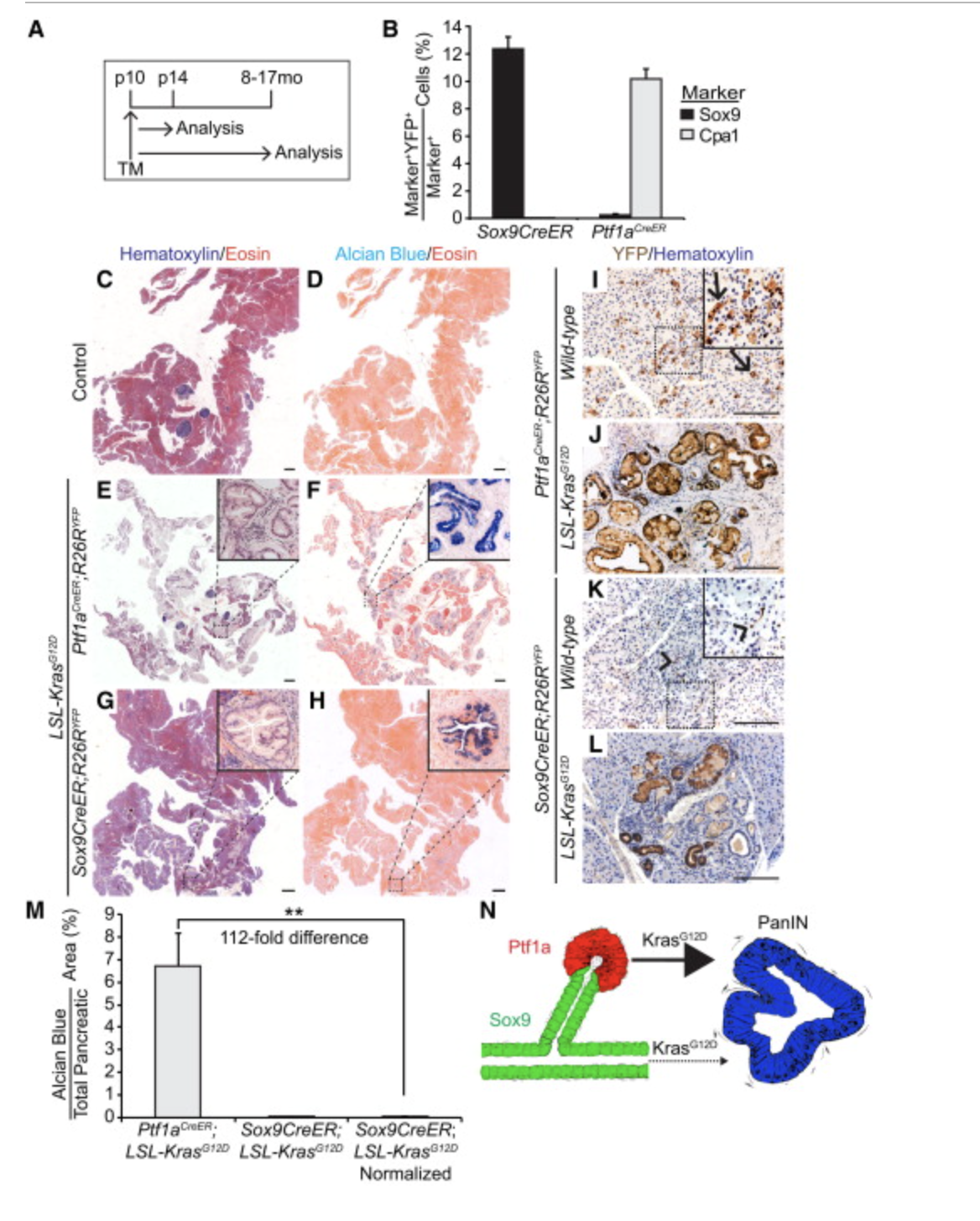
How do the mouse models in figure 2 work on basic level?
CRE-ER under PTF1A or SOX9
TM induces ER change, causing CRE to enter the nucleus
Activates onco-KRAS
Labels active cells YFP
Where is ptf1a expressed?
Acinar cells
Where is Sox9 expressed?
ductal cells

What is the main point of figure 2?
Acinar cells form PanINs more than ductal/CACs with oncogenic KRAS
What is Alcian blue staining for?
PanIN lesion that produce acidic mucins
(NOT ACINAR CELLS)
What does CPA1 do?
Acinar marker
(acidic)
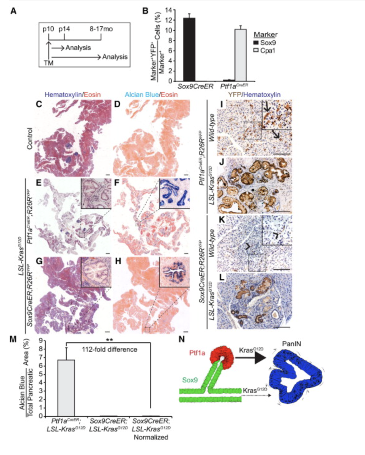
Which cells look better in figure 2 ptf1a or sox9?
sox9 (ductal) look less cancerous
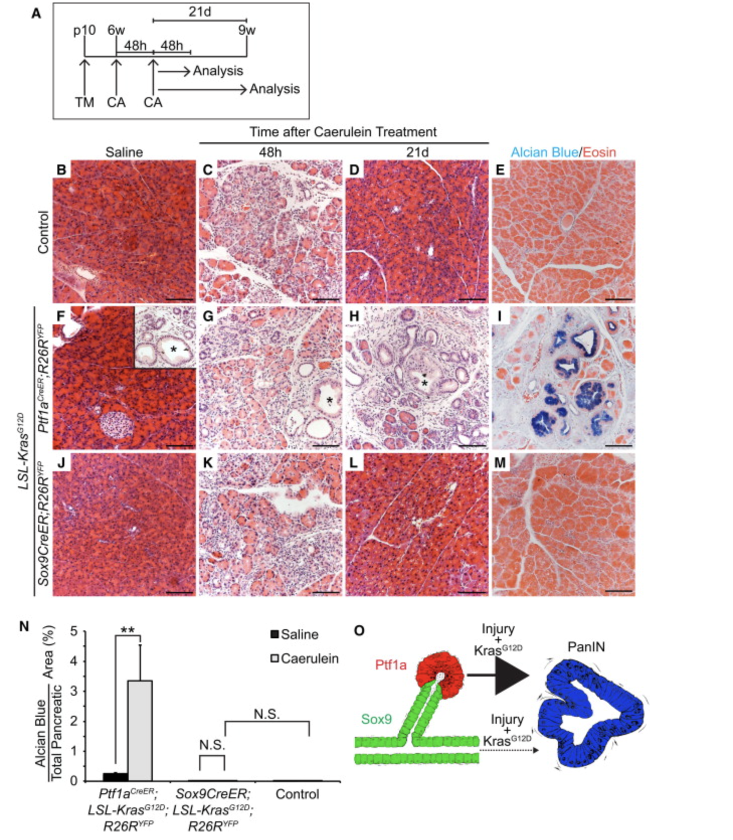
In figure 3 what does caerulein (CA) do?
induce acute pancreatitis
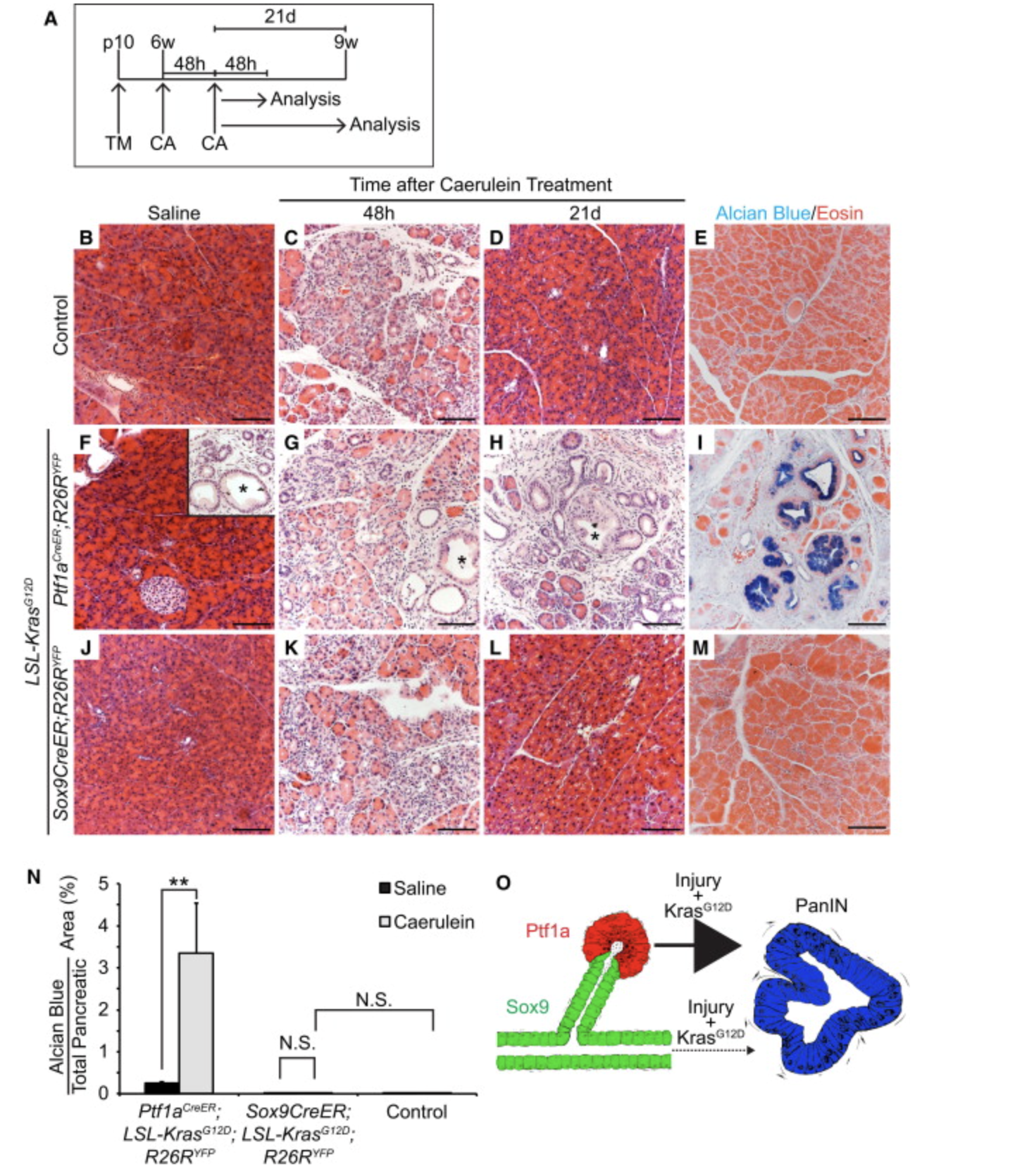
What is the take-away from figure 3?
PanIN formation in acinar cells, not ductal/CACs
(kras-g12d mediated)
What is
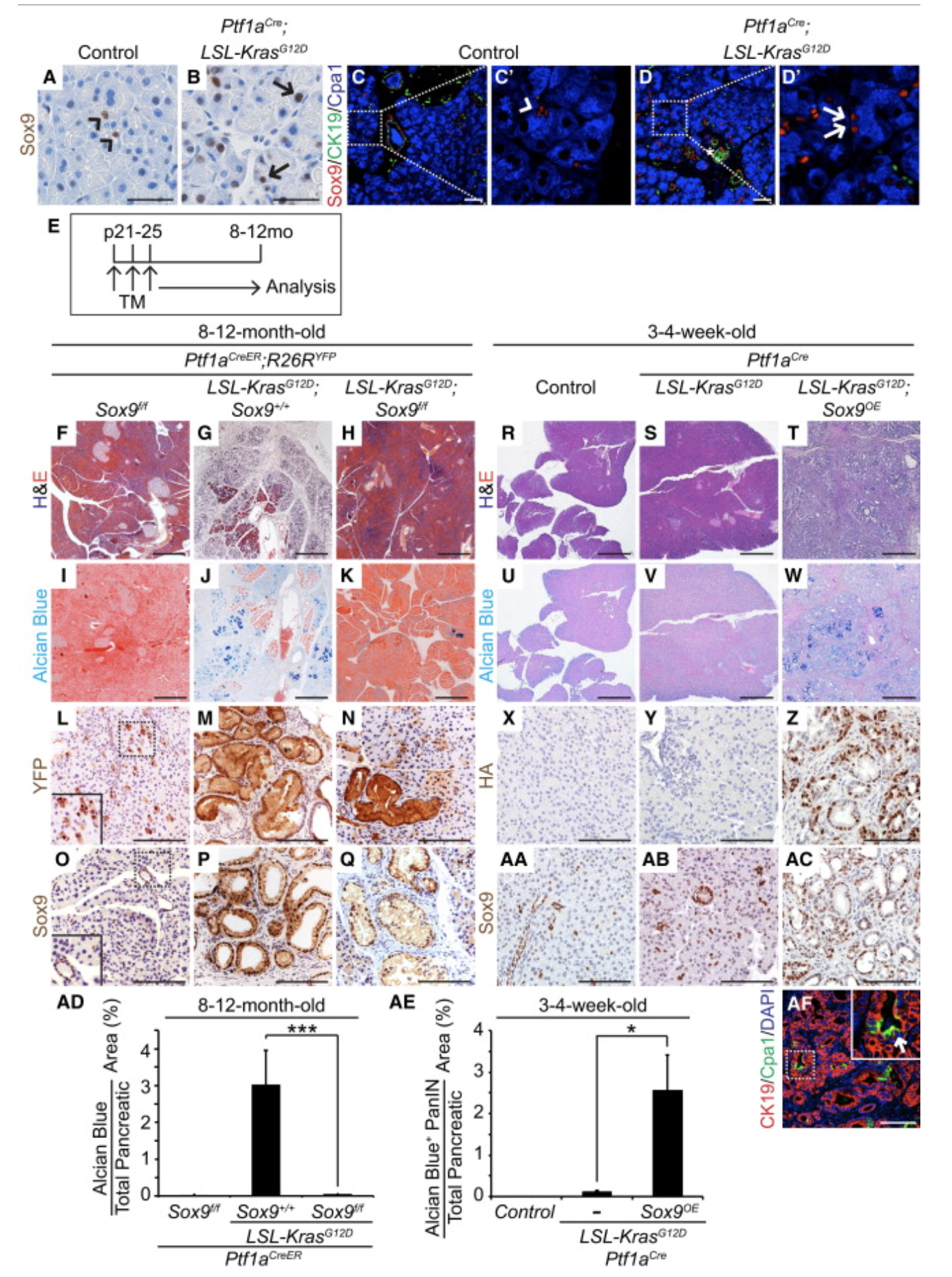
What model do they use for figure 4?
ptf1a acinar model
(ptf1a-lslKRAS)
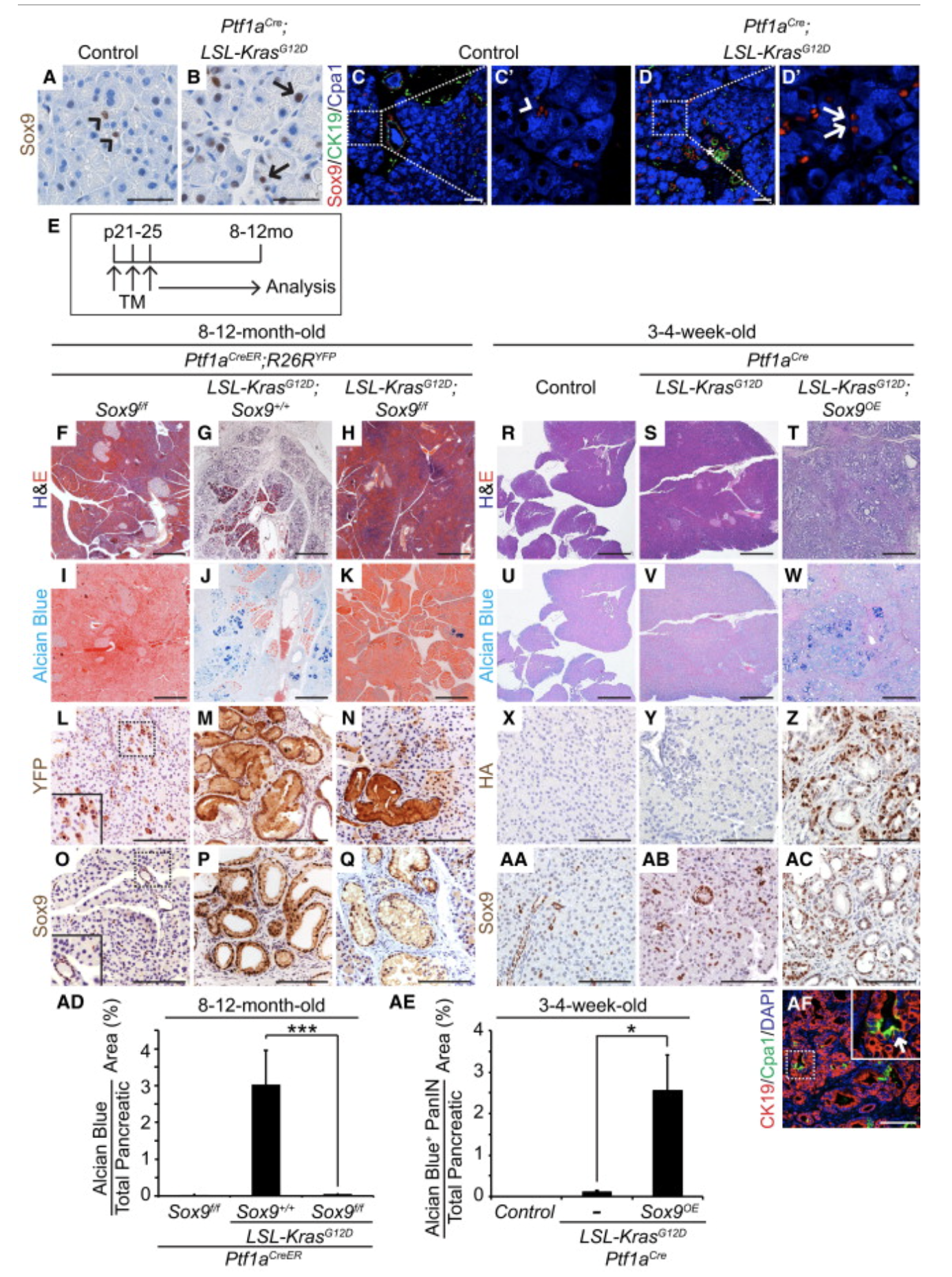
What is the middle pictures in figure 4 left side?
sox9+ cells look worse than sox9- cells
(sox9+ MORE yellow)

What is the middle pictures in figure 4 right side?
sox9 OE cells look worse than sox9 WT cells
(more yellow)
What is Ck19 a marker of?
ductal
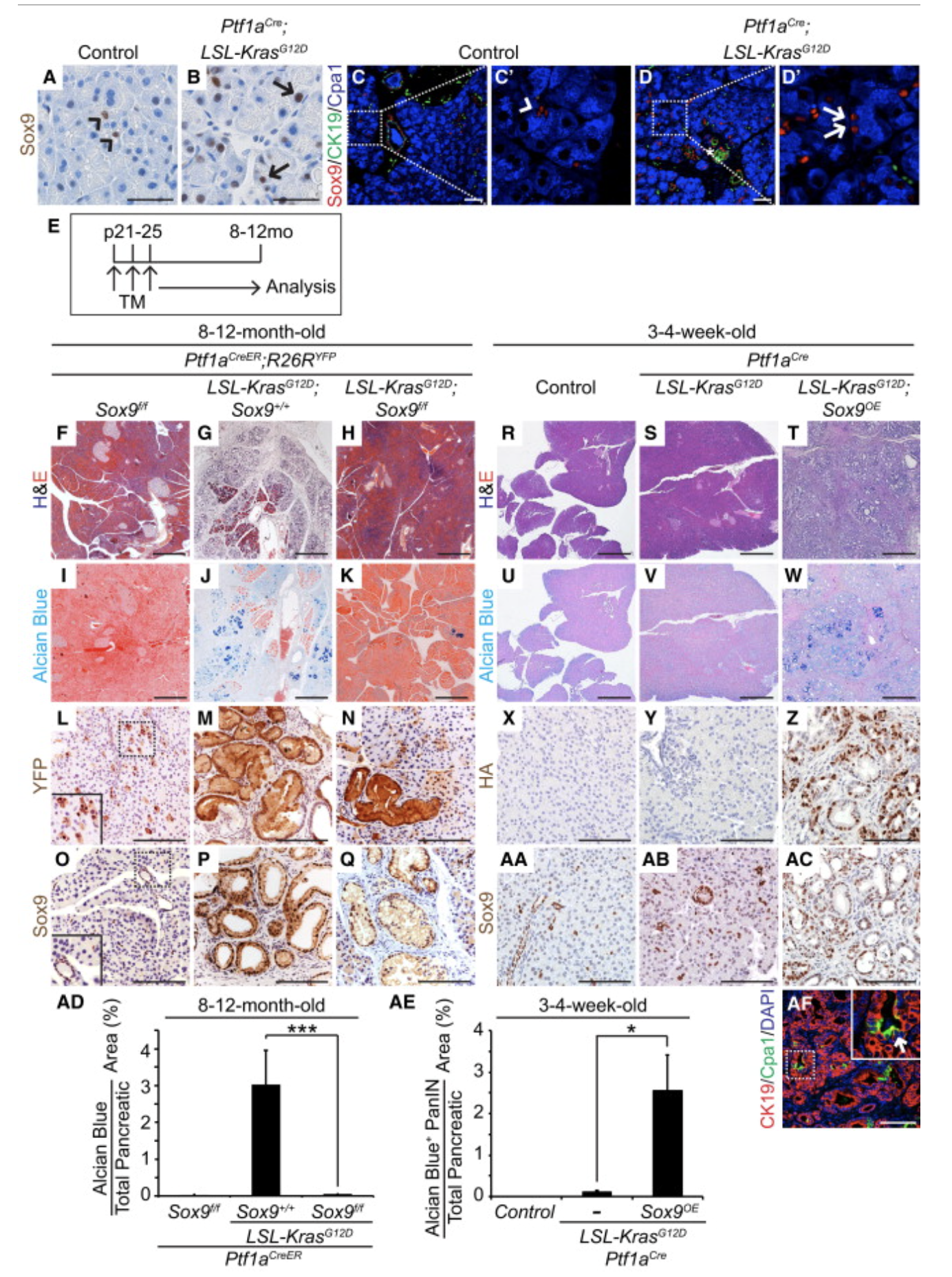
What is the point of panel AF in figure 4?
Ductal identity (CK19)
Acinar identity suppression (CPA1)
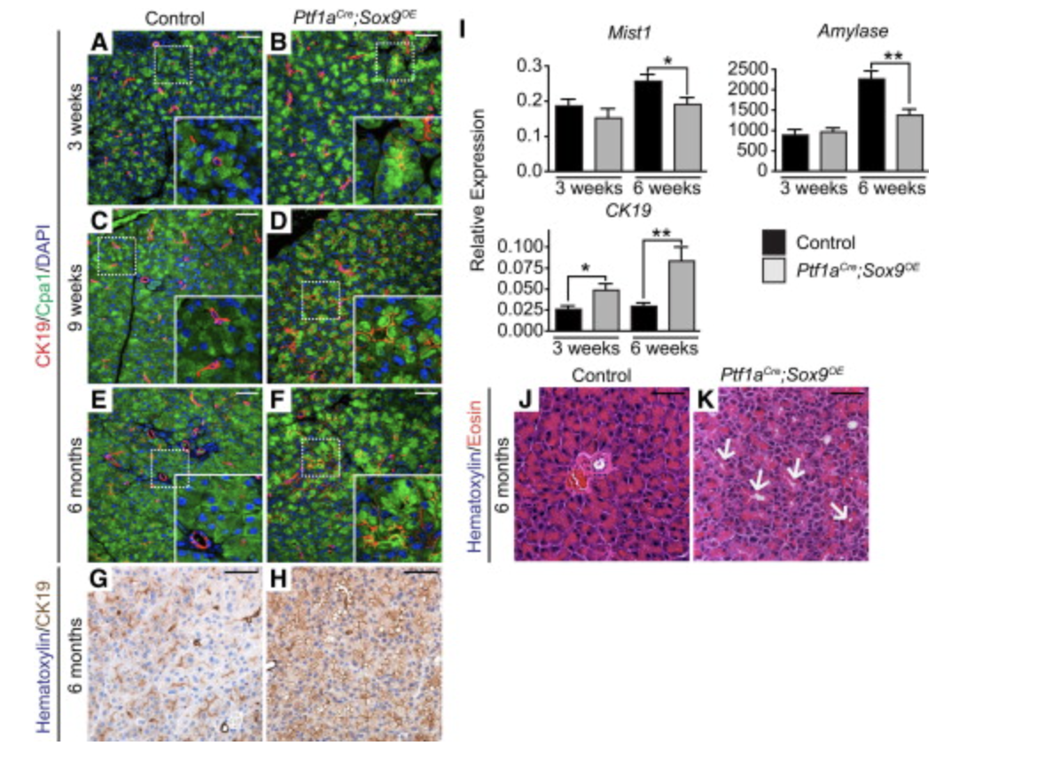
What is the model for figure 5?
Sox9OE

What is going on in figure 5?
Sox9 induces a partial ductal transition

Comparing left images to right whats going on in figure 5?
increase of ck19 expression when sox9 OE
(more ductal Identity when sox9 OE)
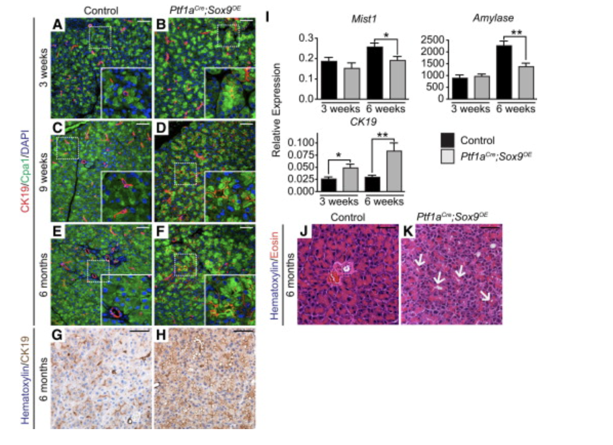
What is going on in the RT-qPCR side of figure 5?
Amylase/Mist1 down indicating lower acinar ID
CK19 up indicating higher ductal ID

What is going on in J/K of figure 5?
still look like acinar not fully duct like
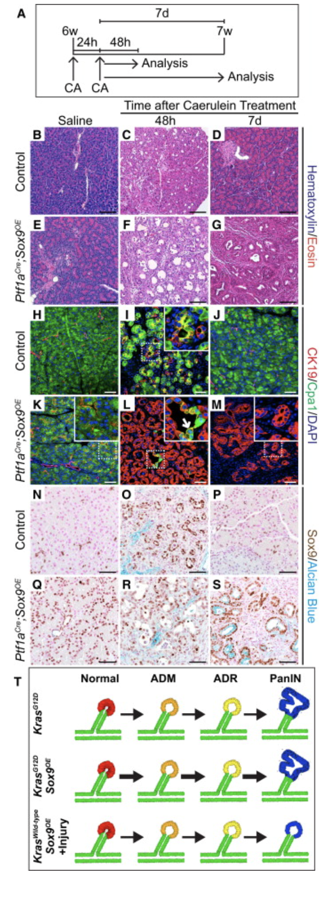
What is the model for figure 6?
Sox9OE under control of ptf1a
CA pancreatitis treatment

What is going on in figure 6?
pancreatitis injury causes ductal reprogramming
What is ADR?
acinar ductal reprograming (figure 6)
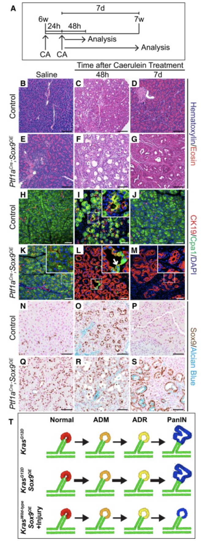
What is the healing going on in figure 6?
pancreatitis heals with ductal tissue not acinar tissue

What is the model for figure 7?
ptf1a model for everything
ptf1a+sox9 or ptf1a+sox9+kras
TM activates kras
Later CA causes pancreatitis
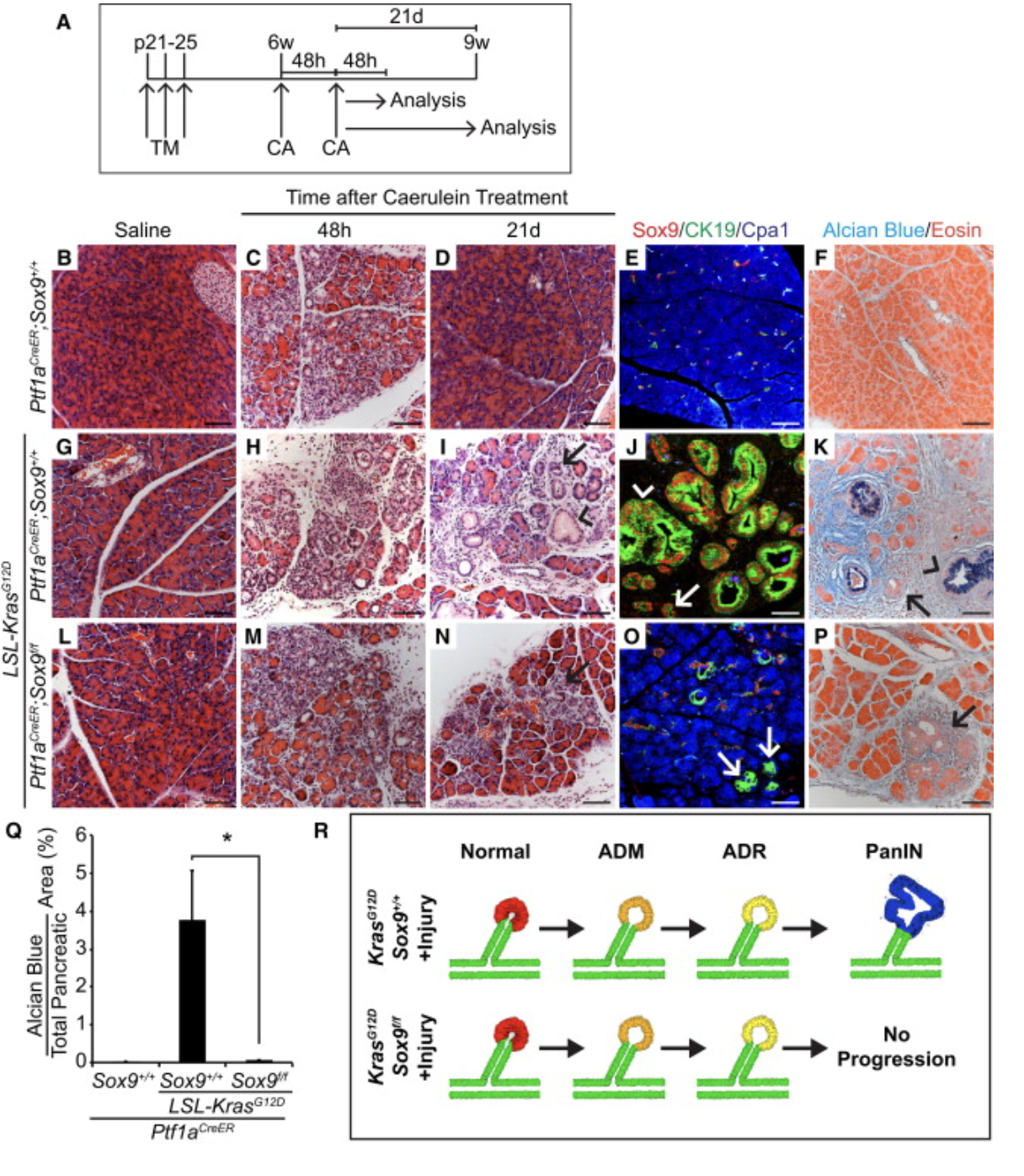
What is the big differentiator for figure 7 compared to figure 1 with regard to PDAC formation in the ptf1a/kras?
it happens much faster here 6 wk’s compared to 8-12 months
What is the big point in figure 7?
ptf1a+sox9 does not cause cancer (9wks)
ptf1a+Kras does not cause cancer (9wks)
ptf1a+Kras+sox9 DOES cuase cancer after 9 weeks