finger & thumb pathology & image analysis
 Call Kai
 Learn
Learn Practice Test
 Spaced Repetition
 Match
Match Flashcards
 Knowt Play
1/42

There's no tags or description
Looks like no tags are added yet.
Name | Mastery | Learn | Test | Matching | Spaced | Call with Kai | Chat |
|---|
No analytics yet
Send a link to your students to track their progress
43 Terms
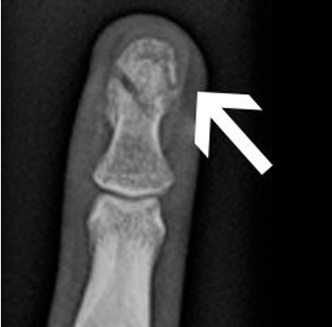
What type of fracture is shown in the image?
Tuft or burst fracture
What type of fracture is “a fracture of the fine cancellous bone at the distal tip of the phalanx”?
Tuft or burst fracture
What is a “tuft or burst fracture”?
a fracture of the fine cancellous bone at the distal tip of the phalanx
What are the causes of a tuft or burst fracture?
direct blow or a crush injury, lacerations, hyperextension, hyperflexion, rotation
What are the complications of a tuft or burst fracture?
Rare, but can have joint stiffness, numbness and hypersensitivity, and nail deformities
What is the radiographic appearance of a tuft or burst fracture?
comminuted fractures of the distal phalanx
What are the technical factor considerations of a tuft or burst fracture?
Neither additive or subtractive. No technical factor change.
What is the prognosis/treatment of a tuft or burst fracture?
Ice and splinted
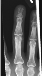
What fracture does the image show?
tuft or burst fracture
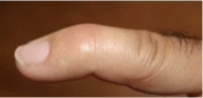
What type of fracture is shown in the image?
Baseball (Mallet) fracture
What is a Baseball (Mallet) fracture?
Injury to the thin tendon that straightens the end joint of a finger or thumb
What type of fracture is “Injury to the thin tendon that straightens the end joint of a finger or thumb”?
Baseball (Mallet) fracture
What are the causes of Baseball (Mallet) fracture?
unyielding object (like a ball) strikes the tip of a finger or thumb and forces it to bend further than it is intended to go
What is the complication of a baseball (mallet) fracture?
Infection
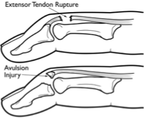
What type of fracture is shown in the image?
Baseball (Mallet) fracture
What is the radiographic appearance of a Baseball (Mallet) fracture?
a piece of the distal phalanx bone breaks away with the tendon
What is the technique for a Baseball (Mallet) fracture?
No manual exposure factor change
What is the Prognosis/Treatment for a Baseball (Mallet) fracture?
Splint or Surgical repair for larger fracture
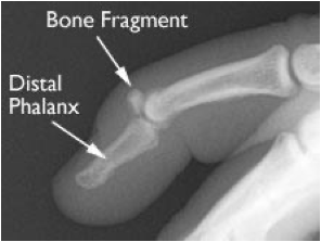
What fracture is shown in the image?
Baseball (Mallet) fracture
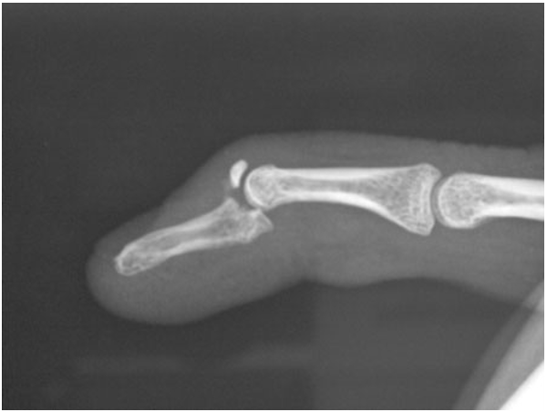
What fracture is shown in the image?
Baseball (Mallet) fracture
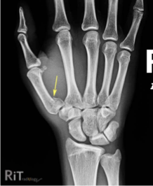
What fracture is shown in the image?
Bennett’s fracture
What is a Bennett’s fracture?
Fracture of the base of the thumb
What is a “Fracture of the base of the thumb” called?
Bennett’s fracture
What is the cause of a Bennett’s fracture?
forced abduction of the first metacarpal
What is a complication of a Bennett’s fracture?
Osteoarthritis
What is the radiographic appearance of a Bennett’s fracture?
two-piece fracture of the base of the thumb metacarpal
What is the technical factor consideration of a Bennett’s fracture?
Neither additive or subtractive. No technical factor change.
What is the prognosis/treatment of a Bennett’s fracture?
Spica cast or surgery
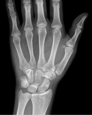
What type of fracture is shown in the image?
Bennett’s fracture
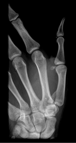
What type of fracture is shown in the image?
Bennett’s fracture
What are the acceptable EI values for fingers?
100-300
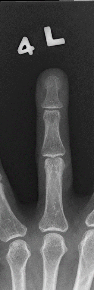
What projection is this finger x-ray?
PA
What deems a PA finger x-ray acceptable?
side marker placed clear of anatomy of interest
Entire digit from fingertip to distal portion of the adjoining metacarpal
No soft tissue overlap from adjacent digits
No rotation
Equal concavity of both sides of the phalangeal bodies
Equal amount of soft tissue on both sides of the phalanges
Fingernail, if seen, centered over the distal phalanx
Open interphalangeal (IP) and MCP joint spaces
Bony trabecular detail and surrounding soft tissues
What deems an oblique finger x-ray acceptable?
side marker placed clear of anatomy of interest
Entire digit, including the distal portion of the adjoining metacarpal
Digit rotated at a 45-degrees, demonstrated by the concavity of the elevated side of the phalangeal bodies
No superimposition of the proximal phalanx or MCP joint by adjacent digits
Open interphalangeal (IP) an MCP joint spaces
Bony trabecular detail and surrounding soft tissues
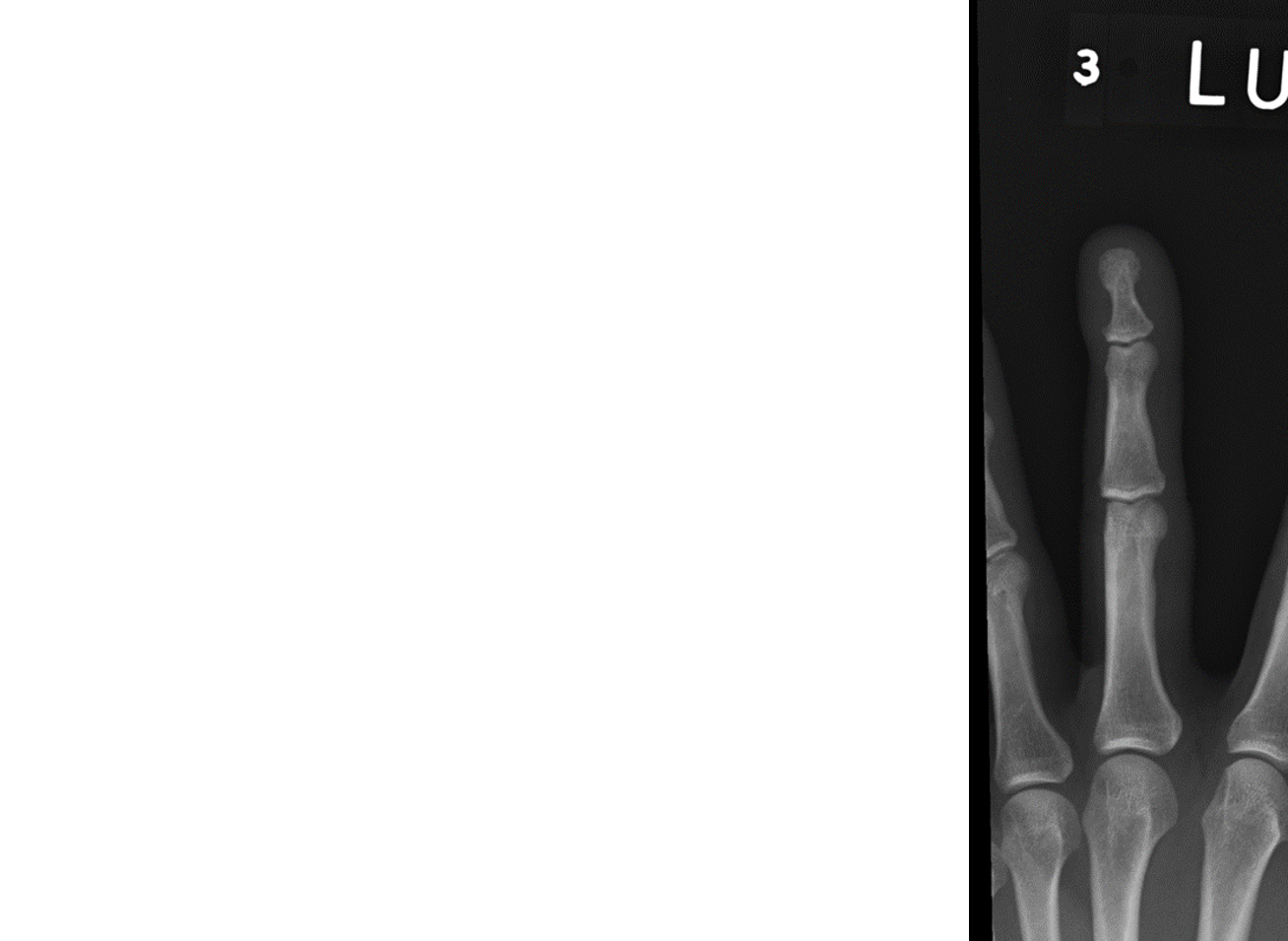
What projection is this finger x-ray?
Oblique
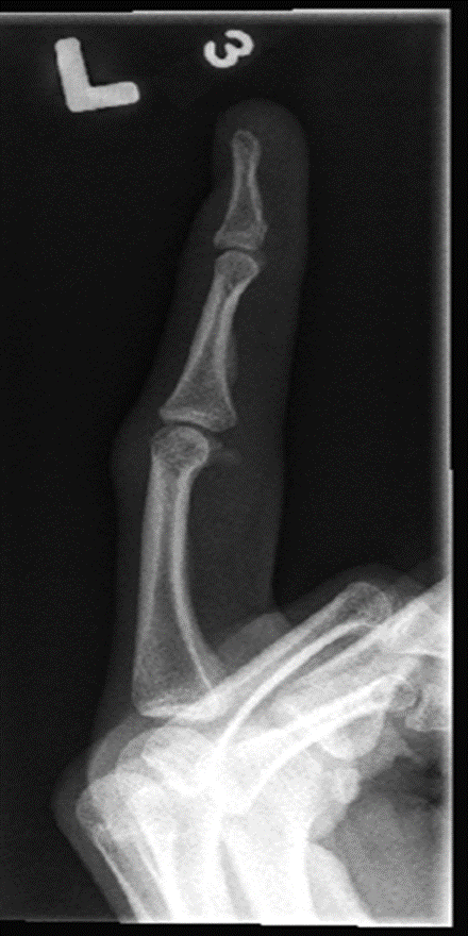
What projection is this finger x-ray?
Lateral
What deems a lateral finger x-ray acceptable?
side marker placed clear of anatomy of interest
Entire digit from fingertip to distal portion of the adjoining metacarpal
No rotation:
Fingernail in profile, if visualized and normal
Concave, anterior surfaces of the phalanges
No superimposition of the proximal phalanx or MCP joint by adjacent digits
Open interphalangeal (IP) joint spaces
Bony trabecular detail and surrounding soft tissues
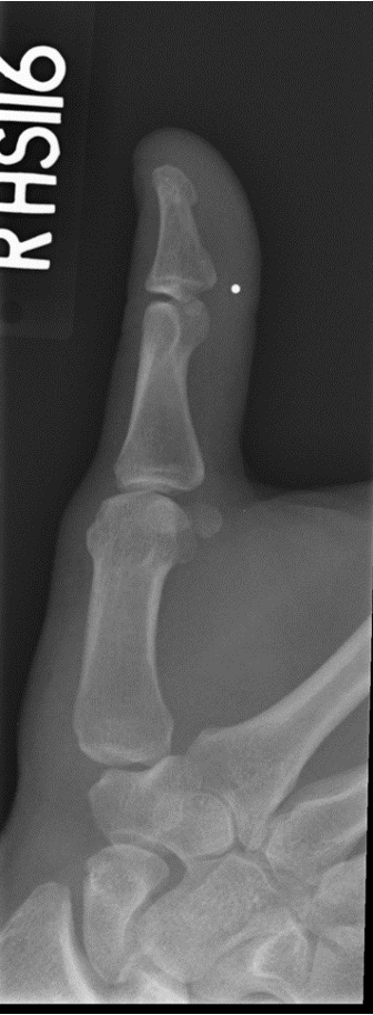
Which x-ray projection + structure is shown?
Thumb oblique
What deems an oblique thumb x-ray acceptable?
side marker placed clear of anatomy of interest
Area from the distal tip of the thumb to the trapezium
Proper rotation demonstrated by concave surface of elevated side of the proximal phalanx and metacarpal
Open interphalangeal (IP) and MCP joint spaces
Bony trabecular detail and surrounding soft tissues
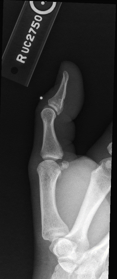
Which x-ray projection + structure is shown?
Thumb - Lateral
What deems a lateral thumb x-ray acceptable?
side marker placed clear of anatomy of interest
Area from the distal tip of the thumb to the trapezium
No rotation:
Concave anterior surface of the proximal phalanx and metacarpal
Thumbnail, if visualized and normal, in profile
Open interphalangeal (IP) and MCP joint spaces
Bony trabecular detail and surrounding soft tissues
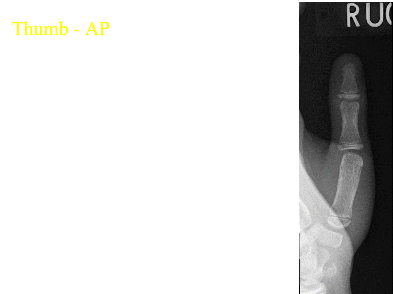
Which x-ray projection + structure is shown?
Thumb - AP
What deems a AP thumb x-ray acceptable?
side marker placed clear of anatomy of interest
Area from the distal tip of the thumb to the trapezium
No rotation:
Concavity of the phalangeal and metacarpal bodies
Equal amount of soft tissue on both sides of the phalanges
Thumbnail, if visualized, in the center of the distal thumb
Overlap of soft tissue profile of the palm over the midshaft of the first metacarpal
Open interphalangeal (IP) and MCP joint spaces without overlap of bones
Bony trabecular detail and surrounding soft tissues