4. Joints
1/55
There's no tags or description
Looks like no tags are added yet.
Name | Mastery | Learn | Test | Matching | Spaced | Call with Kai |
|---|
No analytics yet
Send a link to your students to track their progress
56 Terms
What is the definition of a joint?
A joint, also known as an articulation or arthrosis, is the point of contact between two surfaces (articular) for example bone-bone, bone-cartilage and bone-teeth that allows for a degree of movement.
Why are joints considered the weakest points in the skeleton?
Joints are considered the weakest points in the skeleton due to their structure; however, they are designed to resist forces like crushing and tearing, allowing for movement without compromising stability.
How are joints structurally classified?
Joints are classified based on the presence or absence of a cavity between surfaces and the type of connective tissue present in the joint.
What are the three types of structural classifications of joints?
Fibrous Joints: No cavity, connected by fibrous connective tissue, and allow very little movement.
Cartilaginous Joints: No cavity, bones held together by hyaline cartilage or fibrocartilage, and allow very little movement.
Synovial Joints: Have a cavity, an articular capsule, ligaments, and allow a wide range of movement.
How are joints functionally classified?
Joints are classified according to the degree of movement allowed by a specific structure
What are the three types of functional classifications of joints?
Synarthrosis: An immovable joint, allowing no movement.
Amphiarthrosis: A slightly movable joint.
Diarthrosis: A freely movable joint, where the structure determines the type of movement allowed.
What are the three types of fibrous joints?
The three types of fibrous joints are sutures, syndesmoses, and gomphoses.
Describe the structure of sutures
Sutures are characterized by tightly bound bones connected by a thin layer of dense fibrous connective tissue (CT) called the sutural ligament. They are found only between the bones of the skull, and the edges of the joining bones are wavy and interlocking.
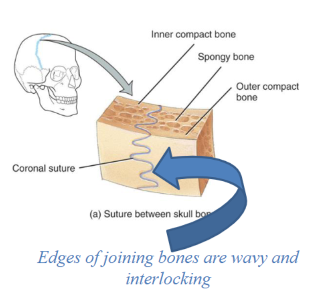
Describe the function of sutures
The primary function of sutures is to provide stability and protection to the brain by being immovable (synarthrosis). They also allow for growth as the skull expands with brain development.
Describe the structure of syndesmoses
Syndesmoses have a greater distance between the two bones compared to sutures and are connected exclusively by ligaments. The connective tissue can be present as a bundle (ligament) or as a sheet (interosseous membrane). The amount of movement allowed depends on the length of the connecting fibers.
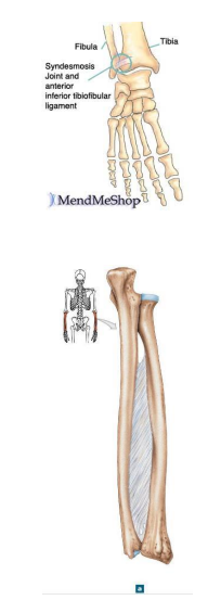
Describe the function of syndesmoses
Syndesmoses allow for slight movement (amphiarthrosis), which can vary based on the length of the fibers connecting the bones. For instance, short fibers provide little movement (as in distal tibiofibular articulation), while longer fibers allow for more significant movement (as in interosseous membrane between radius and ulna).

Describe the structure of gomphoses
Gomphoses are peg-in-socket joints, with the only example being the articulation of a tooth with its socket. The teeth are held in place by periodontal ligaments.
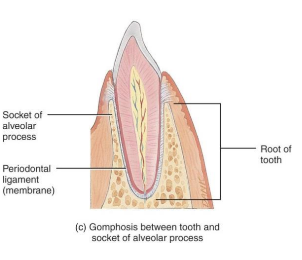
Describe the function of gomphoses
The function of gomphoses is to provide stability for teeth in their sockets, making them immovable (synarthrosis), which is important for maintaining the integrity of the dental structure.
What are the two types of cartilaginous joints?
The two types of cartilaginous joints are synchondroses and symphyses.
Describe the structure of synchondroses.
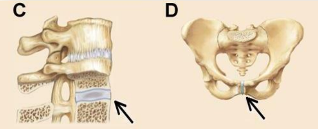
Synchondroses are cartilaginous joints where the connecting material is hyaline cartilage. Some synchondroses (e.g epiphyseal plate) may become ossified in adults, resulting in synostosis. Examples include the epiphyseal plate in growing bones (A) and the joint between the first rib and the sternum (B).
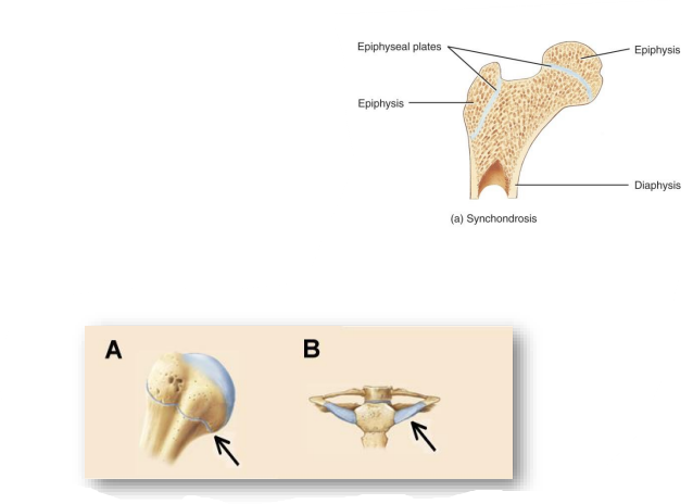
Describe the function of synchondroses.
Synchondroses are classified functionally as synarthrosis, meaning they are immovable joints. This immobility is essential for stability and support, especially during growth.
Describe the structure of symphyses .
Symphyses are cartilaginous joints where the connecting material is made of fibrocartilage, often in the form of a disc.
This fibrocartilage resists both tension and compression, acting as a shock absorber between the articulating bones.
The ends of the articulating bones are covered by hyaline cartilage to reduce friction during movement.
Describe the function of symphyses .
Symphyses are classified functionally as amphiarthrosis, which means they allow slight movement. They act as shock absorbers, resisting both tension and compression, making them particularly suited for midline joints like the pubic symphysis and intervertebral discs.
What are most movable joints classified structurally?
Synovial joints
How are all synovial joints classified functionally?
All synovial joints are classified as diarthroses
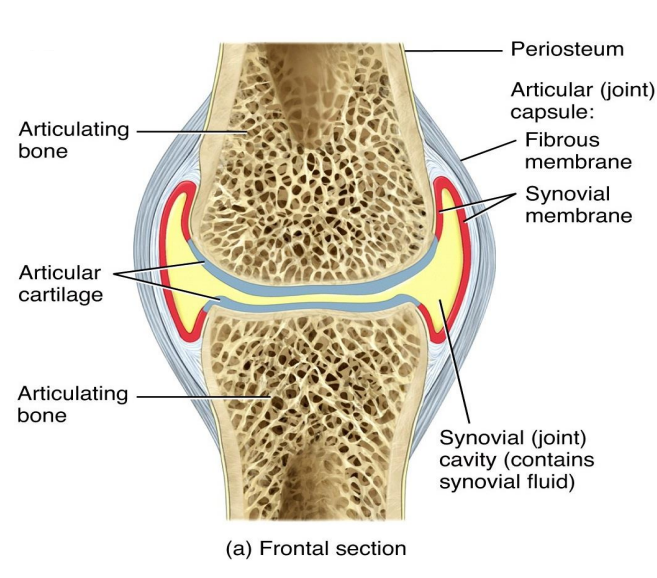
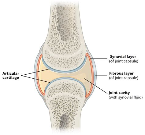
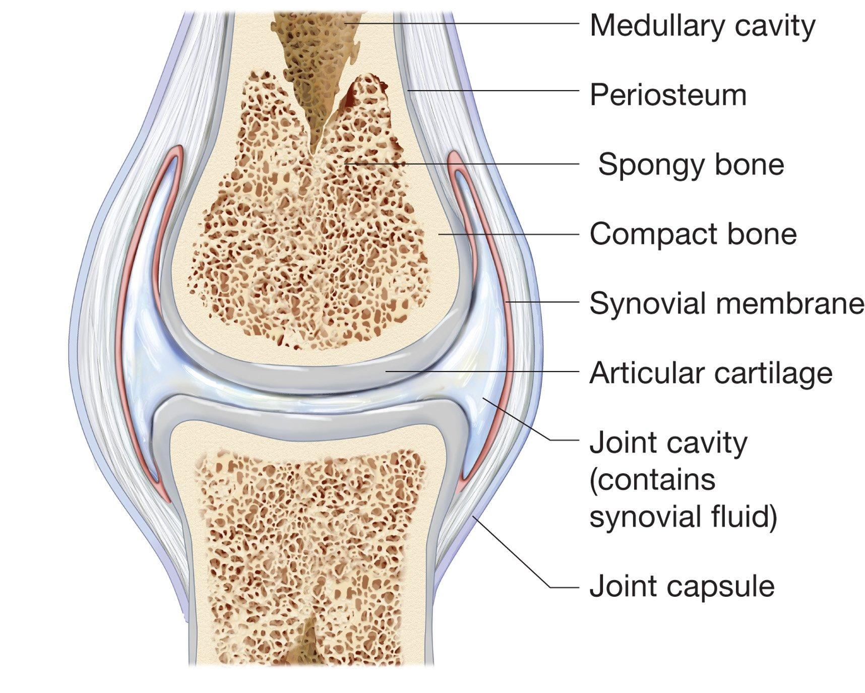
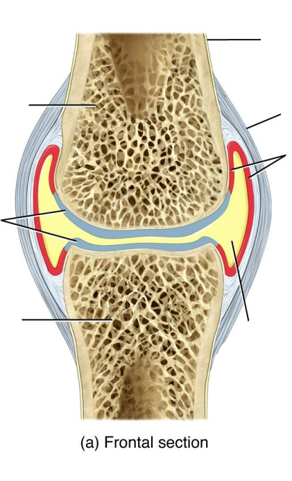
What is the synovial cavity?
The synovial cavity is a space found within synovial joints that separates the articulating surfaces of the bones. It is unique to synovial joints.
What is synovial fluid?
Synovial fluid is a fluid found within the synovial cavity and articular cartilage. It resembles a raw egg white

What are functions of synovial fluid?
Synovial fluid provides lubrication between the articulating surfaces of the bones, reducing friction during joint movement.
As articular cartilage is avascular (lacking blood vessels), it relies on synovial fluid for its nutrient supply. The fluid permeates the cartilage, delivering essential nutrients and oxygen to the chondrocytes (cartilage cells) and facilitating the removal of metabolic waste products.
Prolonged immobility can lead to the thickening of synovial fluid, resulting in joint stiffness.
What type of cartilage covers the articular surfaces in synovial joints, and what are its functions?
The articular surfaces are covered by hyaline cartilage, which reduces friction and absorbs compressive forces, acting as a cushion to prevent bone crushing.
What is the articular capsule, and what are its two layers?
The articular capsule encloses the joint cavity and consists of two layers: the fibrous capsule (outer layer) and the synovial membrane (inner layer)
What is the structure that encloses the joint cavity?
The joint cavity is enclosed by a two-layered structure separated by fatty deposits anteriorly and posteriorly known as the articular capsule.
What additional structures does the joint cavity contain?
Patella ("knee cap"), ligaments, menisci, and bursae.
How does the thickness of the articular capsule vary around the joint cavity, particularly in the front and at the sides?
The articular capsule is thinner in the front and at the sides compared to other areas. This variation in thickness allows for increased flexibility and a greater range of motion in the joint.
What is the structure and function of the fibrous capsule in synovial joints?
The fibrous capsule is the outer layer of the articular capsule in synovial joints. It is made of dense irregular connective tissue rich in collagen, providing flexibility and strength to the joint, preventing the bones from being pulled apart. The fibrous capsule is continuous with the periosteum

What is the structure of the synovial membrane?
The synovial membrane is the inner layer of the articular capsule that lines the joint cavity and all internal joint surfaces not covered by cartilage. It is composed of areolar connective tissue enriched with elastic fibers, allowing it to be flexible and supportive.
What is the function of the synovial membrane?
Production of synovial fluid
Define different part of synovial joints

What are bursae, and what is their primary function in the body?
Bursae (meaning in Latin sac) are small, fibrous sac-like structures filled with synovial fluid that alleviate friction between moving structures such as bones, tendons, muscles, ligaments, and skin. It is not part of the joint. Examples of bursae are subacromial bursa and subscapular bursa (see picture)
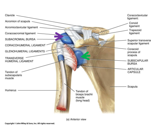
How do bursae contribute to joint movement?
Bursae act like lubricated cushions or "ball bearings" that reduce friction between moving structures during joint movement, thereby facilitating smooth motion and preventing wear and tear on surrounding tissues.
What are tendon sheaths, and what is their primary function in the body?
Tendon sheaths are tube-like bursae that wrap around tendons of muscles (like a bun around a hot dog), where these are in close proximity to bone such as in fingers, wrists and ankles in order to reduce friction (only occur here). It is not part of joint
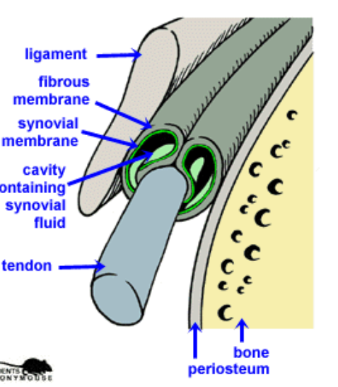
Where do tendon sheaths typically travel?
Tendon sheaths typically travel through joint cavities or are crowded together within narrow canals.
What are the six subtypes of synovial joints?
Plane (Gliding), hinge, pivot, condyloid, saddle and ball-and-socket
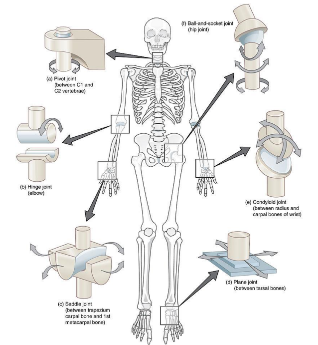
What movement is allowed by a plane (gliding) joint, and what is the typical structure of this joint?
Plane (gliding) joints have flat or slightly curved articular surfaces that allow side-to-side and back-and-forth movements. These joints are non-axial, meaning there is no rotation around an axis. They are commonly found in the intercarpal and intertarsal joints, as well as between the sternum and ribs (2nd to 7th ribs).
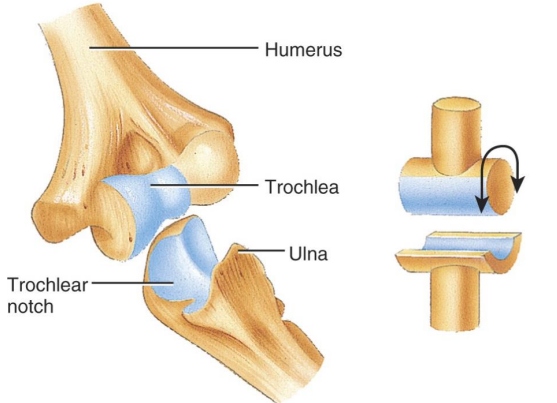
What movement is allowed by a hinge joint, and what is the typical structure of this joint?
Hinge joints consist of a convex surface of one bone fitting into a concave surface of another, allowing movements in one plane (monoaxial). The primary movements are angular opening and closing movements (flexion/extension). Examples include the elbow joint (humerus and ulna), ankle and knee joint and interphalangeal joints.
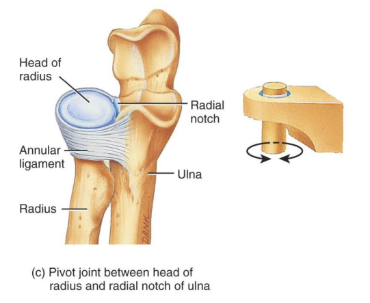
What movement is allowed by a pivot joint, and what is the typical structure of this joint?
Pivot joints allow rotation around a single axis (monoaxial). A rounded or pointed bone surface fits into a ring formed by another bone and a ligament. Examples include the proximal radio-ulnar joint and the atlanto-axial joint in the vertebral column (joint between the atlas and the axis).
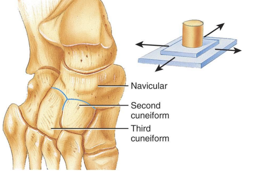
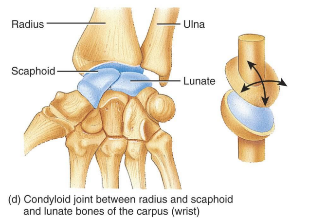
What movement is allowed by a condyloid joint, and what is the typical structure of this joint?
Condyloid joints allow flexion/extension and abduction/adduction, making them biaxial joints. A convex oval-shaped bone fits into an oval depression. Examples include the wrist joint (radius and scaphoid/lunate) and the metacarpophalangeal joints (knuckles).
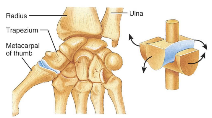
What movement is allowed by a saddle joint, and what is the typical structure of this joint?
Saddle joints have a concave and convex surface that interlock, resembling a rider sitting on a saddle. They allow flexion/extension and adduction/abduction (biaxial movement). An example is the carpometacarpal joint of the thumb, which gives it a wide range of motion, including opposition.
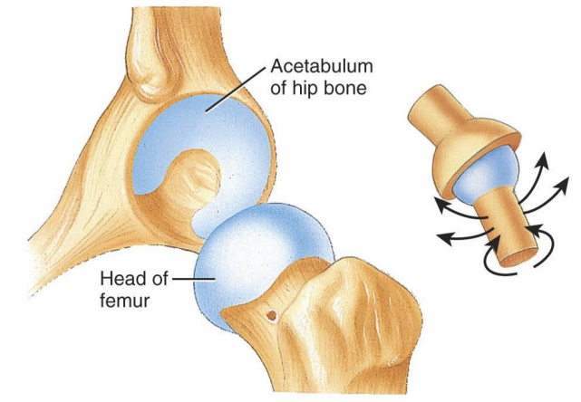
What movement is allowed by a ball-and-socket joint, and what is the typical structure of this joint?
Ball-and-socket joints consist of a ball-like surface of one bone fitting into a cup-like depression of another bone, allowing multi-axial movement. This joint permits rotation, flexion/extension, abduction/adduction, and circumduction. Examples include the shoulder (humero-scapular joint) and the hip joint.
What happens to the synovial fluid when pressure is applied to a synovial joint, and what occurs when the pressure is released?
When pressure is applied to a synovial joint, synovial fluid is squeezed out of the articular cartilage, creating a lubricating film. Once the pressure is released, the fluid flows back into the cartilage, maintaining smooth and efficient joint movement.
How do synovial joints function as lubricating devices, and why is lubrication important for their movement?
Synovial joints function as lubricating devices by allowing bones to move across one another with minimal friction. This lubrication is critical because, without it, the articular surfaces would wear away and overheat due to friction during movement.
What are the normal effects of aging on joints, and how do these changes impact joint function?
With normal aging, several changes occur in the joints that impact their function:
Decreased production of synovial fluid: This leads to less lubrication in the joints, increasing friction and causing joint stiffness.
Thinning of articular cartilage: The wear and thinning of cartilage make joints more prone to damage, leading to pain and reduced mobility.
Shortening of ligaments: Ligaments lose elasticity and shorten over time, reducing flexibility and further contributing to joint stiffness.
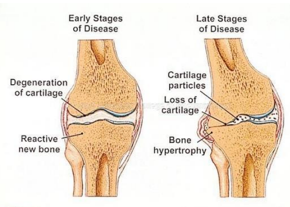
What is osteoarthritis, and how does it affect the joints, particularly in elderly people?
Osteoarthritis is a degenerative disease of the joints, commonly referred to as "wear and tear" arthritis. It primarily affects the articular cartilage, which softens, cracks, and eventually erodes over time. This condition is frequently seen in the elderly due to the cumulative damage on the joints. As the cartilage deteriorates, bony spurs (osteophytes) may develop, further impeding movement and causing pain and stiffness in the affected joints.
How can regular stretching and aerobic exercise help counteract the effects of aging on joints?
Regular stretching and aerobic exercise help maintain joint flexibility and the full range of motion. Mild pressure on hyaline cartilages forces some nutrient rich synovial fluid into the cartilage helping in maintenance
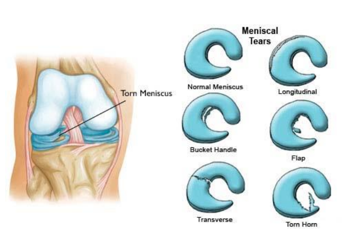
Which joint structure does torn cartilage occur commonly?
Torn cartilage can occur menisci and also in other joints
What is a dislocation (or luxation), and what are its common symptoms?
Dislocation, or luxation, occurs when the bones in a joint are forcibly pushed out of alignment. It is typically accompanied by sprains and can cause severe pain, swelling, and immobility. A subluxation is a partial dislocation, where bones return to their normal position more easily
What is a sprain, and why do partly torn ligaments heal slowly?
A sprain is the forcible twisting or wrenching of a joint, leading to a stretch or tear of ligaments. Partly torn ligaments heal slowly due to poor vascularization (limited blood supply), which delays the healing process. In cases of ruptured ligaments, surgery may be required
What is bursitis, and what are some common causes?
Bursitis is the inflammation of a bursa, which can result from physical blows, friction, arthritis, or infection. It often leads to swelling due to increased fluid in the bursa. An example of bursitis is student's elbow, where repetitive stress causes inflammation in the elbow
What is tendinitis, and how is it typically caused?
Tendinitis is the inflammation of tendons, commonly caused by physical blows, friction, arthritis, or infection. An example is trigger finger, where the tendons in the finger become inflamed, causing pain and difficulty in movement
What is synovitis, and with which condition is it often associated?
Synovitis is the inflammation of the synovial membrane, leading to excess fluid production inside the joint capsule. It is commonly associated with rheumatoid arthritis, causing joint swelling, pain, and stiffness
What is tenosynovitis, and how does it affect tendons?
Tenosynovitis is the inflammation of the tendon or tendon sheath, often caused by repetitive use or friction. This condition can lead to pain, swelling, and restricted movement in the affected area
What is arthritis, and what are some of the common forms and symptoms associated with this condition?
Arthritis refers to a group of more than 100 inflammatory or degenerative joint disorders. Common symptoms include joint pain, stiffness, and swelling. Two notable forms are:
Osteoarthritis: A degenerative "wear and tear" arthritis where the cartilage softens, cracks, and erodes over time, leading to joint pain and limited mobility.
Rheumatoid arthritis: A chronic autoimmune condition that causes persistent inflammation in the joints, often resulting in severe pain, joint deformity, and loss of function