AUTONOMIC NERVOUS SYSTEM
 Call Kai
 Learn
Learn Practice Test
 Spaced Repetition
 Match
Match Flashcards
 Knowt Play
1/20

There's no tags or description
Looks like no tags are added yet.
Name | Mastery | Learn | Test | Matching | Spaced | Call with Kai | Chat |
|---|
No analytics yet
Send a link to your students to track their progress
21 Terms
NERVOUS SYSTEM DIAGRAM
Nervous System
Central Nervous System
Peripheral Nervous System
Somatic Nervous System
Autonomic Nervous System
PARASYMPATHETIC SYSTEM
rest and digest
deaccelerate
motor neurons in brain stem and sacral area of spinal cord
ganglia close to innervated organ
long post-synaptic ganglion axons
short pre-synaptic ganglion axons
SYMPATHETIC SYSTEM
fight or flight
accelerate
motor neurons in lumbar and thoracic area of spinal cord
ganglia close to spinal cord
long pre-synaptic ganglion axons
short post-synaptic ganglion axons
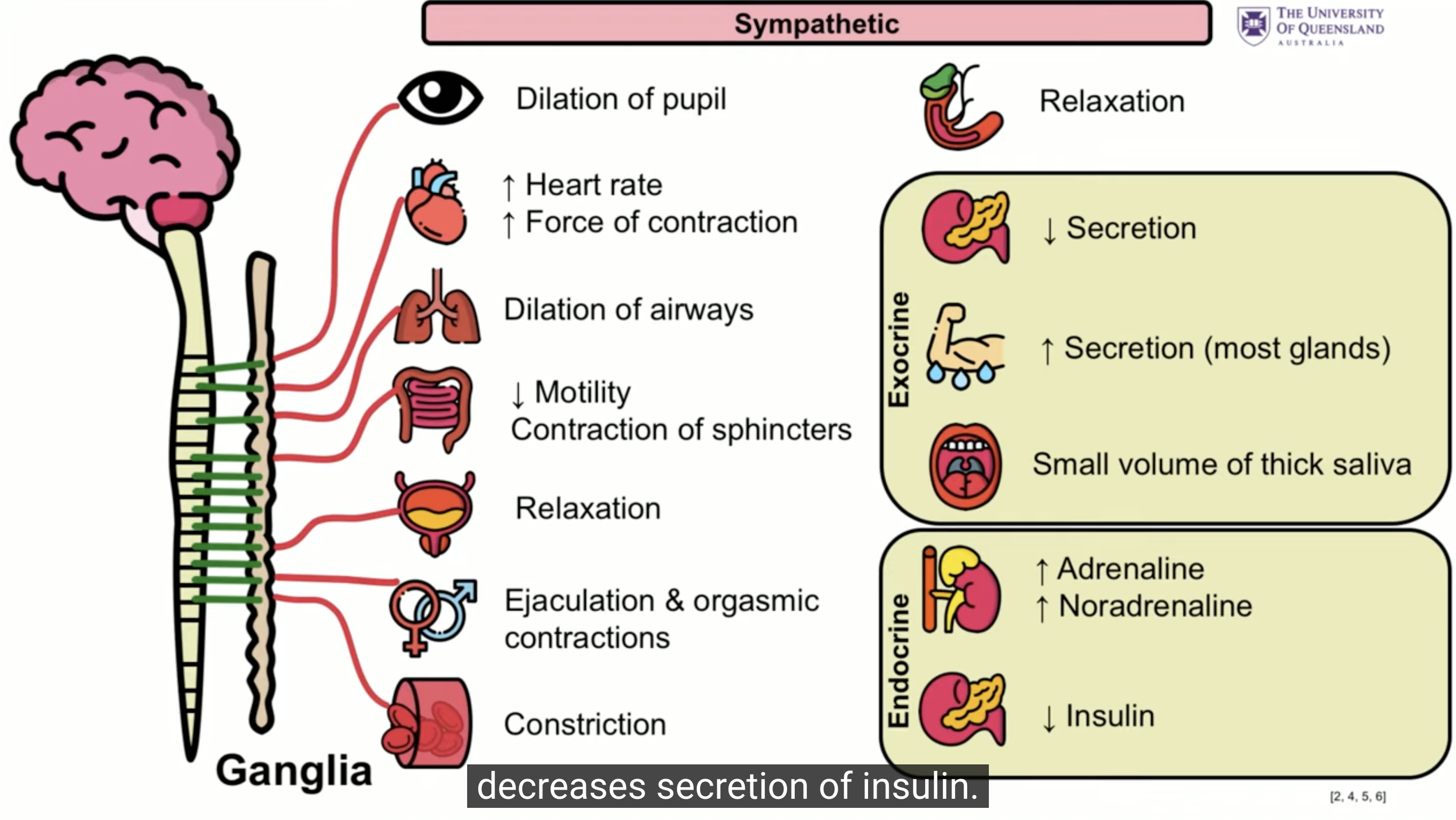
SYMPATHTIC SYSTEM CHARACTERISTICS
pupil dilation
increase heart rate and contraction
blood vessel constriction
increase sweat secretion
airway dilation
decrease motility
decreased bladder and GI activity
adrenaline and noradrenaline secretion
PARASYMPATHETIC SYSTEM CHARACTERISTICS
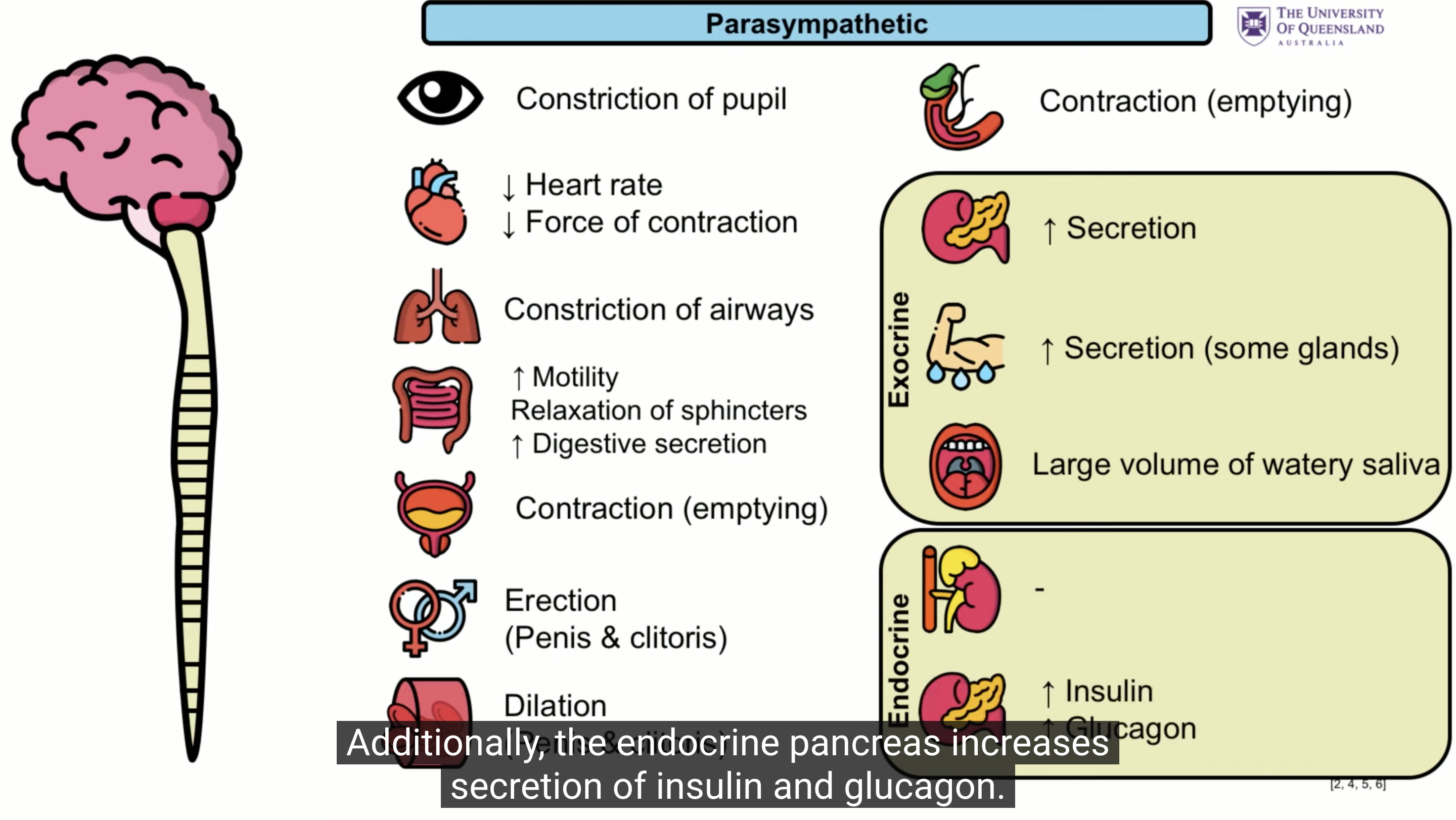
constriction of pupils
decrease heart rate and contraction
airway constriction
increase motility
blood vessel dilation
bladder emptying
insulin and glucagon increase
AUTONOMIC GANGLIA
Are a cluster of nerve cell bodies that bridge the central motor neurons and the effector organs.
CNS → Autonomic ganglia via pre-ganglionic fibres
Autonomic ganglia → Effector organs via post-ganglionic fibres
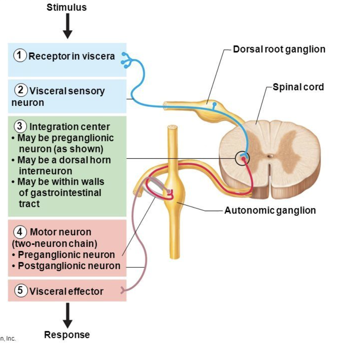
AUTONOMIC GANGLIA PATHWAY
Stimulus
Receptor in viscera (internal organ)
Visceral sensory neuron
Integration center
Motor neuron (pre or post-ganglionic neuron)
Visceral effector
Response
AXON TERMINALS
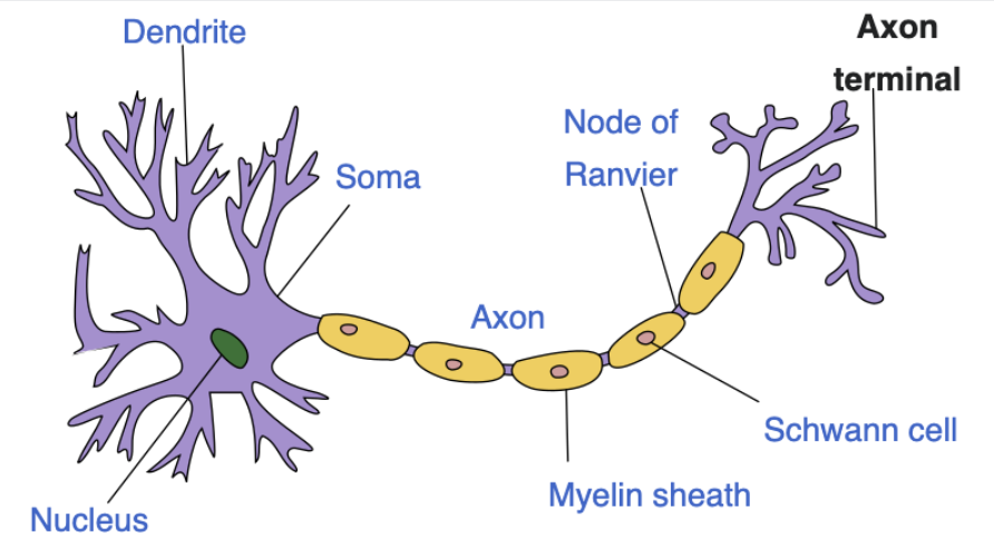
Axon terminals or ‘boutons’ are found at the terminal ends of axons where synapses occur with other neurons/cells.
SYMPATHETIC VARISCOCITIES
Variscocities = ‘string of beads’ where post-ganglionic axons terminate on the effector organ.
Two types of smooth visceral muscles:
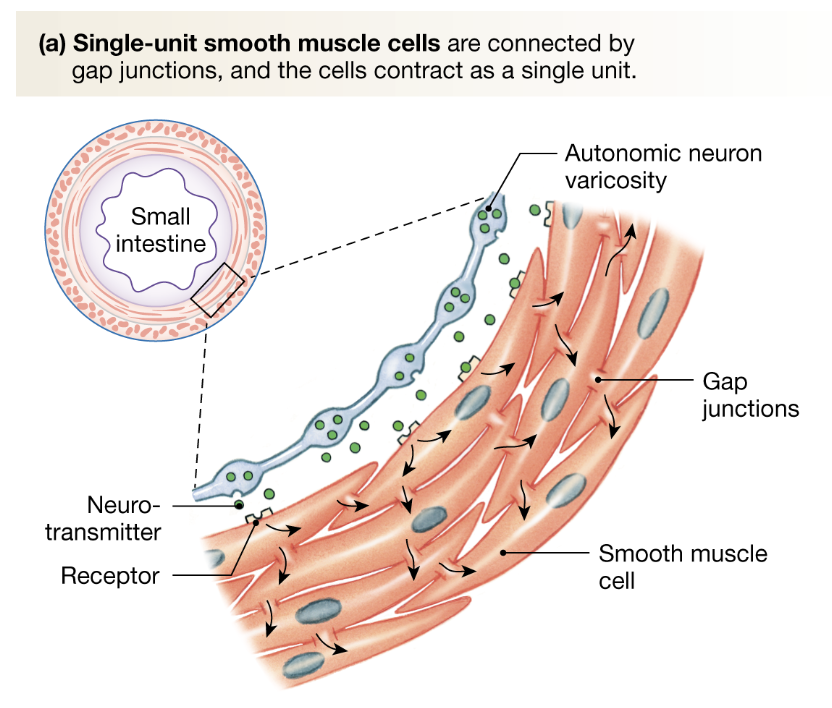
Single-unit smooth muscle cells
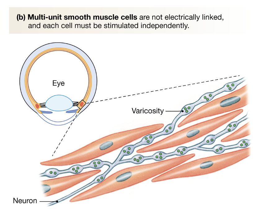
Muli-unit smooth muscle cells
SINGLE UNIT SMOOTH MUSCLE CELLS
Line walls of GI tract
gap junctions
enable electrical signals to be propagated to adjacent cells
results in synchronised depolarisation and contraction as a single unit
MULTI-UNIT SMOOTH MUSCLE CELLS
in your eye
each cell must be individually innervated
GANGLION CELL INNERVATION
Boutons release ACh which activates nicotonic ganglionic receptors
These are ion channels that result in post-synaptic potential
The potential activates the ganglia
Ganglia sends signals via post-synaptic fibres to effector organ
Smooth muscle cells activated by varicosities
CHOLINERGIC SIGNALS
Choline pumped into post-ganglionic nerve endings
Choline converted to ACh
ACh stored in vesicles
When nerve terminal stimulated by AP, calcium enters terminal
Calcium causes vesicle to move towards pre-synaptic membrane
Fusion, neurotransmitters released into synaptic cleft
Neurotransmitters act on ACh receptors = response
ACh also prohibits further release
MUSCARINIC CHOLINERGIC RECEPTORS
G-protein coupled receptors
M1 - CNS
M2 - Cardiac muscle
M3 - GI tract smooth muscle
Stimulated by ACh
Stimulates/inhibits function of target cell
NICOTONIC CHOLINERGIC RECEPTORS
Ligand gated ion channels
N1 - Skeletal muscle
cholinergic signalling in neuromuscular junction
N1 - Neuronal
cholinergic signalling in the CNS
Stimulated by ACh or nicotine
Causes rapid depolarisation of cell
ADRENERGIC RECEPTORS
Tyrosine enters post-ganglionic terminal
Tyrosine converted to dopamine
Dopamine converted to noradrenaline
Noradrenaline stored in vesicles
When activated by AP, calcium enters terminal
Calcium causes vesicle to synpase on pre-synpatic membrane
Transmitters released into synaptic cleft
Transmitter acts on alpha and beta receptors
Negative feedback prohibits further release
ALPHA RECEPTORS
A1 - GI tract
vasoconstriction
A2 - CNS
Auto-inhibition
Greater affinity for noradrenaline
G-protein coupled receptors
BETA RECEPTORS
B1 - Cardiac muscle cells
increased cardiac muscle contractility
B2 - Muscle blood vessels
vasodilation and bronchodilation
greater affinity for adrenaline
G-protein coupled receptors
PRE-GANGLIONIC FIBRES (PARASYMPATHETIC + SYMPATHETIC)
release ACh
acts on nicotonic receptors located on post-ganglionic neurons at ganglion
POST-GANGLIONIC FIBRES (PARASYMPATHETIC)
Parasympathetic varicosities release ACh onto muscarinic receptors on post-synaptic target organs
M2 receptors are activated by ACh release during parasympathetic activation.
POST-GANGLIONIC FIBRES (SYMPATHETIC)
Acts primarily by releasing adrenaline/noradrenaline to act on adrenergic receptors
ATP can be co-released to activate post-synaptic voltage gated calcium channels (faster smooth muscle contraction)
Can act through cholinergic pathways on certain organs and dopaminergic pathways in renal system
Has a neuroendocrine component.