Coronary & Conductance Systems
 Call Kai
 Learn
Learn Practice Test
 Spaced Repetition
 Match
Match Flashcards
 Knowt Play
1/50

There's no tags or description
Looks like no tags are added yet.
Name | Mastery | Learn | Test | Matching | Spaced | Call with Kai | Chat |
|---|
No analytics yet
Send a link to your students to track their progress
51 Terms
Name the anterior sulci of the heart
Coronary sulcus
Anterior interventricular sulcus
Name the posterior sulci of the heart
Coronary sulcus
Posterior interventricular sulcus
What runs through the anterior interventricular sulcus
Great cardiac vein
Anterior interventricular branch of the left coronary artery
What runs through the coronary sulcus
Right coronary artery
Coronary sinus (with great & small cardiac vein)
Circumflex branch of the left coronary artery
(coronary sulcus runs almost completely around top of heart) (2 arteries, 2 veins & a sinus)
What runs through the posterior interventricular sulcus
Posterior interventricular branch of the right coronary artery
Middle cardiac vein
What decides if a heart is right/left side dominant
Whether the posterior interventricular artery branches from the right coronary artery (right side dominant) or the circumflex branch of the left coronary artery (left side dominant)
Are right or left side dominant hearts more common - what percentages
Right side are more common (90%)
Blood supply to SA node
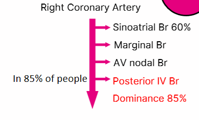
Sinu-atrial nodal branch of the right coronary artery
What is the typical branching pattern of the major coronary arteries
Generally 2 marginal & 1 diagonal
Right marginal branch of right coronary artery
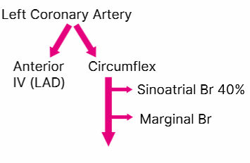
Left marginal branch of circumflex branch
Diagonal branch of anterior interventricular branch
Branches of the right coronary artery
Branches of the left coronary artery
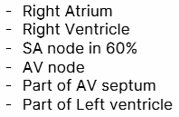
What does the right coronary artery supply
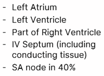
What does the left coronary artery supply
Which branch would be most detrimental to stop working
anterior interventricular branch of the left coronary artery (Left anterior descending LAD)
The LAD supplies blood to:
Most of the left ventricle, which is the heart's main pumping chamber.
The anterior 2/3 of the interventricular septum, including the bundle branches, which are crucial for electrical conduction.
Parts of the right ventricle.
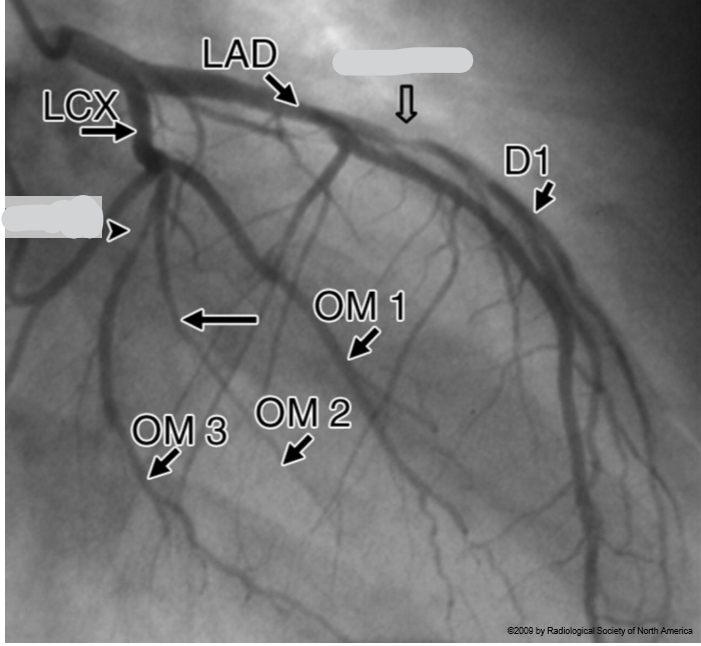
What do the arrows point towards
stenosis
treatment for stenosis of the coronary arteries.
percutaneous intervention (stent)
Through what vessel is a stent fed to get to a coronary artery
Femoral artery
myocardial infarction
heart attack
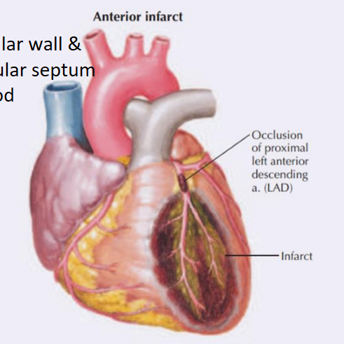
Occlusion of what causes an anterior infarct
left anterior descending branch aka anterior interventricular branch of the coronary artery
What does an anterior infarct affect
Left ventricular wall & interventricular septum without blood
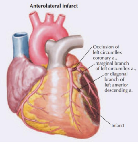
Occlusion of what causes an anterolateral infarct
left circumflex coronary artery
marginal branch of the left circumflex artery
or
Diagonal branch of the left anterior descending artery
What does an anterolateral infarct impact
Lateral wall of the left ventricle
The posterior wall (if the left is dominant)
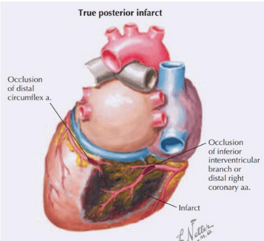
Occlusion of what causes a true posterior infarct
Distal circumflex artery
&
Inferior interventricular branch of the coronary artery
(all arteries on the post. surface of the heart)
What does a true posterior infarct affect
Posterior left ventricle
Occlusion of what causes a diaphragmatic / inferior infarct
Right coronary artery
What does a diaphragmatic / inferior infarct affect
Posterior wall of the left ventricle
The interventricular septum (if the right is dominant)
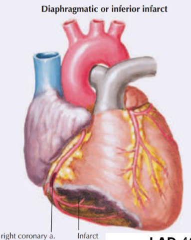
What are the 3 most common artery occlusions that cause a myocardial infarction. What is their percentage occurrence in a myocardial infarction
Left circumflex: 15-20%
Right coronary: 30-40%
Left anterior descending (LAD): 40-50%
What vessels are used to create a coronary artery bypass graft (main & 2 alternative)
Superficial veins of lower limb - e.g. short saphenous vein (from great saphenous vein) (or internal thoracic/radial artery)
Alternative treatment to coronary artery bypass graft
Stent placement using Coronary angioplasty
What are the small coronary veins called
Anterior cardiac veins
Thesbian veins
Where do the anterior cardiac veins drain into
Directly into the right atrium
Thesbian veins are aka
venae cordis minimae
What do thesbian veins drain
drain blood directly from the myocardium into the atria and ventricles.
The anterior interventricular vein along its course becomes what
The great cardiac vein
What veins run in the posterior interventricular sulcus
The middle cardiac veins
The great, middle & small cardiac veins drain their blood into _________ which drains into _______
The coronary sinus
The right atrium
What are the components of the conducting system
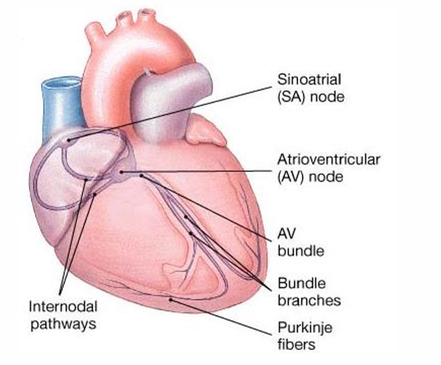
Where in the heart does the AV bundle run
The interventricular septum
What conductor does the septomarginal trabeculum come off
Atrioventricular node → Atrioventricular bundle → Right bundle branch → Septomarginal trabecula
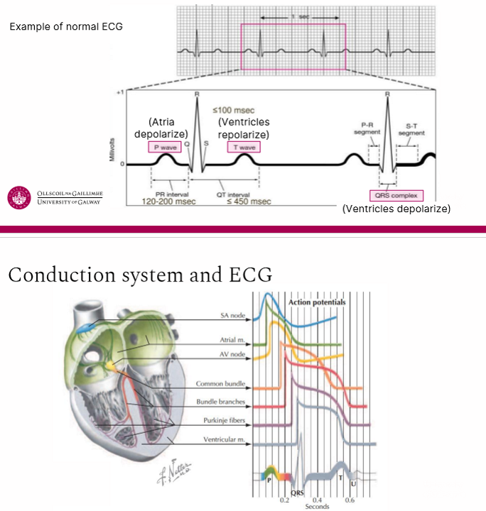
What are the steps of impulse conduction through the heart
SA node activity and atrial activation begin
Stimulus spreads across the atrial surfaces and reaches the AV node (takes 50 msec)
There is a 100 msec delay at the AV node. Atrial contraction begins.
The impulse travels along the interventricular septum within the AV bundle and the bundle branches to the Purkinje fibres (takes 25 msec)
The impulse is distributed by Purkinje fibres and relayed throughout the ventricular myocardium. Atrial contraction is completed and ventricular contraction begins. (50 msec)
What activities in the heart cause what parts of the ECG
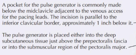
How do endocardial leads of a pacemaker reach the heart
Introduced via the subclavian(/brachiocephalic) vein, through the vena cava to the right atrium
Where is the pulse generator placed in a pacemaker
What fibres of the ANS are involve in cardiac innervation
Post-ganglionic fibres
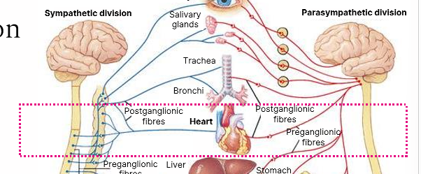
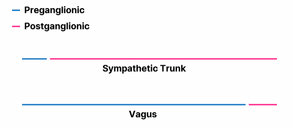
What nerves are associated with preganglionic & postganglionic fibres
the sympathetic trunk provides sympathetic innervation
the vagus nerves provide parasympathetic innervation
What in the cardiac system is the ANS responsible for
The Autonomic Nervous System is directly responsible for regulating heart rate, force of contraction and cardiac output
Effect of parasympathetic stimulation on heart
Parasympathetic decrease heart rates, reduces force of contraction and constricts Coronary Arteries
Effect of sympathetic stimulation on heart
Sympathetic increase heart rate and increase force of contraction
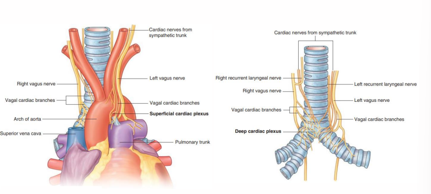
Branches from parasympathetic (Vagus) and sympathetic (Trunk) systems contribute to the cardiac plexus. The plexus has how many parts
Cardiac plexus has two parts – Superficial and Deep
Visceral Afferents travel from the _____ to the ______ carrying what
Visceral Afferents travel from the heart to the CNS carrying information regarding Cardiac Homeostasis (Blood Pressure / Biochemistry) and Pain
Learn this?