Neural Induction
1/69
There's no tags or description
Looks like no tags are added yet.
Name | Mastery | Learn | Test | Matching | Spaced | Call with Kai |
|---|
No analytics yet
Send a link to your students to track their progress
70 Terms
Define neural induction
Neural induction is the process by which embryonic cells in the ectoderm make a decision to acquire a neural fate rather than give rise to other structures such as epidermis or mesoderm
What major process characterises early development in Xenopus?
Gastrulation, involving complex tissue movements.
Where does gastrulation begin in Xenopus embryos?
It begins with a small involution of the smooth surface opposite the sperm entry site.
What is the name of the region where the initial involution occurs?
The involuting marginal zone (IMZ), also known as the dorsal lip of the blastopore (DLB).
What tissue lies beneath the neurogenic region during Xenopus gastrulation?
Mesoderm.
What key outcome does gastrulation establish in Xenopus embryos?
The formation of the three germ layers.
What is neural induction in Xenopus development?
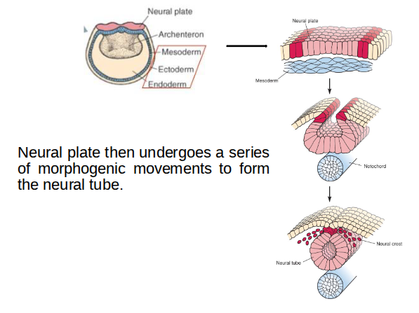
The process that converts the neurogenic region into the neural plate.
Neural plate then undergoes a series of morphogenic movements to form the neural tube.
Which part of the embryo specifies cell fate?
In early development, the part of the embryo that specifies cell fate is the marginal zone, particularly the dorsal lip of the blastopore (Spemann’s organiser).
More generally:
Cell fate is specified by the organiser region, which sends signals that direct neighbouring cells to adopt particular developmental pathways.
Did Spemann/Mangold discover the organiser?
Ethel Brown Harvey - The production of new hydranths in Hydra (Cnidaria, class Hydrozoa) by the insertion of small grafts (1909)
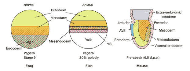
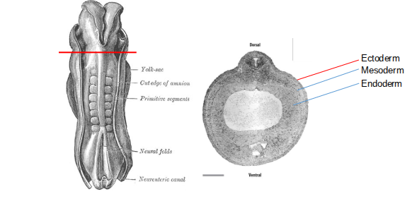
Germ Layers in Frog, Fish and Mouse
1. Ectoderm- skin & nervous system
2. Mesoderm- musculoskeletal, vasculature, reproductive & excretory systems
3. Endoderm- gut, lungs, liver, thyroid
Early Development of the Human Embryo
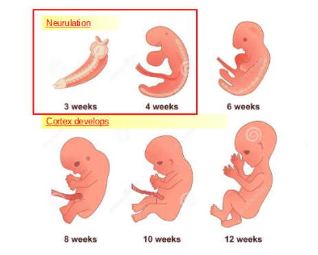
When does the neural tube appear in human development, and from which germ layer is the central nervous system derived?
The neural tube appears during the fourth week after fertilisation, and the central nervous system develops from the ectoderm layer.
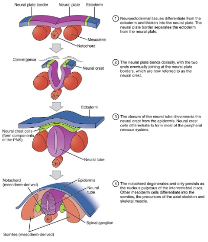
How does neurulation occur in mammals, and what structures arise from the neural tube, neural crest, and mesoderm?
In mammals, chemical signals cause the ectoderm to form a neural plate, which folds into the neural tube. The entire CNS develops from the neural tube, the PNS arises from the neural crest, and mesodermal somites form bone and muscle.
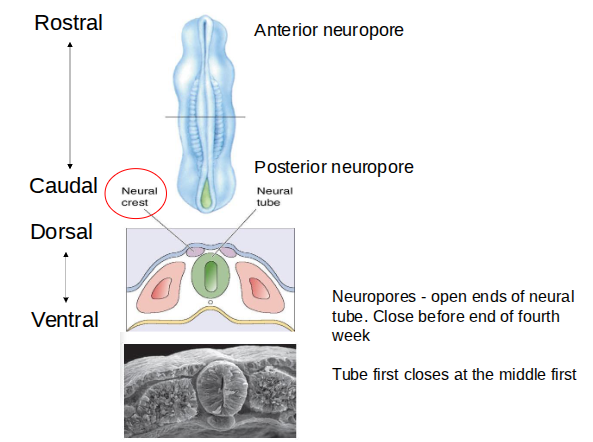
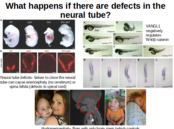
What happens if there are defects in the
neural tube?
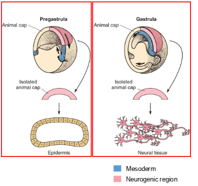
When does ectoderm acquire neural fate, and how is neural tissue induced?
Ectoderm acquires neural fate during gastrulation: animal cap ectoderm removed before gastrulation forms epidermis, whereas the same tissue taken during gastrulation forms neuronal tissue. Neural tissue is induced by signals (from the organiser region) that convert ectoderm into neural tissue.
How is mesoderm induced in Xenopus embryos?
Mesoderm is induced through interactions between the animal and vegetal regions, where a diffusible signal from the vegetal cells instructs adjacent animal cells to form mesoderm.
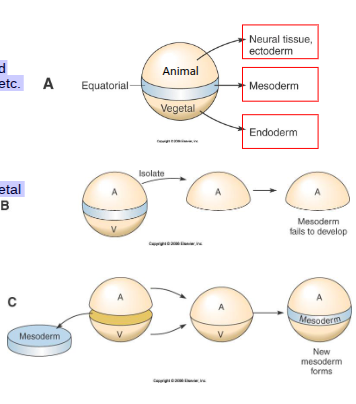
What are the developmental fates of the animal, vegetal, and marginal regions in a Xenopus embryo?
The animal region forms neural tissue and ectoderm, the vegetal region forms endoderm, and the mesoderm arises from the region between the animal and vegetal halves, giving rise to muscle, blood, and bone.
What does the animal cap assay show about mesoderm induction?
It shows that mesoderm formation requires contact with vegetal cells, as isolated animal caps do not form mesoderm unless exposed to a vegetal-derived, diffusible signal.
Is the animal cap assay useful for studying neural induction, and why?
No. Isolated animal caps fail to form both mesoderm and neuroectoderm, so they cannot model the normal induction events that occur during embryonic development.
What does the animal–vegetal combination experiment show regarding neural fate?
It shows that combining animal and vegetal tissues produces both mesoderm and neural tissue, suggesting that inductive signals are present in these regions.
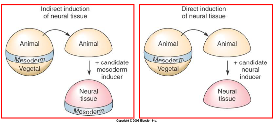
What challenge arises when testing for neural inducers in vivo?
The organiser contains mesoderm, making it difficult to separate factors that induce neural fate directly from those acting indirectly via mesoderm.
What is needed to identify a direct neural inducer?
A factor that triggers neural markers in animal caps without inducing mesoderm-specific markers.
Why must mesoderm inducers be distinguished from neural inducers in experiments?
Because some signals may induce neural tissue only as a secondary effect of mesoderm formation, masking the identity of true neural inducers.
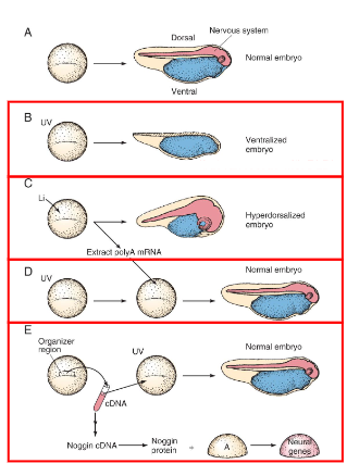
What happens to Xenopus embryos treated with UV light, and how does lithium treatment differ?
UV treatment ventralises embryos, whereas lithium treatment dorsalises them.
How was mRNA used to rescue ventralised Xenopus embryos?
mRNA from dorsalised embryos was injected into ventralised embryos and was able to restore dorsal structures.
What strategy was used to identify Noggin from the organiser region?
Researchers prepared cDNA from the organiser, injected RNA pools into ventralised embryos, and progressively tested smaller pools until the Noggin-encoding clone was found.
How was it confirmed that Noggin directly induces neural tissue?
Adding Noggin to animal caps or ventralised embryos showed that it directly activates neural genes.
Isolation of Animal Caps: The animal cap (AC) is a piece of embryonic ectoderm isolated from the blastula stage embryo. When cultured alone, this tissue normally develops into undifferentiated epidermis.
Application of Purified Noggin Protein: Purified Noggin protein (or mRNA encoding it) was added directly to these isolated animal caps in a simple culture medium.
Absence of Mesoderm Induction: The researchers confirmed that the Noggin treatment did not induce the formation of mesodermal tissue (e.g., muscle or notochord), which is typically required for neural induction in the intact embryo.
Expression of Neural Markers: The treated animal caps were then assayed for the expression of specific neural marker genes (such as NCAM,
βbeta
𝛽
-tubulin, and nrp-1) using techniques like RNase protection assays and in situ hybridization.
Differentiation and Patterning: The results showed robust expression of these neural-specific genes, confirming that the ectoderm had differentiated into neural tissue. Further analysis showed the induced tissue could even differentiate into mature neurons and display some anterior-posterior and dorsal-ventral patterning, such as the non-overlapping expression of dorsal brain (cpl-1) and ventral brain (etr-1) markers.
What method was used to begin identifying Chordin?
Differential screening of Xenopus dorsal lip cDNA
How was the embryonic cDNA library screened for Chordin candidates?
Using RNA probes from dorsalised and ventralised embryos to find clones expressed in dorsalised but not ventralised embryos
How were positive candidate clones tested further?
By assessing whether they could induce a neural axis in ventralised blastulae and by checking their tissue expression patterns using in situ hybridisation.
Who identified the Follistatin–Activin pathway in neural induction, and when?
Doug Melton’s group identified it in April 1994 (Hemmati-Brivanlou et al.).
What molecule was isolated in a screen for mesoderm inducers, and what type of protein is it?
Activin, a member of the TGF-β family that signals via a two-part receptor.
What happens in an animal cap assay when a truncated Activin receptor is used?
Mesoderm does not form, and instead of epidermis, the tissue develops neural characteristics.
What does blocking Activin signalling in animal caps reveal about neural induction?
Blocking Activin causes neural induction even without a neural inducer
Activin induces non-neural fates: Activin typically induces mesoderm and endoderm at different concentrations, but in the absence of other signals, it promotes the formation of epidermis in animal caps.
Neural fate is the default: When activin signaling is inhibited using a dominant-negative activin receptor (a mutant receptor that blocks the signaling pathway), the isolated animal cap cells autonomously differentiate into neural tissue. This neural tissue expresses general markers of the central nervous system.
Epidermis is an induced fate: This finding demonstrated that the formation of epidermis is an induced process, requiring the presence of signals like Activin/BMP4, rather than the intrinsic or default state of the ectoderm.
A common strategy: These results, along with similar experiments in Drosophila, suggested a common evolutionary strategy for nervous system formation in vertebrates and invertebrates, involving the inhibition of a conserved signaling pathway (BMP/Activin) to promote neural development.
What hypothesis was proposed to explain the effect of blocking Activin signalling?
That Activin (or a similar molecule) normally inhibits neural induction; therefore, neural fate can arise through inhibition of an inhibitor.
What molecule was identified as the “inhibitor of the inhibitor” in neural induction?
Follistatin, which is known to bind and block Activin.
Where is Follistatin expressed during early Xenopus development?
In the dorsal lip of the blastopore (DLB) and the axial mesoderm.
What effect does Follistatin mRNA have when injected into embryos?
It can induce a secondary body axis.
What does Follistatin protein do in animal cap assays?
It induces neural tissue from the animal cap.
What do intact animal caps isolated before gastrulation normally develop into when cultured?
epidermis
What happens when animal cap cells are dissociated before gastrulation and then cultured?
They express neuronal markers and develop neural characteristics
Why do dissociated animal cap cells adopt a neural fate?
Because cell–cell signalling is disrupted, removing inhibitory signals that normally prevent the default neural state.
What does adding BMP4 to dissociated animal cap cells cause them to become?
epidermis
What does the BMP4 experiment suggest about BMP4’s role?
BMP4 acts as a neural inhibitor.
What overall conclusion does this support about neural fate?
Neural tissue is the default state; inhibition of BMP signalling allows neural induction
What is the Default Hypothesis/Model of neural induction?
It states that ectodermal cells naturally adopt a neural fate unless they receive signals from neighbouring cells; during normal development, BMPs inhibit this default neural fate and instead specify epidermis on the ventral side of the embryo.
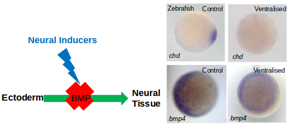
How do neural inducers promote neural fate during early development?
They act by blocking BMP signalling, which normally inhibits neural induction.
Which tissue releases BMP antagonists during gastrulation?
The involuting mesoderm.
What does Chordin bind to, and with what effect?
Chordin binds BMP4 with high affinity, preventing it from exerting anti-neural effects
How does Noggin interact with BMP4?
Noggin binds BMP4 with even greater affinity than Chordin, blocking BMP signalling.
Which BMPs and related molecules does Follistatin bind?
Follistatin binds BMP7 and Activin
What is the overall outcome of these BMP antagonists acting around the midline?
They sequester free BMPs, lifting BMP inhibition and allowing neural tissue to form
Are these neural-inducing signalling molecules evolutionarily conserved?
Yes, they are conserved from Xenopus to mammals.
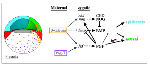
Can neural induction be fully explained by the Default Hypothesis/Model?
No. In addition to BMP inhibition, neural induction also involves Wnt/β-catenin signalling blocking BMP4 transcription in dorsal ectoderm, organiser-derived Wnt antagonists (Frzb1, Cerberus, Dkk1), and pre-gastrula FGF signalling, all of which contribute to establishing neural fate.
Neural induction is complex interaction of signalling pathways
Zebrafish Embryonic Fate Map
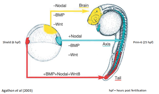
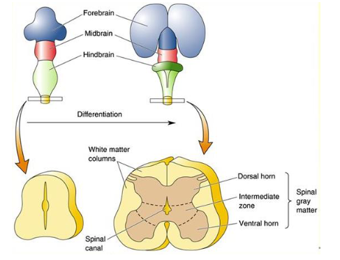
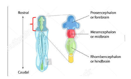
How does the rostral end of the neural tube differentiate during the 3-primary vesicle stage?
It forms three vesicles at the rostral (head) end, with this regionalisation influenced by a gradient of Wnt protein
Differentiation to the 5-secondary vesicle stage
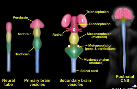
Differentiation of the prosencephalon (vesicle 1 - forebrain)
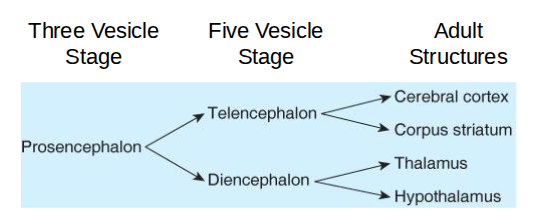
What are the major structures of the telencephalon, and what are their functions?
Cerebral cortex: Processes sensory and motor functions.
Basal telencephalon: Includes the basal ganglia (striatum) and amygdala.
Corpus callosum: White matter axons linking the two cerebral hemispheres.
What are the main components of the diencephalon and their roles?
Thalamus: Relays sensory and motor signals to the cortex.
Hypothalamus: Links the nervous system to the endocrine system.
Internal capsule: White matter axons connecting the cortex, thalamus, brain stem, and spinal cord.
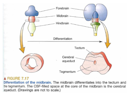
Differentiation of the mesencephalon (vesicle 2 - midbrain)
The CSF-filled cerebral aqueduct connects rostrally (towards head) with the third
ventricle and caudally (towards tail) with the 4th ventricle.
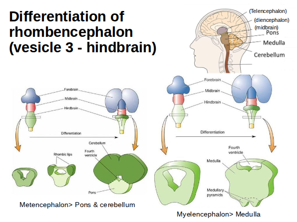
Differentiation of rhombencephalon (vesicle 3 - hindbrain)
Differentiation of the spinal cord (caudal neural tube)