chp. 12: transport across cell membranes
 Call Kai
 Learn
Learn Practice Test
 Spaced Repetition
 Match
Match Flashcards
 Knowt Play
1/107

There's no tags or description
Looks like no tags are added yet.
Name | Mastery | Learn | Test | Matching | Spaced | Call with Kai | Chat |
|---|
No analytics yet
Send a link to your students to track their progress
108 Terms
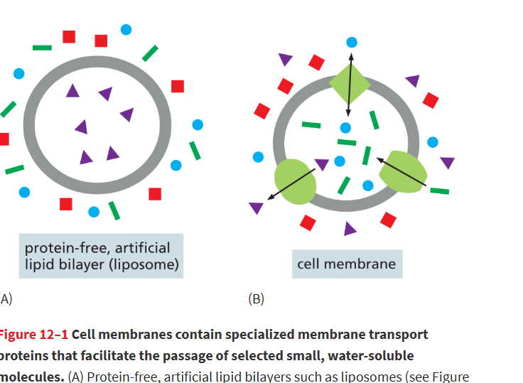
membrane transport proteins
Any transmembrane protein in lipid bilayer that provides a passageway for the movement of select substances across a cell membrane.
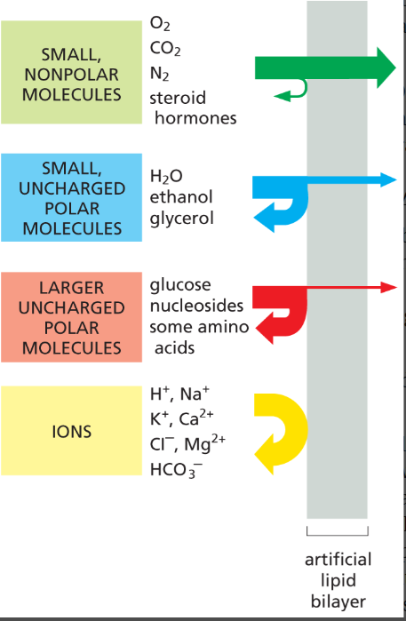
What can and can’t cross a lipid bilayer?
Can cross easily: small, nonpolar molecules (O₂, CO₂, steroid hormones)
Cross slowly if small polar and uncharged: water (H₂O), ethanol, glycerol
Hardly cross: larger uncharged polar molecules (glucose, some a.a, nucleosides)
Cannot cross: all ions (Na⁺, K⁺, Cl⁻, H+)
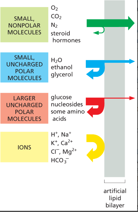
what determines the rate a solute crosses a membrane through simple diffusion?
size and solubility
simple diffusion
The movement of molecules from a region of higher concentration to a region of lower concentration directly through the lipid bilayer, without needing proteins or energy.
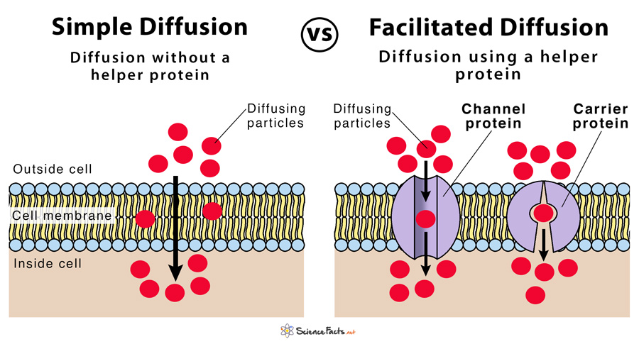
what r the 2 main classes of membrane transport proteins
transporters
channels
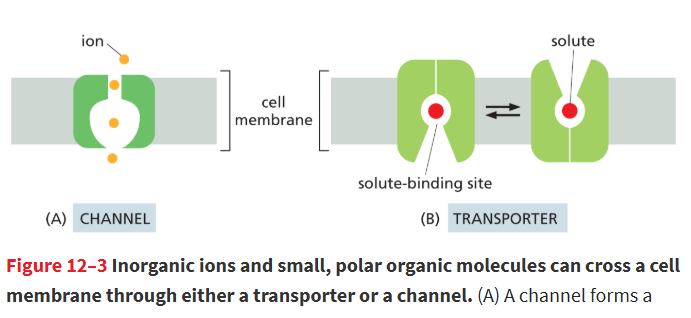
transporters
only binds specific molecules or ions at dedicated binding sites, like enzymes binding substrates. This gives them high selectivity, but they transport more slowly than channels
undergo a series of conformational changes to move solutes across the membrane.
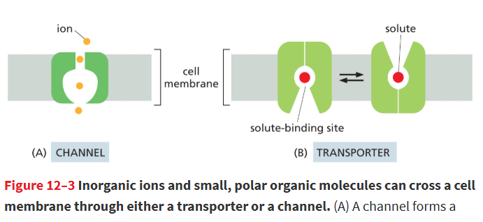
channels
are selective based on size and electric charge; only ions/molecules of the appropriate size and charge can pass through when the channel is open.
form a pore through the bilayer and exist in open or closed states; opening is controlled by external stimuli or cellular conditions.
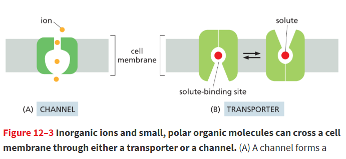
Why do cells need membrane transport proteins?
Many substances like sugars, amino acids, ions, and metabolites cross lipid bilayers very slowly or not at all, so transport proteins provide selective pathways for these molecules.
What is the general structure of membrane transport proteins?
They are multipass α-helical proteins. Multiple helices form a hydrophilic pathway through the membrane, allowing selective transport of ions or small molecules. Channels work by size/charge; transporters use specific binding and conformational changes.
Which ion is most abundant outside a mammalian cell?
Na+ (sodium)
Which ion is most abundant inside a mammalian cell?
K+ (Potassium)
Why are ion concentrations inside a cell different from outside?
Cells maintain different internal and external ion concentrations to survive and function properly.
positive and neg. charges must be balanced inside/outside of cell to prevent it from dying
membrane potential
Voltage difference across a membrane due to a slight excess of positive ions on one side and of negative ions on the other.
when the anions/cations r balanced it’s called the resting membrane potential, but it does not = 0!
passive transport
The spontaneous movement of a solute down its concentration gradient across a cell membrane via a membrane transport protein, such as a channel or a transporter.
doesnt use energy
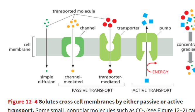
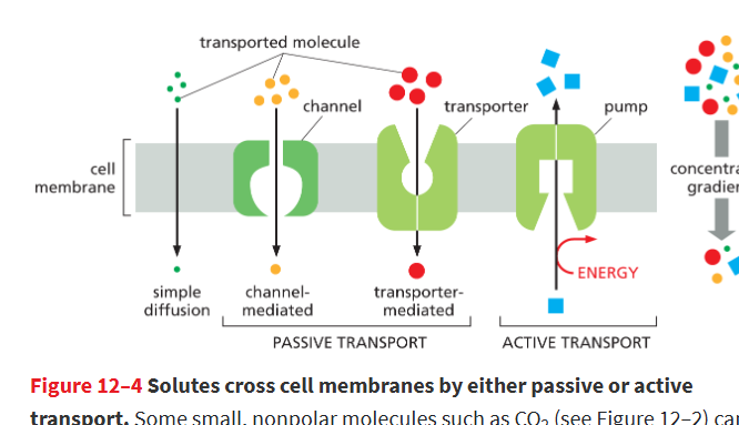
active transport
The movement of a solute across a membrane against its electrochemical gradient; requires an input of energy, such as that provided by ATP hydrolysis.
carried out by special types of transporters called pumps
if a molecule is electrically charged, the direction of passive transport depends on..
concentration gradient and membrane potential
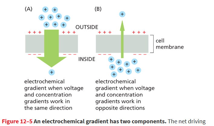
electrochemical gradient
Driving force that determines which way an ion will move across a membrane; consists of the combined influence of the ion’s concentration gradient and the membrane potential.
the cystolic side of membrane is usually at a neg. potential relative to extracellular side
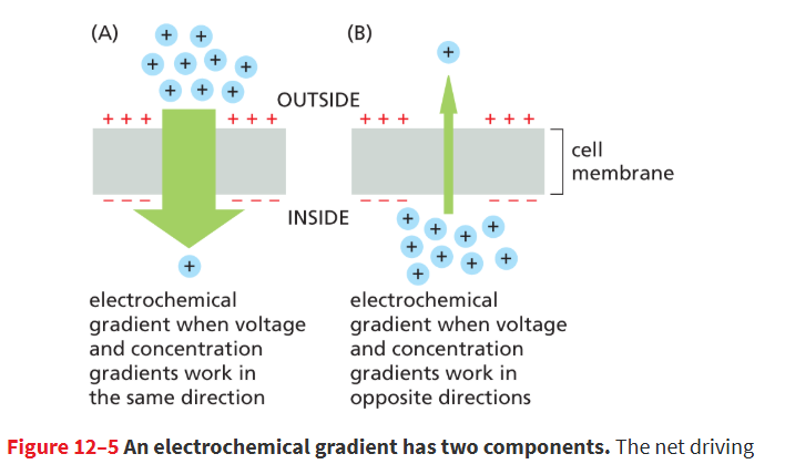
What happens when voltage and concentration gradients act in the same direction? Give an example.
They create a strong electrochemical gradient.
Example: Na⁺, which is higher outside the cell and positively charged, so it tends to enter the cell.
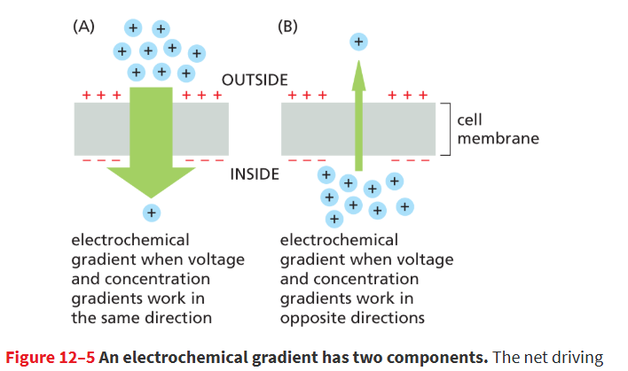
What happens when voltage and concentration gradients oppose each other? Give an example.
They create a small electrochemical gradient.
Example: K⁺, which is more concentrated inside the cell while the inside is negatively charged, so there is little net movement.
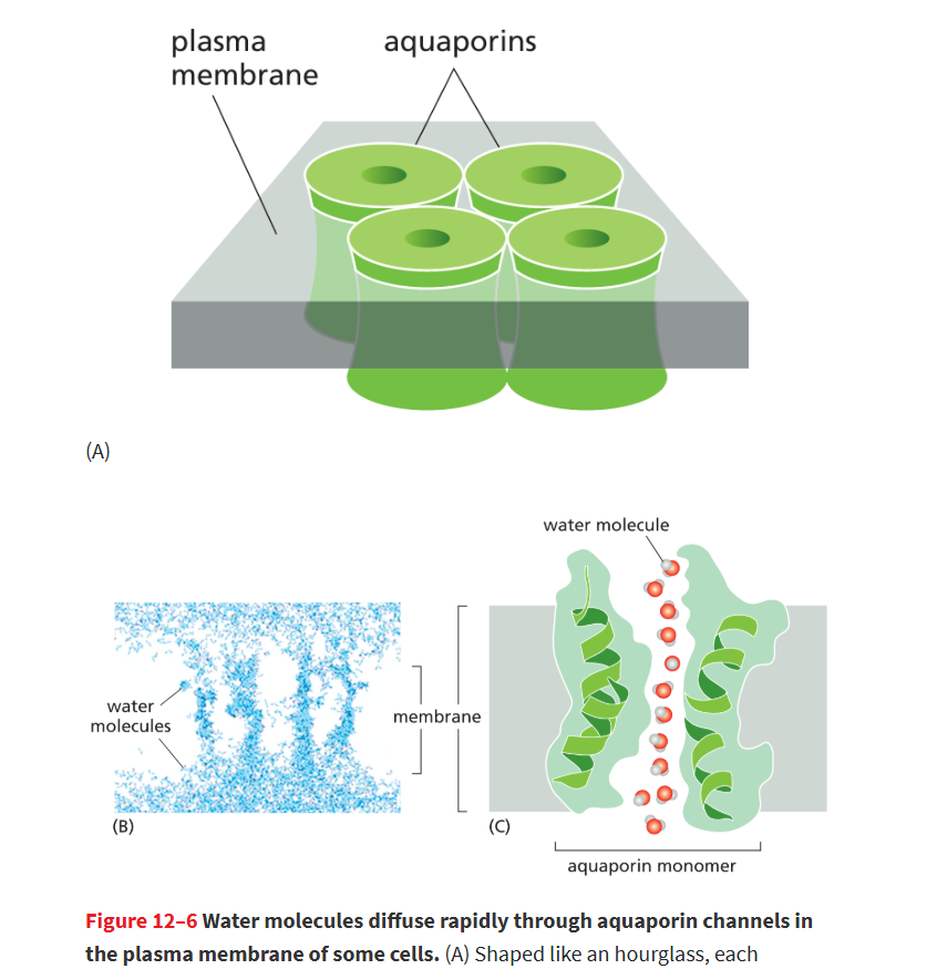
aquaporins
Channel that facilitates the transport of water, but not ions, across cell membranes; those found in the plasma membrane greatly increase a cell’s permeability to water.
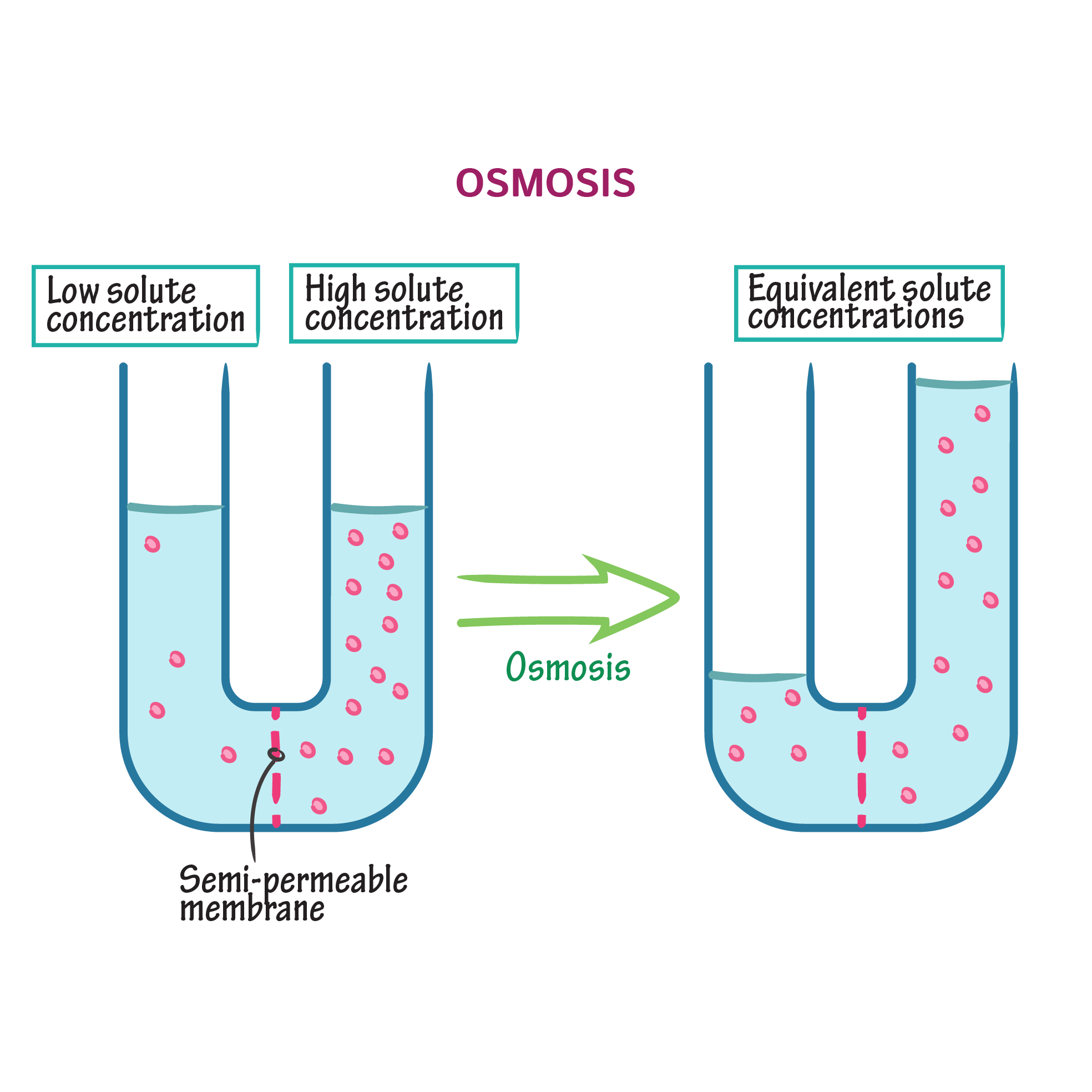
osmosis
Passive movement of water across a cell membrane from a region where water conc’n is high (conc’n of solutes low) to low conc’n of water ( conc’n of solutes is high)
can make a cell swell
turgor pressure
force that builds as water flows into plant and yeast cells by osmosis
drives the expansion of cells that underlie plant growth and maintains rigidity of plant stems and leaves.
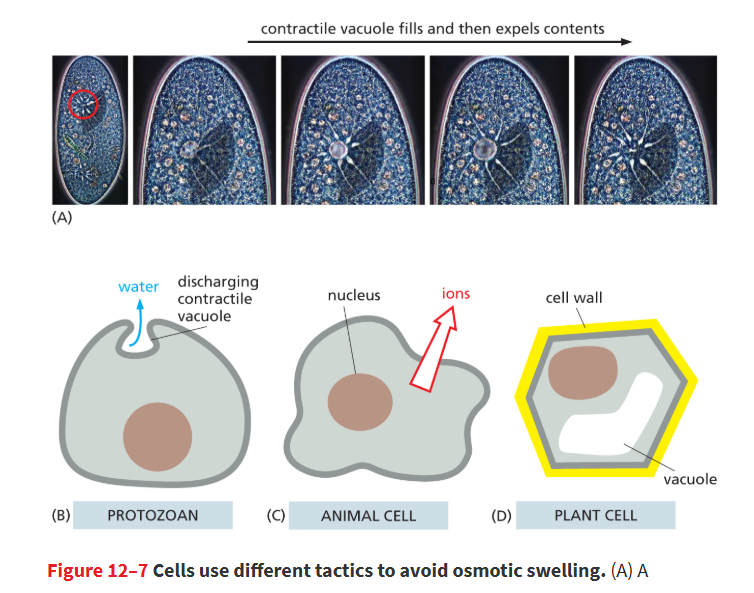
How does water cross cell membranes?
By slow diffusion through the lipid bilayer or rapidly through aquaporin channels.
Cells usually have a higher solute concentration (higher osmolarity) than the outside environment, creating an osmotic gradient that pulls water inside cell
How do different cells prevent swelling from osmosis?
Protozoa: contractile vacuoles pump out water
Animal cells: pump out ions (like Na⁺)
Plant cells: rigid cell wall creates turgor pressure
Which type of membrane transport protein can perform both active and passive transport?
Only transporters can move a solute against its concentration gradient. Some transporters also allow passive transport of molecules down their concentration gradient.
what r the usual concentrations of Na+ and K+
Na+ is the most plentiful positively charged ion outside the cell, while K+ is the most plentiful inside
selectivity of transporters
they r highly selective and each cell membrane has a diff transporter for particular membranes
example:
- Plasma membrane: imports nutrients (sugars, amino acids, nucleotides)
Lysosome membrane: imports H⁺ to acidify and exports digestion products
Mitochondrial inner membrane: imports pyruvate and exports ATP
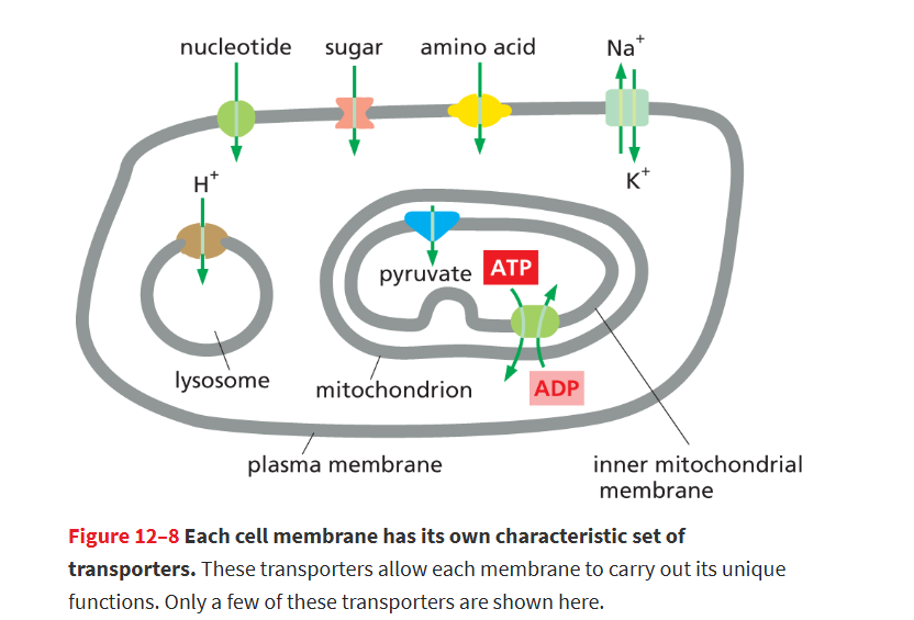
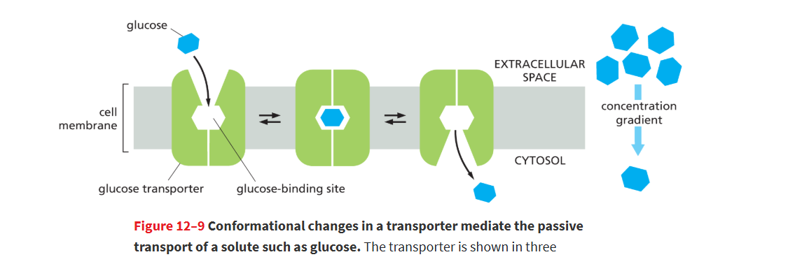
How does the glucose transporter work in passive transport?
glucose transporter is polypeptide chain that crosses the membrane 12x
Transporter changes conformation randomly
Glucose binds on the side with higher concentration
It is released on the side with lower concentration
Net transport depends only on the glucose concentration gradient, cuz glucose is uncharged, so the electrochemical gradient = 0
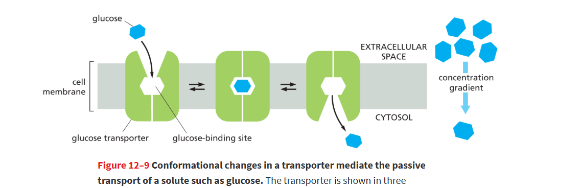
How does the glucose transporter move glucose in and out of cells?
The transporter changes shape and binds glucose on the side with higher concentration.
After a meal: more glucose outside → glucose moves into cells.
When blood glucose is low: glucagon causes liver cells to make glucose → glucose moves out of cells.
transmembrane pumps
Transporter that uses a source of energy (active transport), such as ATP hydrolysis or sunlight, to actively move a solute across a membrane against its electrochemical gradient.
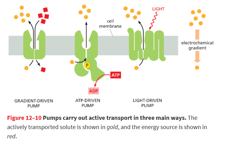
they can carry out active transport in 3 ways:
gradient-driven pumps
ATP-driven pump
light-driven pump
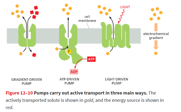
What are the three main types of pumps in active transport?
Gradient-driven pumps: use the downhill movement of one solute to move another uphill
ATP-driven pumps: use ATP hydrolysis for energy
Light-driven pumps: use sunlight energy (mostly in bacteria)
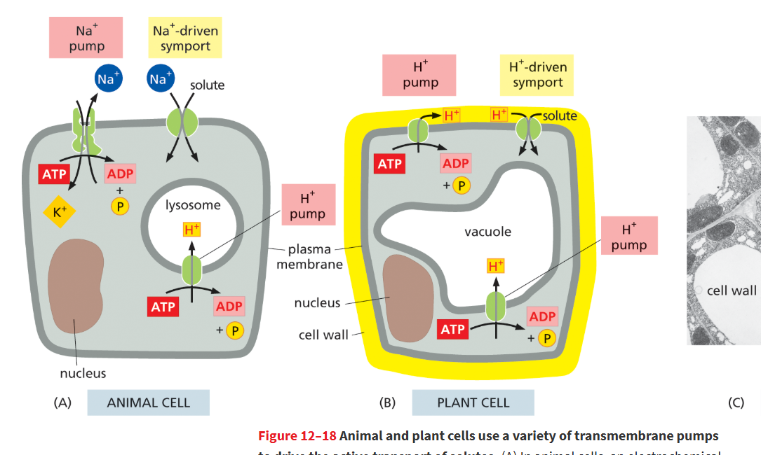
Why is the ATP-driven Na⁺ pump important in animal cells?
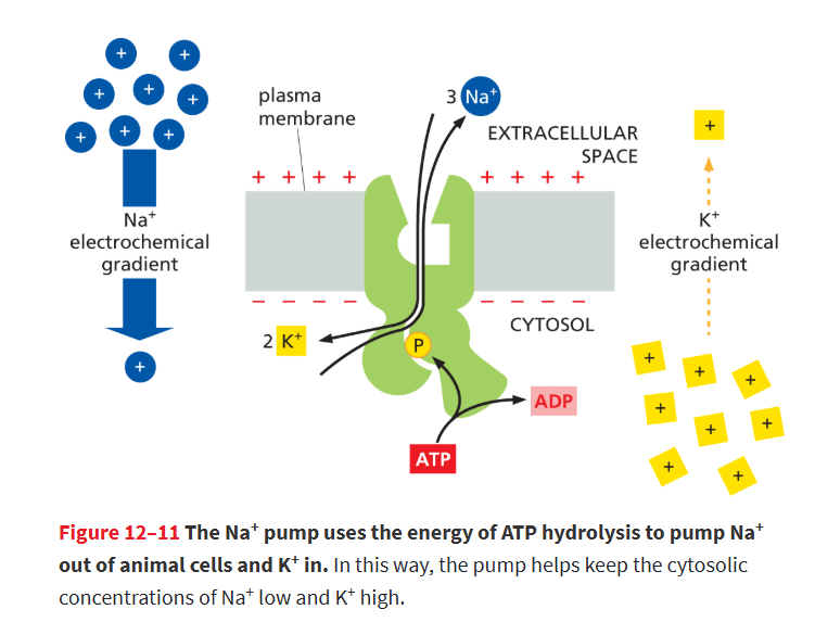
It pumps Na⁺ out of the cell, creating a Na⁺ gradient that powers many gradient-driven pumps to transport other substances into the cell.
Na+ pump (or Na+–K+ ATPase)
Transporter found in the plasma membrane of most animal cells that actively pumps Na+ out of the cell and K+ in using the energy derived from ATP hydrolysis.
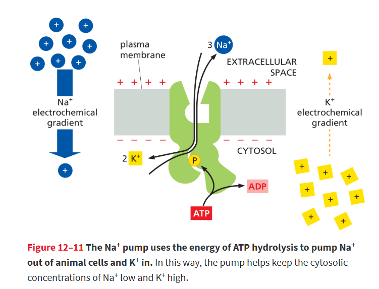
results in 3 Na⁺ out of the cell and 2 K⁺ into the cell.
contributes to the electrochemical gradient of the membrane , one (-) charge inside the membrane each turn of the pump.
this pump uses 25% of ALL of ur ATP!
How many ions move during one cycle of the Na⁺–K⁺ pump?
3 Na⁺ out of the cell and 2 K⁺ into the cell.
contributes to the electrochemical gradient of the membrane , one (-) charge inside the membrane each turn of the pump.
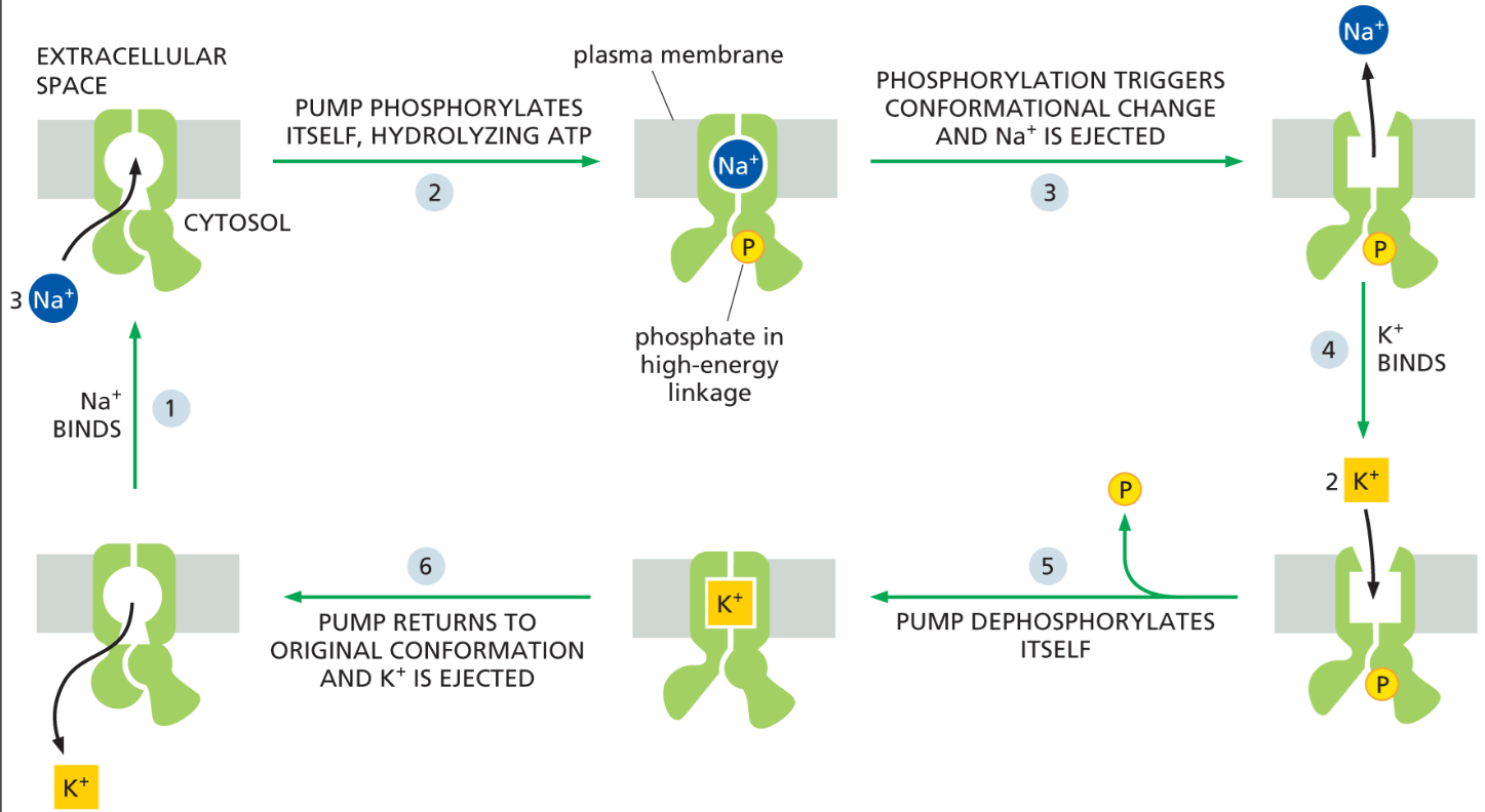
How does ATP power the Na⁺–K⁺ pump?
ATP hydrolysis adds a phosphate to the pump, causing conformational changes that move Na⁺ out and K⁺ in.
It maintains low Na⁺ and high K⁺ inside cells and creates ion gradients used for many other transport processes.
a⁺–K⁺ pump = 3 Na⁺ out / 2 K⁺ in
this pump uses 25% of ALL of ur ATP!
What gradient does the Na⁺ pump create across the plasma membrane?
It keeps Na⁺ much lower inside the cell and K⁺ much higher inside
this creates a large Na⁺ electrochemical gradient, which stores energy that cells use for transport and other processes.
The high Na⁺ concentration outside the cell is like water behind a dam, storing energy that can drive other transport processes when Na⁺ flows back into the cell.
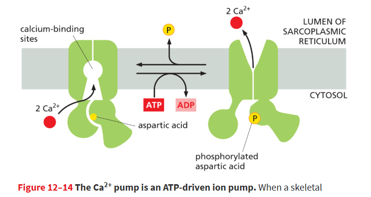
Ca2+ pump (or Ca2+ ATPase)
An active transporter that uses energy supplied by ATP hydrolysis to actively expel Ca2+ from the cell cytosol.
maintain low Ca2+ conc’n in cytosol, and high Ca2+ conc’n outside of cell (like Na+)
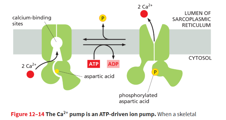
Why do cells keep cytosolic Ca²⁺ levels very low?
Ca²⁺ binds to proteins and changes their activity, acting as an important intracellular signal.
increased Ca2+ can trigger muscle contraction, fertilization, and nerve cell communication.
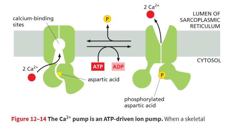
Where is the Ca2+ pump found?
are found in both the plasma membrane and the endoplasmic reticulum (ER).
In muscle cells, a specialized ER called the sarcoplasmic reticulum (SR) stores Ca²⁺.
remove Ca²⁺ from the cytosol into the ER lumen for storage.
gradient-driven pumps
an active transport pump where one molecule moving down its gradient provides the energy to mover another molecule moving against its gradient.
includes:
symport
antiport
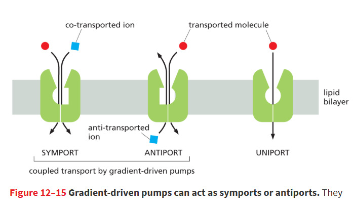
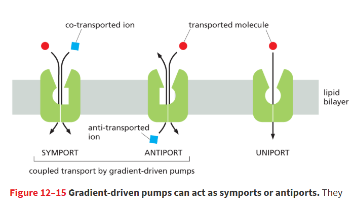
the 2 types of gradient-driven pumps
Symport- pump moves pair of solutes in same direction
antiport- moves them in opposite directions
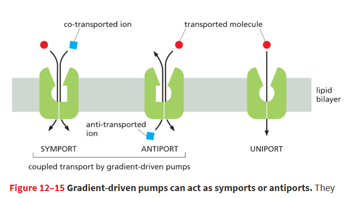
uniports
move only a single type of solute across the membrane at a time
facilitates passive diffusion down its conc’n gradient, so not a pump (no energy)
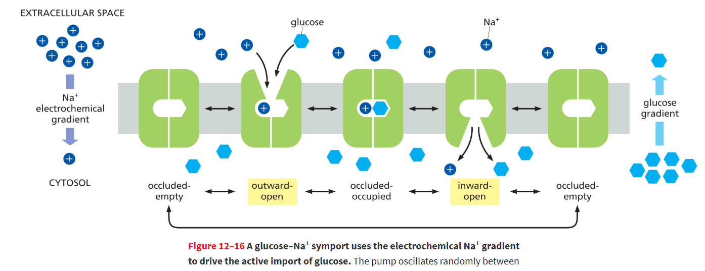
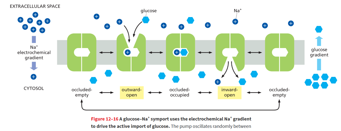
Na⁺–glucose symport
Uses the Na⁺ electrochemical gradient to actively import glucose into epithelial cells.
There is a steep Na⁺ electrochemical gradient across the apical membrane: high outside (gut lumen), low inside (cytosol).
When Na⁺ moves down its gradient into the cell, it “drags” glucose along, even if glucose concentration is higher inside the cell than outside.
Cooperative binding: The transporter only works when both Na⁺ and glucose bind, preventing Na⁺ from leaking uselessly into the cell.
found in apical domain of plasma membrane, facing gut lumen
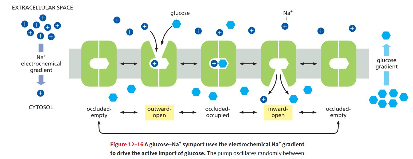
steps of Na+ glucose pump
Outward-open state: Transporter faces gut lumen; Na⁺ binds first (high outside).
Glucose binds to the outward-open transporter.
Occluded state: Both solutes are trapped inside the transporter, inaccessible from either side.
Inward-open state: Transporter opens to the cytosol; Na⁺ and glucose are released.
Transporter returns to outward-open state empty, ready for another cycle.
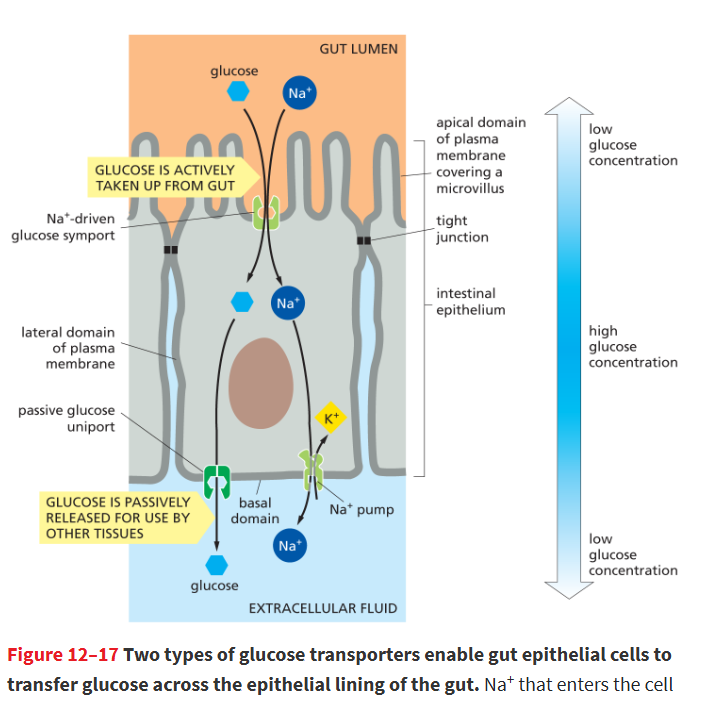
where is the Na+-glucose pump vs glucose uniport located?
Na+-glucose symport located in apical domain of plasma membrane, facing the gut lumen
glucose uniport is located in the basal/lateral domains of plasma membrane, facing extracellular fluid
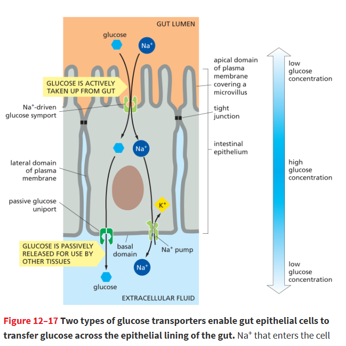
How is the Na⁺ gradient maintained in epithelial cells?
Na⁺ pumps in the basal and lateral membranes expel Na⁺, keeping cytosolic Na⁺ low and the electrochemical gradient steep for continued symport.
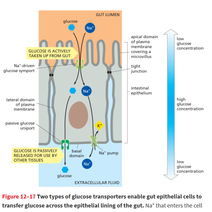
How do gut epithelial cells release glucose to other tissues?
They have glucose uniports on the basal and lateral membranes, which passively release glucose down its concentration gradient.
located in basal/lateral domains of plasma membrane
what r the 2 types of transporters in gut epithelial cells?
if only had Na+-glucose symporter, glucose would never be released, so it needs another'
Glucose–Na⁺ symport (cotransporter)
Location: Apical membrane (facing the gut lumen)
Function: Actively imports glucose into the cell using the Na⁺ electrochemical gradient.
Mechanism: Na⁺ moves down its gradient and “drags” glucose along against its concentration gradient.
Special feature: Cooperative binding—both Na⁺ and glucose must bind for transport to occur.
Glucose uniport (facilitated diffusion transporter)
Location: Basal and lateral membranes (facing the blood and neighboring cells)
Function: Passively releases glucose from the cytosol into the blood down its concentration gradient.
Mechanism: Does not use energy; glucose flows according to its gradient.
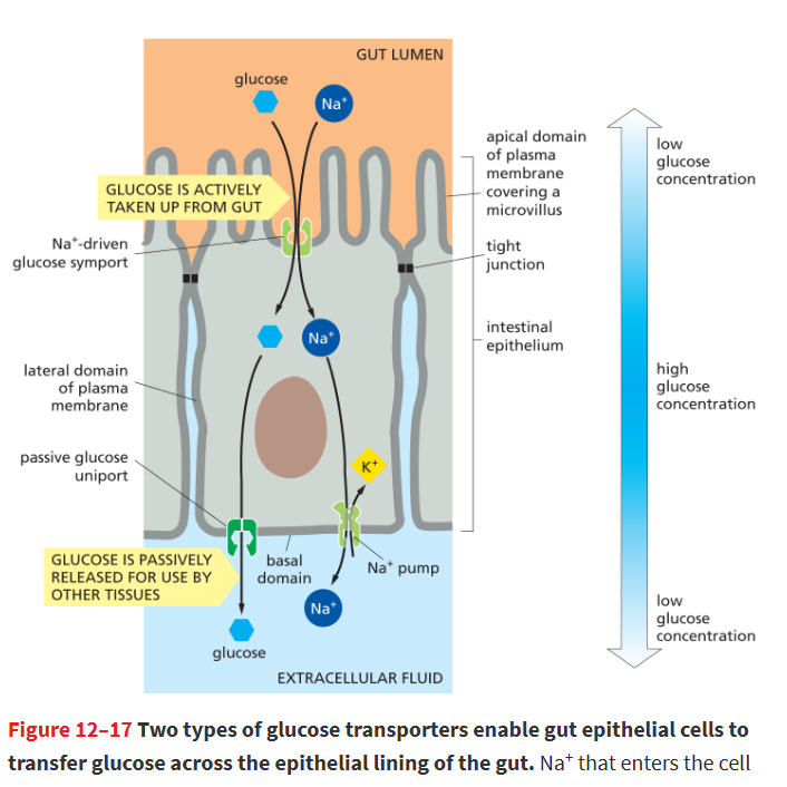
how r the 2 types of glucose transporters kept segregated?
kept in proper domains by a diffusion barrier formed by tight junctions around apex of cell
prevents mixing btwn the 2 membrane domains
allows for directional movement of glucose out of intestine and into bloodstream
What other solutes can be transported using the Na⁺ electrochemical gradient?
Other sugars and amino acids via symports
H⁺ via antiports like the Na⁺–H⁺ exchanger to regulate cytosolic pH.
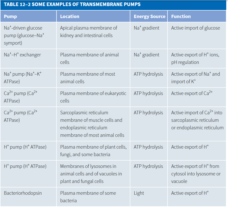
H+ pumps
A protein or protein complex that uses energy supplied by ATP hydrolysis, an ion gradient, or light to actively move protons across a membrane.
plants, bacteria, and fungi don’t have Na+ pumps, so they use this instead to create an electrochemical gradient and import solutes
What energy sources power H⁺ pumps in these cells?
ATP-driven H⁺ pumps (most plants, fungi, bacteria)
Light-driven H⁺ pumps (e.g., bacteriorhodopsin in photosynthetic bacteria)
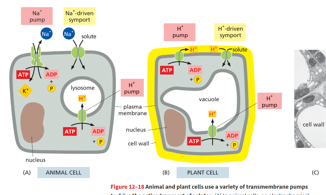
How do H⁺ symports work?
Solutes (like sugars and amino acids) are imported into the cell by coupling their transport to H⁺ moving down its electrochemical gradient.
H⁺ pumps use ATP to move H⁺ out of the cell, creating a steep H⁺ gradient.
H⁺ symports use the H⁺ gradient: H⁺ flows back into the cell down its gradient, and this energy drags solutes (sugars, amino acids) into the cell, even against their own gradient.
Key point: ATP is not used by the symport; it was only used to set up the gradient.
When the glucose–Na+ symport protein is in its outward-open state, which is more likely to occur?
Na+ binds to its binding site.
Because Na+ concentrations are high outside the cell, Na+ readily binds to the transporter in its outward-open state. The transporter must then wait for a rare glucose molecule to bind.
What are ion channels?
Proteins that form hydrophilic pores in cell membranes to allow passive movement of small, water-soluble molecules or ions down electrochemical gradient.
no energy req, substances move down conc’n gradient
examples: gap junctions, aquaporins
selectivity filter
Part of an ion channel that determines which ions the channel can transport; located in the region where the channel is narrowest
only ions of right size and charge r able to pass
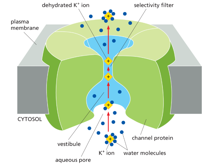
What are the two main properties that distinguish ion channels from simple holes in the membrane?
1) Ion selectivity – only specific ions can pass through.
2) Gating – channels open and close in response to a stimulus.
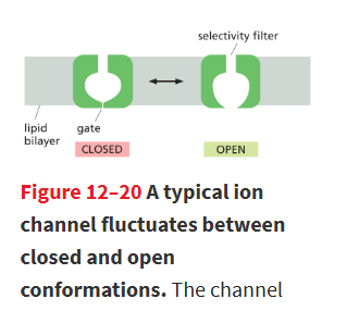
How do ion channels differ from transporters in terms of ion flow?
Channels do not undergo conformational changes for each ion like transporters, so they can transport ions much faster than transporters
channels cant undergo active transport w/ ATP, but transporters can
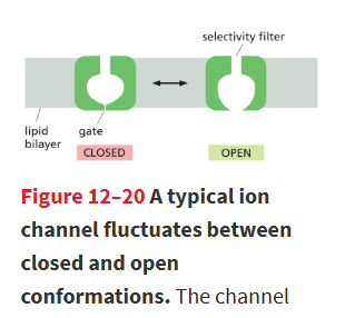
What happens to ions and the membrane potential when an ion channel opens?
Ions move rapidly down their electrochemical gradients, which changes the membrane potential.
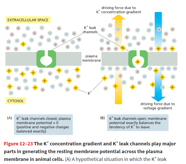
K+ leak channels
Ion channel permeable to K+ that randomly flickers between an open and closed state, allowing K+ to move freely across membrane
largely responsible for the resting membrane potential in animal cells.
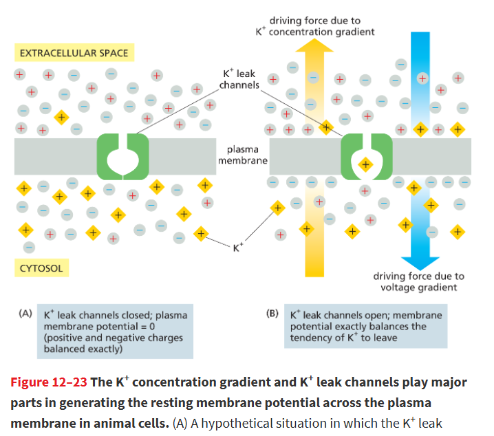
resting membrane potential
Voltage difference across the plasma membrane when a cell is not stimulated.
flow of + and - charges balanced
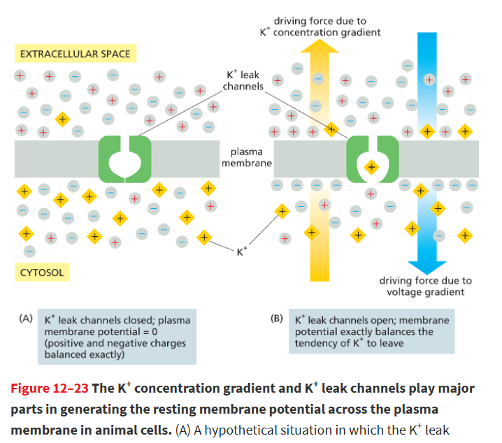
What role do K⁺ leak channels play in membrane potential?
They allow K⁺ to move out of the cell down its concentration gradient, creating a voltage difference (inside negative) until equilibrium is reached.
the exit of K+ from cell is limited by the + chargew on the outside of the membrane
What happens when a cell is stimulated?
Ion channels open, ions move in or out, and the cell’s voltage changes.
also changes membrane’s permeability to ions
ion channels randomly snap btwn open and closed states
follows an “all or none”, must be fully open/closed
What r the 3 ways ion channels r regulated?
mechanically/physically gated channels
ligated gated channels
voltage gated channels
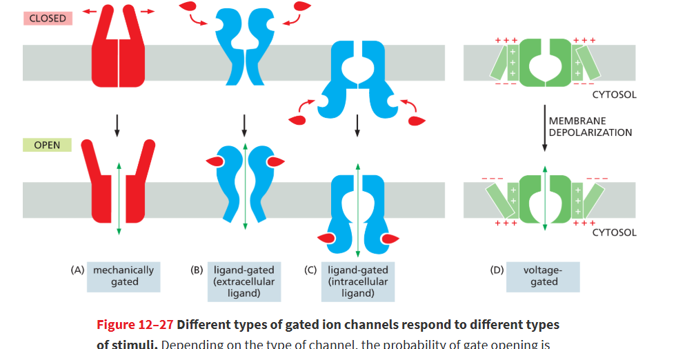
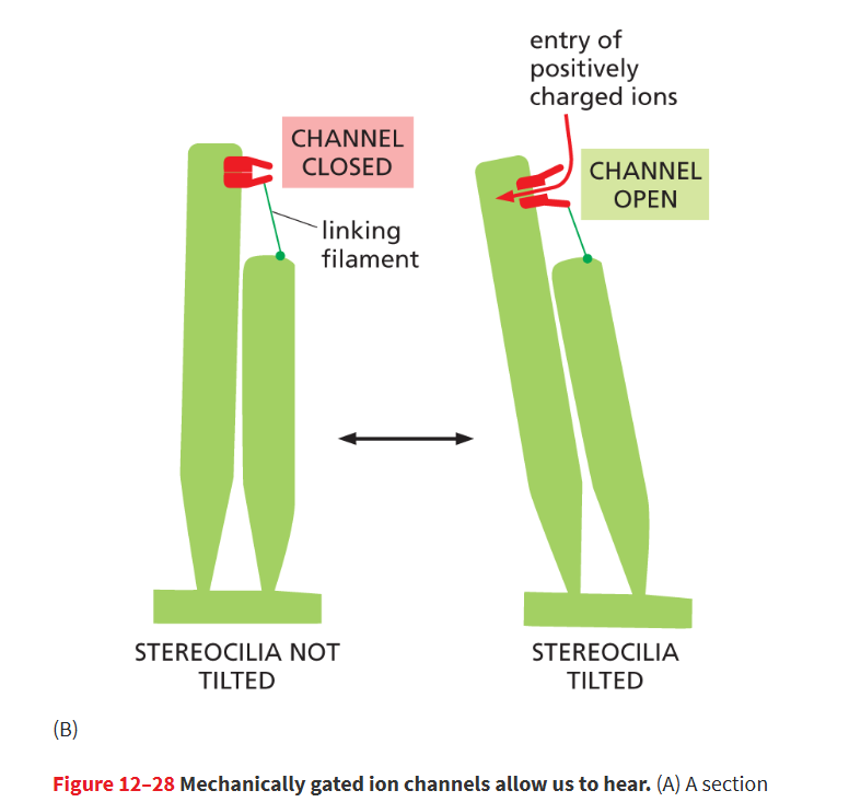
mechanically gated channel
An ion channel that allows the passage of select ions across a membrane in response to a physical perturbation.
ex: auditory hair cells when sound vibrations pull channels open
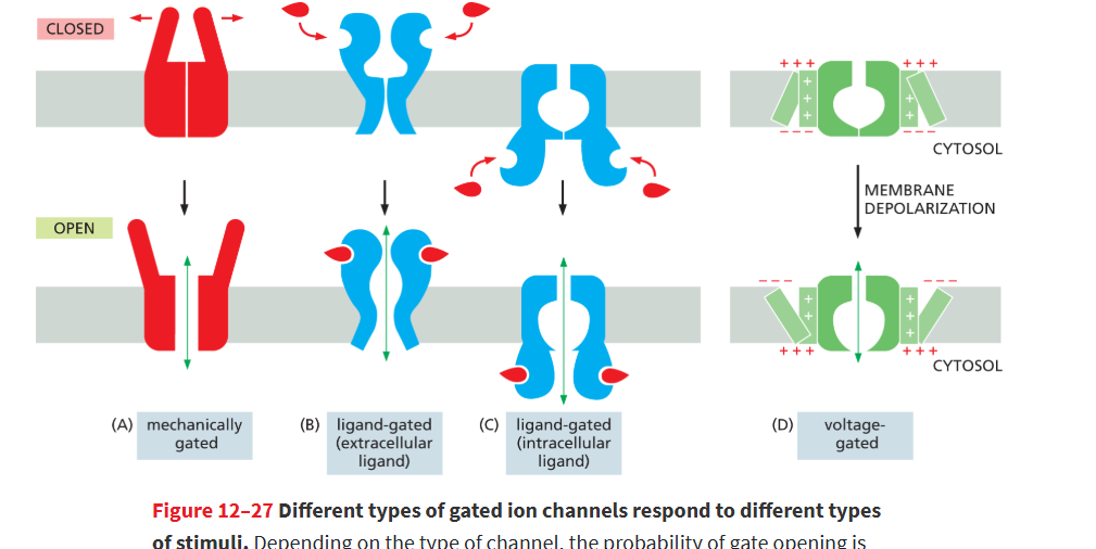
ligand-gated channel
An ion channel that is stimulated to open by the binding of a small molecule such as a neurotransmitter.
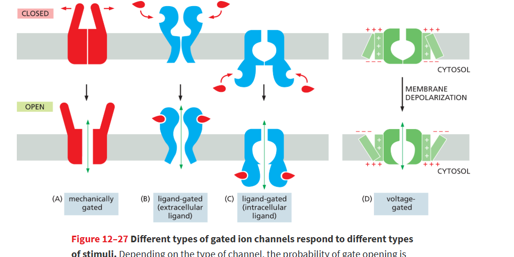
voltage-gated channel
Channel protein that permits the passage of selected ions, such as Na+, across a membrane in response to changes in the membrane potential.
Found primarily in electrically excitable cells such as nerve and muscle cells.
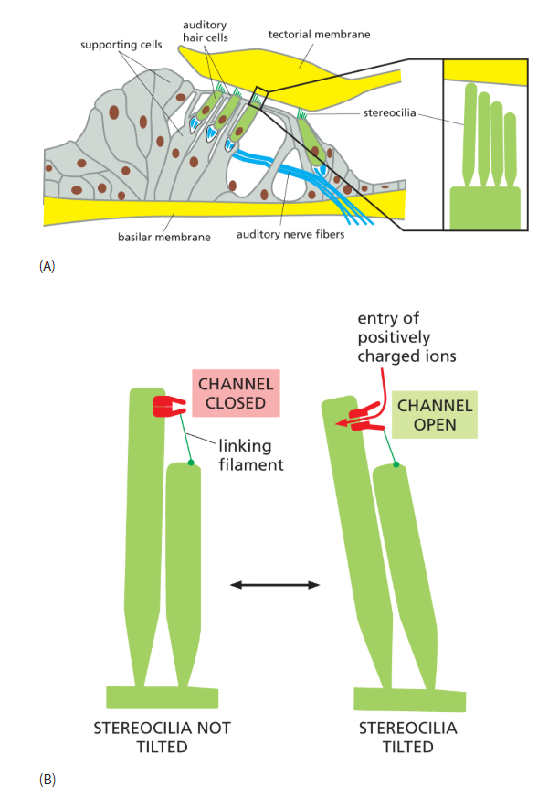
mechanical gated channel and auditory reception
stereocilia of auditory hair cells attached to tectorial membrane, which vibrates due to sound waves
movement of stereocilia open mechanically gated ion channels, resulting in influx of + ions
this activates underlying auditory nerves
piezo channels
a mechanically gated ion channel that responds to pressure
allows us to sense light touch or detect when bladder is full
voltage sensors
are inside of a voltage gated ion channel and r very sensitive to changes in membrane potential. when change in potential exceeds threshold value, channel switches open
change in membrane potential alters the probability that it will open
How do voltage-gated channels contribute to electrical signaling?
Opening one type can change the membrane potential, which can trigger other voltage-gated channels to open, creating a chain reaction.
this is the cause for the ability to generate an electric impulse
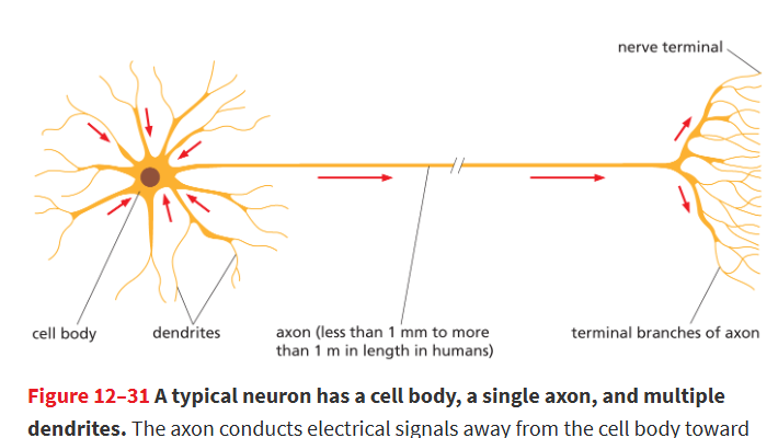
neuron
receive, integrate, and transmit signals in the nervous system
Cell body (soma): nucleus and organelles.
Axon: carries signals away.
Dendrites: receive signals.
they communicate signals through changes in the electrical potential across their plasma membrane.
action potential/nerve impulse
Traveling wave of electrical excitation caused by rapid, transient, self-propagating depolarization of the plasma membrane in a neuron or other excitable cell
can carry a message from one neuron to another
triggered by a sufficiently strong stimulus depolarizes the membrane to the threshold potential
depolarized
A shift in the membrane potential, making it less negative on the inside of the cell.
when a neuron is stimulated, this happens and the membrane potential shifts to a less neg. value.
Na+ rushes in making inside less negative
voltage-gated Na+ channel
Protein in the plasma membrane of electrically excitable cells that opens in response to membrane depolarization, allowing Na+ to enter the cell.
It is responsible for action potentials in these cells.
causes further depolarization (makijjng membrane potential even less -)
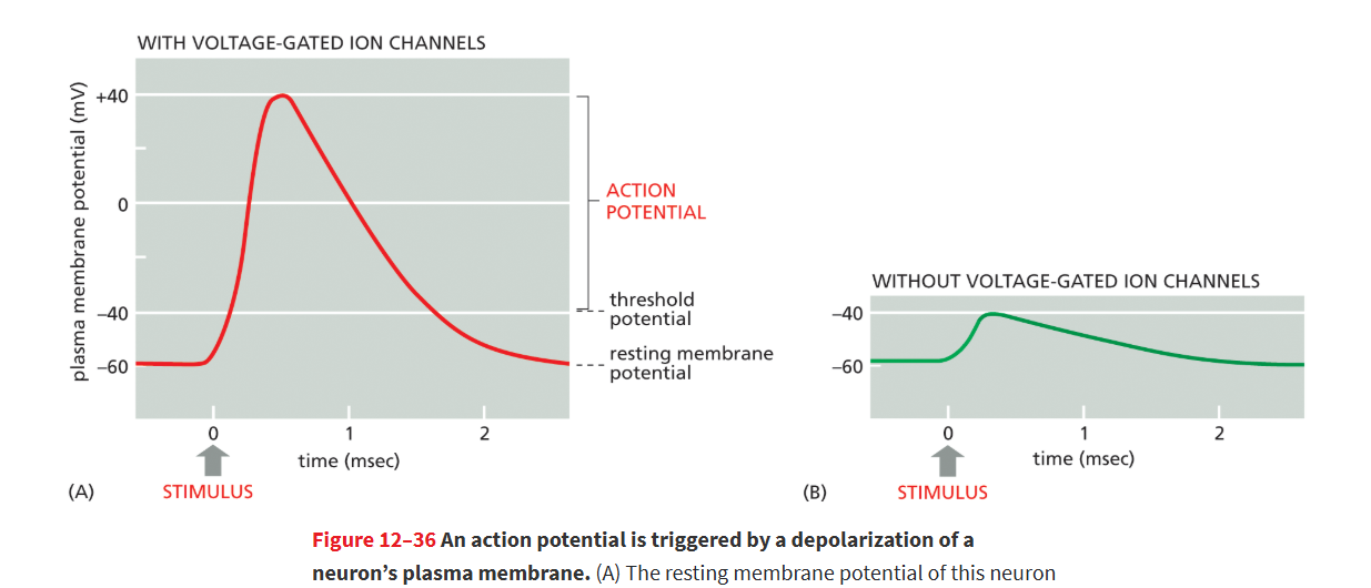
What happens as Na⁺ enters the neuron?
The membrane becomes less negative, opening more Na⁺ channels and causing rapid depolarization to about +40 mV.
Na⁺ entry triggers more Na⁺ channels to open, creating a positive feedback loop and action potential
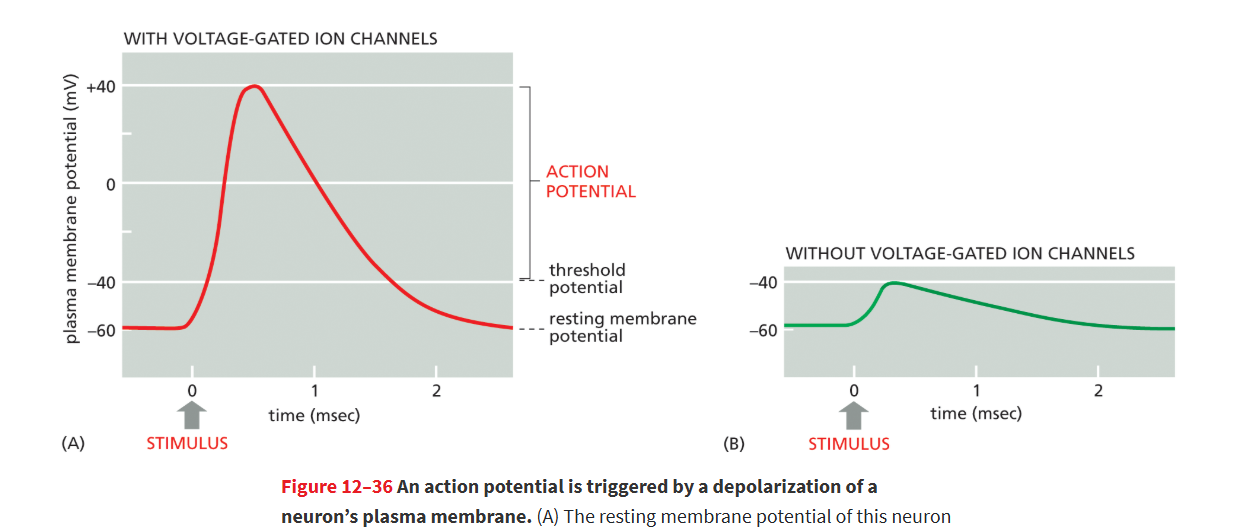
Why does Na⁺ flow into a neuron during depolarization?
Na⁺ flows in because of its electrochemical gradient: high outside concentration pushes it in, and the negative inside of the cell attracts it. During depolarization, the inside becomes less negative, reducing electrical pull, but Na⁺ still enters until the membrane reaches ~+40 mV, where the concentration and electrical forces balance.
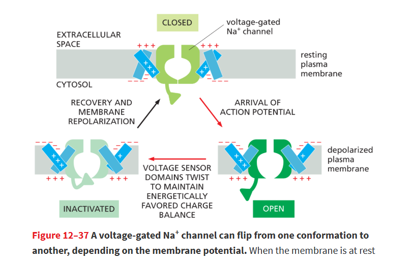
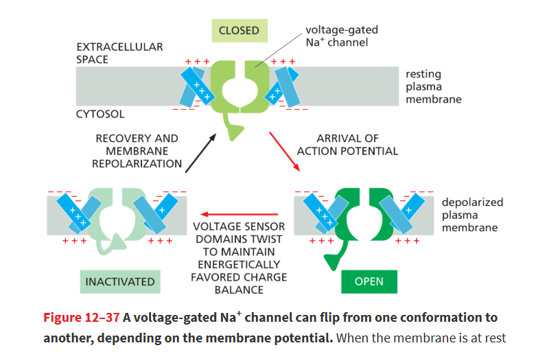
Na+ channel inactivation
Voltage-gated Na⁺ channels have a built-in “timer.”
After opening briefly, they automatically inactivate, closing even though the membrane is still depolarized.
They stay inactivated until the membrane returns to resting potential.
This prevents the neuron from getting “stuck” with Na⁺ channels open.
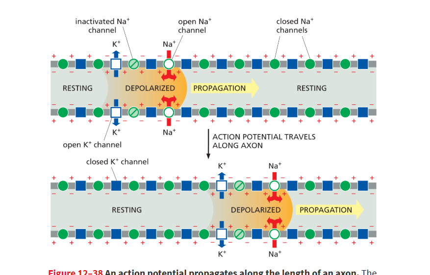
repolarization
Voltage-gated K⁺ channels open
K⁺ flows out of the neuron because:
There’s more K⁺ inside than outside.
The inside is positive (from Na⁺ coming in), which pushes K⁺ out.
This brings the inside back to negative → repolarization.
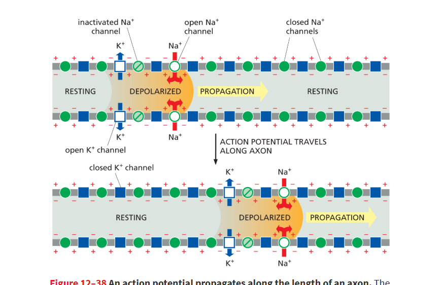
voltage gateed K+ channels
help return depolarized membrane back to its resting potential by having K+ flow out of the neuron to restore the negative membrane potential after the influx of Na
the K+ will flow out cuz there’s more K+ inside than outside, and the inside is positive from the Na+ coming in, which pushes the K+ out
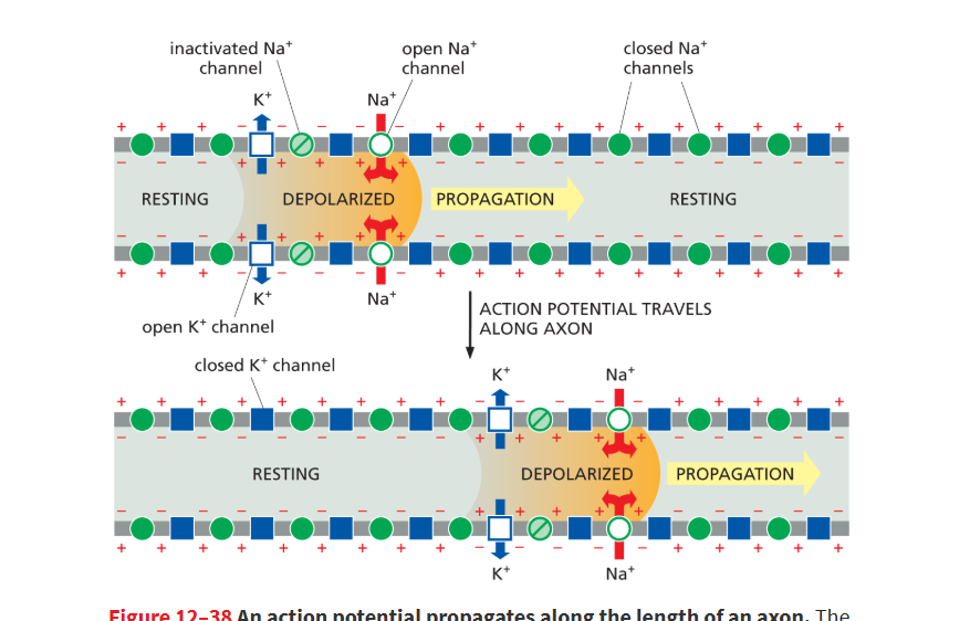
How do Na⁺ and K⁺ cause depolarization and repolarization in a neuron?
Na⁺ channels open → depolarization
Na⁺ rushes into the neuron.
The inside becomes positive.
K⁺ channels open → repolarization
K⁺ flows out of the neuron.
The inside becomes negative again.
This rapid change in voltage (Na⁺ in, then K⁺ out) is what we call an action potential, and it travels down the axon to send the nerve signal.
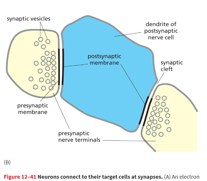
synapse
Specialized junction where a nerve cell communicates with another cell (such as a nerve cell, muscle cell, or gland cell), usually via a neurotransmitter secreted by the nerve cell.
theres a pre and postsynaptic cell separated by a synaptic cleft
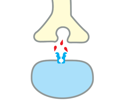
neurotransmitter
Small signaling molecule secreted by a nerve cell at a synapse to transmit information to a postsynaptic cell.
Examples include acetylcholine, glutamate, GABA, and glycine
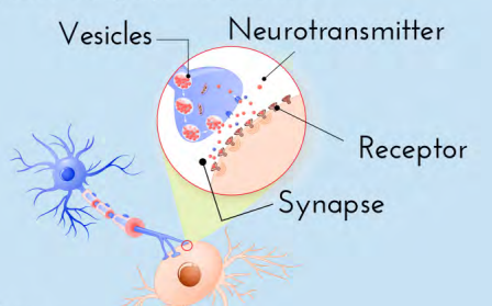
synaptic cleft
The tiny gap separating the pre and post synaptic cells where neurotransmitters travel to send the signal from one cell to another.
electrical signal cannot cross, so its converted to chemical signal as a neurotrasnmitter
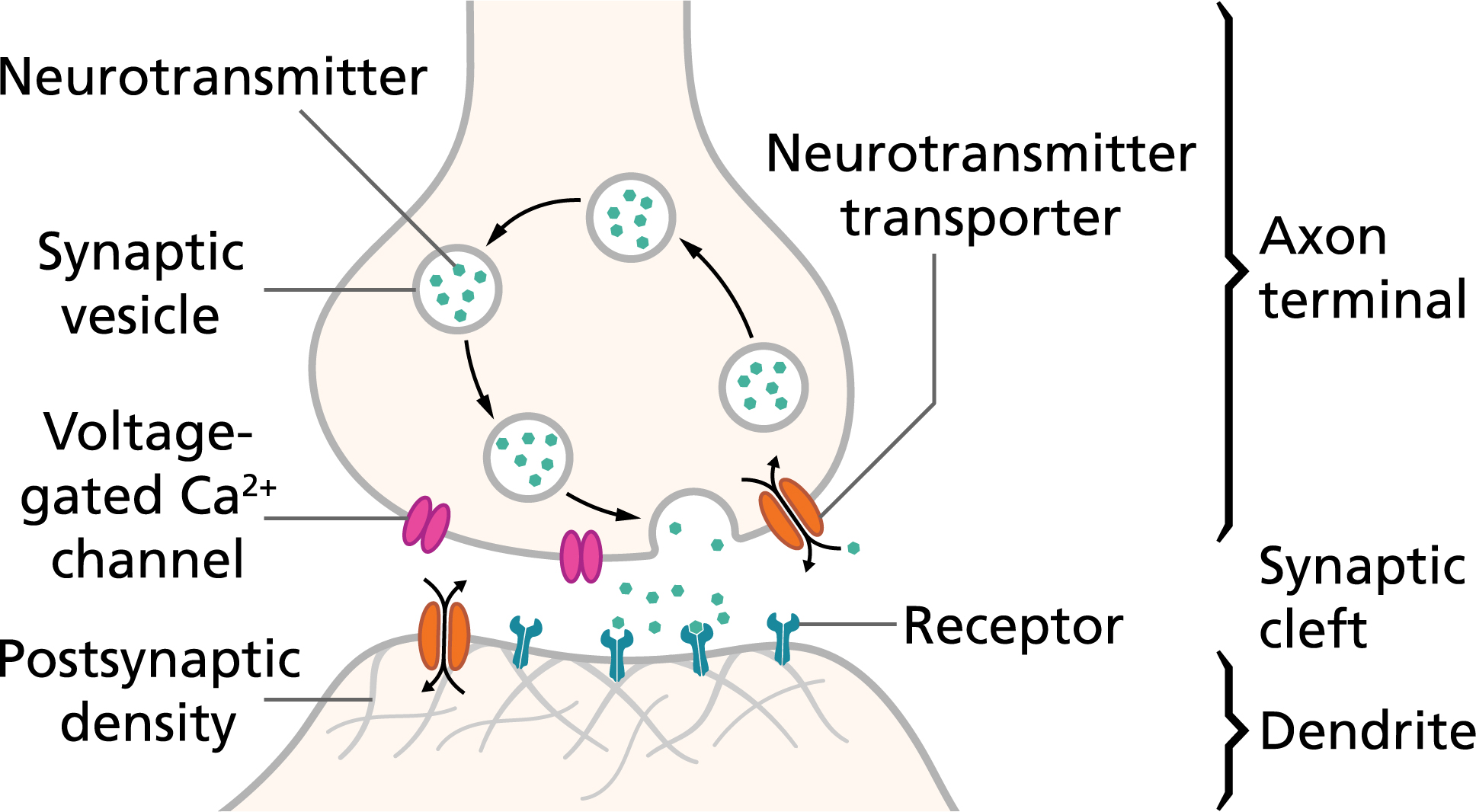
presynaptic cell
The sending neuron that releases neurotransmitters into the synaptic cleft.
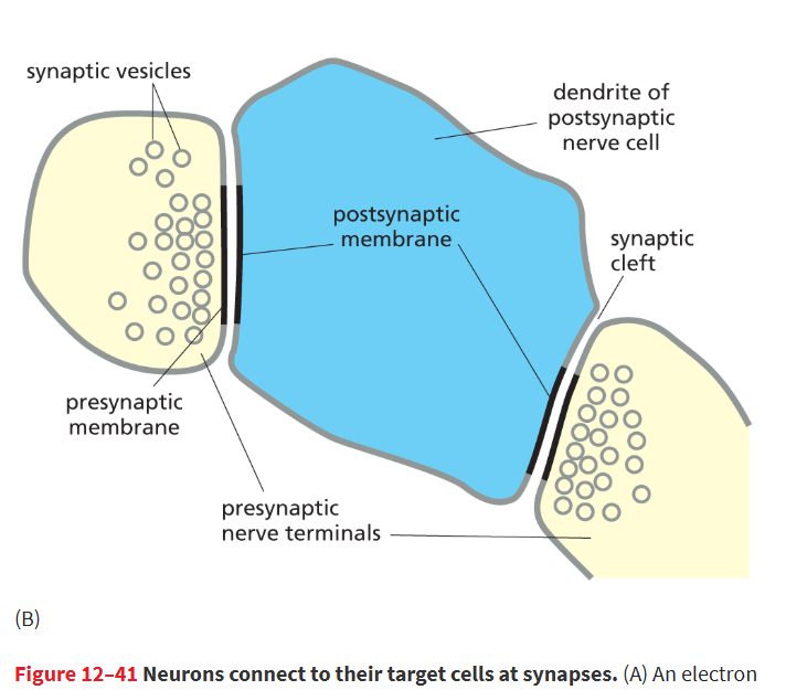
postsynaptic cell
The receiving cell that detects the neurotransmitters and continues the signal.
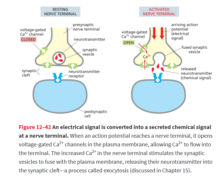
How do voltage-gated Ca²⁺ channels convert an electrical signal into a chemical signal at a synapse?
When an action potential reaches the nerve terminal, voltage-gated Ca²⁺ channels open.
Ca²⁺ rushes into the cell, which triggers synaptic vesicles to fuse with the membrane and release neurotransmitters into the synaptic cleft, sending the signal to the next cell. (exocytosis)
Ca²⁺ entering the neuron is the signal that causes neurotransmitter release.
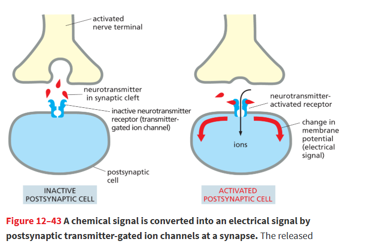
transmitter-gated ion channels
Transmembrane receptor protein or protein complex that opens in response to the binding of a neurotransmitter, allowing the passage of a specific inorganic ion; its activation can trigger an action potential in a postsynaptic cell.
they convert chemical signals back into electrical signals cuz the resulting ion flows alter the membrane potential of postsynaptic cell
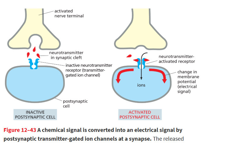
What happens when a neurotransmitter binds to receptors on the postsynaptic membrane?
The transmitter-gated ion channel opens, letting ions move across the membrane and changing the membrane potential.
this changes chemical signal (neurotransmitter) back into an electrical signal
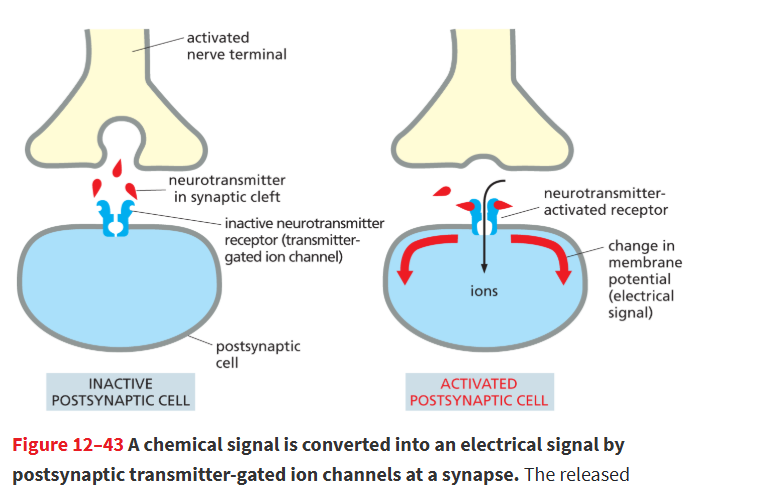
How can a postsynaptic cell trigger an action potential?
If the ion flow depolarizes the membrane enough, it triggers an action potential in the postsynaptic cell.
neurotransmitters trigger ligand gated ion channels in post-synaptic cell membraner and result in membrane depolarization
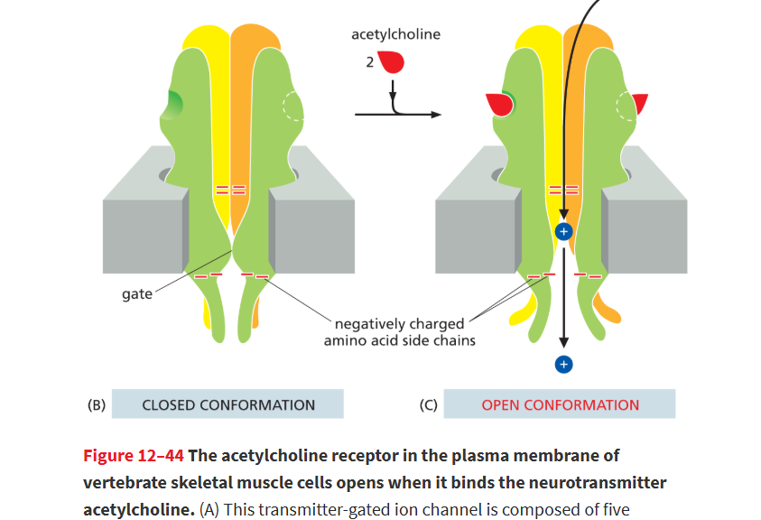
What is an example of a transmitter-gated ion channel?
The acetylcholine receptor, which opens when acetylcholine binds and allows Na⁺ to enter, depolarizing the cell.
influx of Na will trigger release of Ca from the sarcoplasmic reticulum and muscle contraction
neurotransmitters can be inhibitory or excitory
excitory neurotransmitters: when neurotransmitter binds, channels open for influx of Na+ which depolarizes plasma membrane and activates postsynaptic cell, encouraging it to fire action potential
acetylcholine and glutamate
Inhibitory neurotransmitters: when it binds, channels open allowing influx of Cl- to enter cell, this inhibits the postsynaptic cell by making its plasma membrane harder to depolarize
glycine and y-aminobutyric acid (GABA)
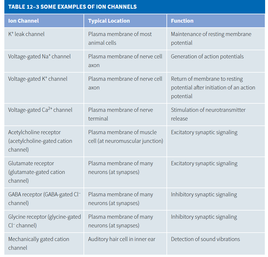
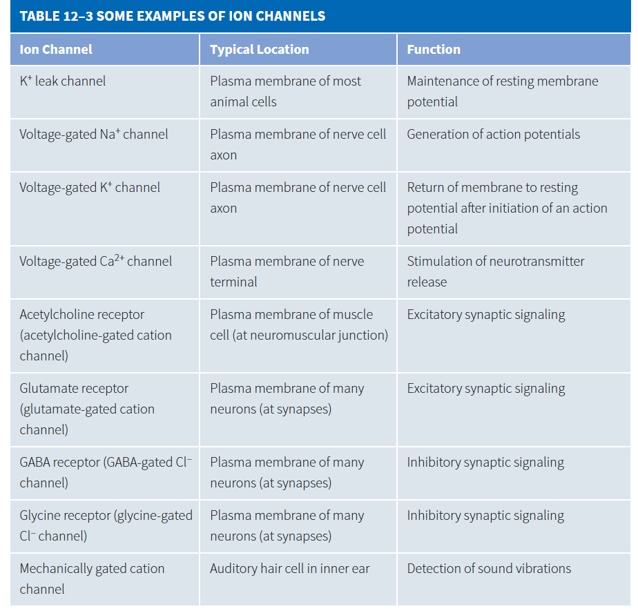
K+ leak channel
maintenance of resting membrane potential
location: plasma membrane of most animal cells
voltage gated Na+ channel
generation of action potentials (depolarization)
location: plasma membrane of nerve cell axon
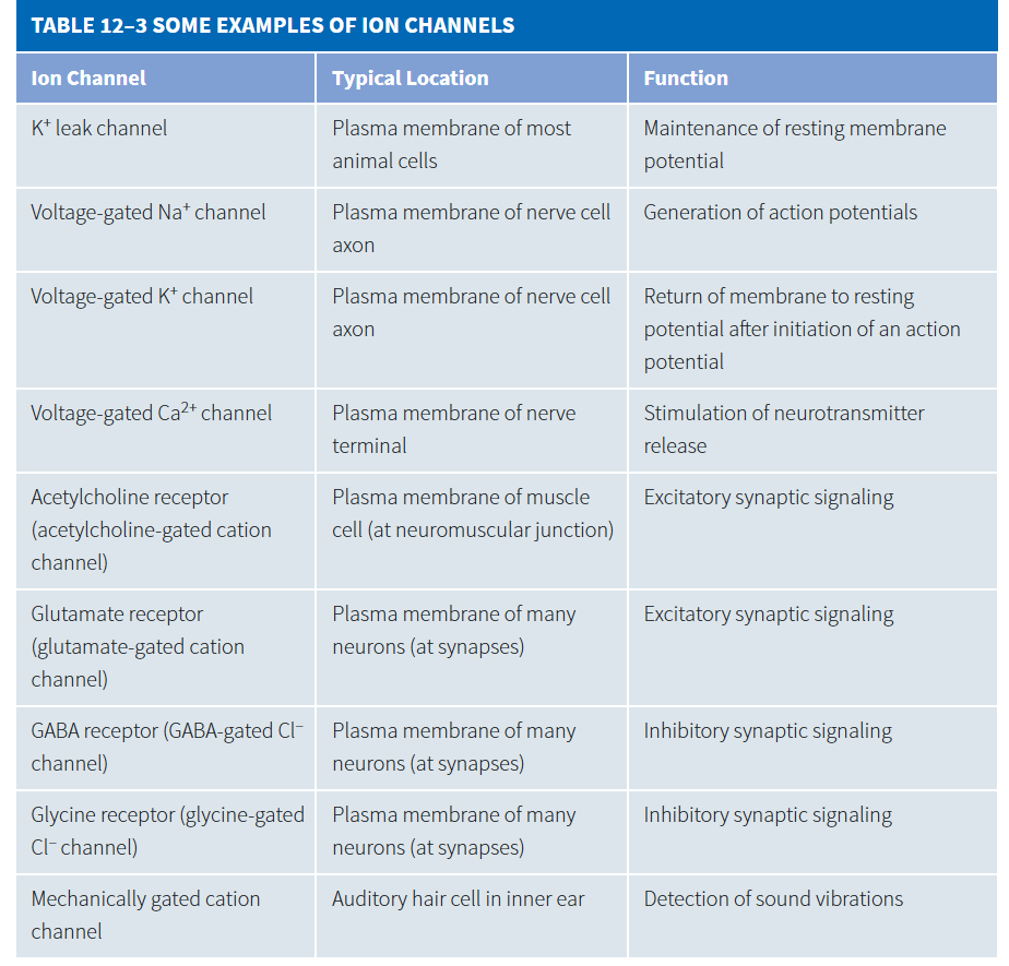
voltage-gated K+ channel
return of membrane to resting potential after initiation of an action potential (repolarization)
location: plasma membrane of nerve cell axon
voltage-gated Ca2+ channel
stimulation of neurotransmitter release
location: plasma membrane of nerve terminal
acetylcholine receptor
excretory synaptic signaling
location: plasma membrane of muscle cell (neuromuscular junction)
glutamate-gated cation channel
excretory synaptic signaling
location: plasma membrane of many neurons (at synapses)