SLP Praxis Review
 Call Kai
 Learn
Learn Practice Test
 Spaced Repetition
 Match
Match Flashcards
 Knowt Play
1/1026

There's no tags or description
Looks like no tags are added yet.
Name | Mastery | Learn | Test | Matching | Spaced | Call with Kai | Chat |
|---|
No analytics yet
Send a link to your students to track their progress
1027 Terms
Speech-Language Pathology Praxis Review
This study set was adapted from the user Anetz's practice set. She stated the following:
"This study set has at least 500 cards gleaned from as many SLP Praxis sets available on Quizlet as of August 2016. Then I added more as I studied various practice Praxis tests. Feel free to import this set into your own account so you can change personalize it.
This comes with no guarantees that it is 100% accurate! Check everything, and good luck!
Btw, I did pass the praxis."
As I have been studying, I have been confirming the information and insuring all information from the Advanced Review of Speech-Language Pathology Fourth Edition has been included. Subject areas that have been confirmed will include a "confirmed" in the subject heading.
NEUROANATOMY AND NEUROPHYSIOLOGY: THE NERVOUS SYSTEM (pg. 25-56)
NEUROANATOMY AND NEUROPHYSIOLOGY: THE NERVOUS SYSTEM- confirmed
Efferent/afferent neurons
transmit info away from brain (motor)/bring info to brain (sensory)
Nerve cells endings
Dendrites - receive signals
Axons - send signal on
Microglia
Scavengers to remove dead cells and other waste in brain
Autonomic nervous system
Components:
Sympathetic nervous system
Parasympathetic nervous system
Sympathetic nervous system
Activates fight or flight response
Parasympathetic nervous system
Returns body to state of relaxation
Pyramidal system vs Extrapyramidal
P: direct motor activation pathway, voluntary movement, directly signals nerve cells in spine or brainstem (fast, skilled, conscious movements)
E: maintains posture and tone for voluntary movements, indirectly regulates LMN movement activity (slow, postural, reflexive movements)
Damage results in involuntary and bizarre movement disorders; indirectly regulates nerve cells in spine or brainstem
Both are motor pathways. E is called that to distinguish it from the pyramidal system that reaches target locations by traveling through the 'pyramids' of the medulla.
Extrapyramidal composition
Subcortical nuclei: red nucleus, substantia nigra, subthalamus, basal ganglia, and their pathways
Pyramidal system componenets
Projection fibers from cortex divide into:
Corticobulbar tract
Corticospinal tract
Corticobulbar tract
Control all voluntary muscles of speech (except respiratory)
Decussate (cross) at brainstem where CN's III-XII transition from UMN to LMN
With the exception of lower muscles of facial expression, all functions of the corticobulbar tract involve inputs from both sides of the brain.
Corticospinal tract
Descend from motor cortex through the internal capsule, continue through midbrain and pons (UMN)
Decussate at the medulla, 80-85%, causing contralateral control
After medulla they are LMN but no longer corticospinal tract
Lower motor neurons
Motor neurons in the spinal and cranial nerves
Peripheral nervous system
Upper motor neurons
Motor fibers within the central nervous system
Cerebrum
Referred to as "gray matter"
Topmost portion of the brain (six layers)
Four lobes in the left and four lobes in the right
Lobes
Frontal
Parietal
Temporal
Occipital
Frontal lobe
Primary motor cortex (motor strip)- located on the precentral gyrus
Supplementary motor cortex
Broca's area
Parietal lobe
Also called the sensory cortex- somesthetic
Supramarginal gyrus- Damage= Conduction, agraphia (writing disorder)
Angular gyrus- Damage=writing, reading and naming impairment; transcortical sensory aphasia
Occipital lobe
Primary visual cortex and association
Temporal lobe
Primary auditory cortex (superior temporal gyrus)
Auditory association area (Heschl's)
Wernicke's (posterior temporal gyrus)
Brodmann's area 4
Primary motor cortex
Brodmann's area 44
Broca's area
Brodmann's area's 3, 1, 2
Somatosensory cortex
Brodmann's area 6
Supplementary motor cortex
Sound movement sequences
If damaged: Apraxia
Supramarginal gyrus
Parietal lobe, Brodmann's 40
Anterior to angular gyrus
Posterior to Wernicke's
Conduction aphasia, receptive aphasia
Language perception and processing
Agraphia
Angular gyrus
Pariental lobe (temporal edge), Brodmann's 39
Posterior to supramarginal gyrus
Transcortical sensory aphasia
Writing, reading, naming
Arcuate fascilicus
Connects Broca's to Wernicke's
Part of speech-production pathway
Anterior cingulate cortex
Brodmanns 24, 32, 33
Autonomic functions, blood pressure, heart rate
Higher-level function role: reward anticipation, decision making, impulse control, emotion
Supplemental motor area
Sound-movement sequences
Damage: Apraxia
Posterior right hemisphere damage result
Impaired facial recognition
Executive function location
Anterior cingulate
Dorsolateral pre-frontal corteces
Attention location
Frontal and parietal lobes
Hippocampus function
Memory
Integrates all sensations into experience
Memory location
Hippocampus
Pre-frontal cortex
Types of memory (7)
Working - ability to hold a given amount of info for immediate processing
STM - retention of info for longer than 30 seconds lasting hours
LTM - retention of info for months/years
Semantic - knowledge, words, meaning
Episodic - recollection specific events, etc
Declarative - facts
Procedural - how to do things, sequencing of task
Thalamus function
All ascending sensations pass through on way to brain except olfactory; Relays motor info
Hypothalamus function
Monitors emotion
Substantia nigra
One of four nuclei in BG
Dopamine secretion
Important for attention
Reticular activating system
CNS's primary mechanism of attention alertness and consciousness
Also maintains states of consciousness (sleep, wake, drowsiness, alertness, excitement)
Brainstem
Connects spinal cord with brain via diencephalon
Internally consists of longitudinal fiber tracts, cranial nerve nuclei, and the reticular formation
Reticular activating system integrates in and out neural activity in brainstem and upper spinal cord
Outwardly key structures include the midbrain, pons and medulla
Diencephalon (Brainstem)
Above midbrain between brainstem and cerebrum
Contains thalamus (regulates incoming sensory; relays motor info) and hypothalamus (integrates automatic nervious system, controls emotions)
Midbrain (Brainstem)
Reflexes
III, IV (Neither involved in speech)
Pons (Brainstem)
Hearing
Balance
V, VII
Medulla (Brainstem)
Automatic bodily functions
VIII-XII (Very important for speech production)
Corpus Striatum parts
Globus Pallidus, Caudate Nucleus, Putamen
Sections of Basal Ganglia - Extrapyramidal - regulate and modify cortically initiated motor movements
Cerebellum
Fine motor coordination
Received neural impulses from other brain centers and helps coordinate and regulate them; e.g."Modulator" of neural activity
Equilibrium, posture, coordinated fine motor movement
Damage to Cerebellum= Ataxia
Basal Ganglia function
Direct input from frontal lobe, facilitates motor movements, inhibits unneeded movements
Spatial-temporal aspects of speech
Damage: unusual body postures, dysarthria, change in body tone, involuntary uncontrolled movments interfere with voluntary speech, walk or other movements (dyskinesias)
Connecting fibers in brain
Projection - cortex to subcortical (cerebellum, BG, brainstem, spinal cord)
Association - connect within a hemisphere (arcuate fasciculus)
Commissural - connect left and right hemispheres (corpus collosum)
Medulla oblongata
Continuation of the spinal cord within the skull forming the lowest part of the brainstem, containing control centers for the heart and lungs.
Meninges of brain
Protective layers of the brain:
Dura mater ("tough mother") - adheres to skull and to arachnoid
Arachnoid ("spider web") - weblike and vascular
Pia mater ("tender mother")- adheres to brain following sulci/gyri; blood enters brain through it
Thrombosis vs embolism
Mneunomic: T not T
Thrombosis - local blockage
Emoblism - TRAVeling, gets lodged elsewhere
External carotid artery
Moves toward face and branches into smaller arteries
Supplies muscles of mouth, forehead and face
Internal carotid artery
Major supplier of blood to brain
Splits into 3 branches: (2 major) Anterior and middle cerebral arteries, and the posterior cerebral artery
Anterior cerebral artery
Supplies middle portion of parietal and frontal lobes, coropus callosum, BG
Damage: cog. deficits, impaired judgement, attn, reasoning, possible paralysis feet/legs
Middle cerebral artery
Supplies entire lateral cortex and major regions of frontal lobe
Damage: strokes, aphasia, reading and writing deficits, contralateral hemiplegia, impaired sensation
Largest branch of internal carotid artery
Posterior cerebral artery
Supplies surfaces of occipital and temporal lobes
Affects vision, reading, writing
Circle of Willis
At base of brain
Formed by two carotids and two vertebral arteries
Gives rise to: three pairs cerebral arteries
Common blood supply to all cerebral arteties so blocks below circle can be circumnavigated
MISC CRANIOFACIAL ANOMALIES
REVIEW FOR COMPLETENESS
Lingual frenulum
Structure at inferior portion of tongue connecting it to mandible/floor of mouth
Anyloglossia
Lingual frenulum too short and tongue tip mobility is reduced
Orofacial myofunctional disorder (OMD)
Tongue may exert some force against front teeth (tongue thrust) or push through teeth
May be deviant swallow
Artic errors involving /s/, /z/, /sh/...
Infancy has normal tongue thrust for sucking, usually thrust decreases after infancy
Tx tongue thrust
OMD
SLP works with team: Dentist, orthodontist, physician
SLP evals: swallow, rest postures, speech
Deviant tongue size impacts what?
NOT:
all speech sounds
stridency deletion
misarticulated fricatives
impair vowel production
DOES: have no sig impact
Tongue part names
Tip (teeth, lips), blade (alveolar ridge), dorsum (hard palate), root (pharyngeal wall)
Deciduous teeth (baby teeth)
Temporary teeth appear around 6-9 mo.
20- ten in each arch
Adults have how many teeth
32- 16 in each arch
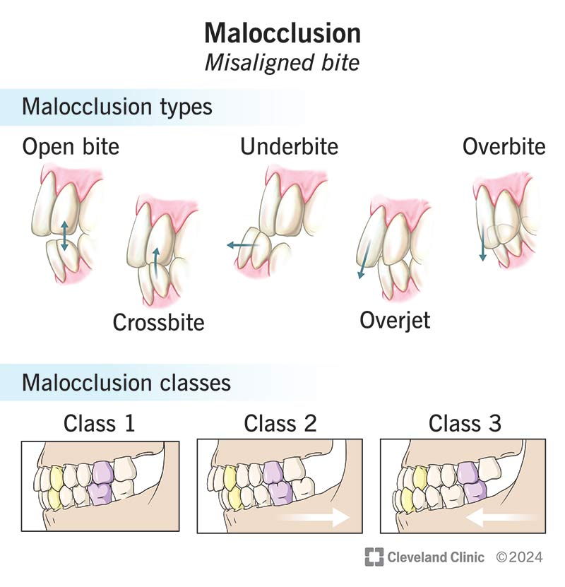
Dental deviation types
Skeletal malocclusion: deviations in shape and dimension of mandible and maxilla
Dental malocclusion: deviations in positioning of individual teeth
Class I - mostly only teeth misaligned; arches ok
Class II -overjet, overbite, maxilla more forward
Class III - underbite, mandible more forward
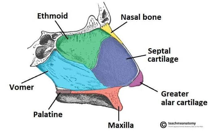
Bones of nasal septum
Ethmoid
Vomer bone
Quadrangular Cartilage of Septum
Hard palate components
Premaxilla- front four teeth
Palatine process- two pieces of bone that grow and fuse at midline
Alveolar process- houses molar, bicuspid, and cuspid teeth
Bones of hard palate that fuse
Premaxilla - front 4 teeth
Palatine process of maxilla
Palatine bone
(Alveolar process of maxilla - side and back teeth - on lateral sides so not referenced in hard palate fusion)
Hard palate fuses in utero
8-9 week
Three pieces move to midline at this time: maxilla and two palatine bones
Lip closure in utero
5-6 weeks
If premaxilla isn't in place in time, then cleft lip, right, left or both.
Soft palate fuses in utero
10-12 weeks
Knits together last
If only a small cleft, it will be in this posterior section
Occult submucous cleft
Type of cleft that is only detectable by x-ray exam or nasopharyngoscopy.
Submucous cleft of the soft palate
Midline deficiency or lack of muscular tissue as well as incorrect positioning of the muscles; Usually also has a bifid uvula present.
Delayed hard palate closure
Surgery first to close soft palate, then later surgery to close hard palate
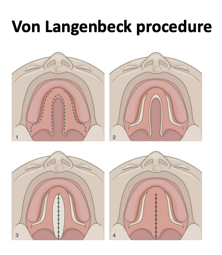
von Langenbeck method
Surgery to repair cleft palate by raising two bipedicled (attached at two ends) flaps of mucoperiosteum and bringing them together and attaching them to close the cleft.
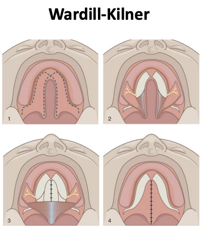
Veau-Wardill-Kilner method
Surgery to repair cleft palate by raising two single-based flaps from palate, bringing together, pushed back close to cleft to lengthen palate, improves VP closure
VPI
Velar Pharyngeal Insuffiency - structural - not enough soft palate there
Velar Pharyngeal Incompetence - functional - muscles don't close velum sufficiently
Adam's apple
Laryngeal prominence on thyroid cartilage
Helps protect VF
Pharyngeal flap
Secondary palatal surgical procedure in which a muscular flap is cut from the posterior pharyngeal wall, raised, and attached to the velum
Openings on either side of the flap allow for nasal breathing, nasal drainage, and production of nasal sounds.
Helps close the VP port and reduces hypernasality.
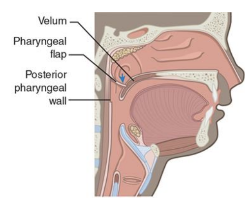
Palatal shelf surgery
Obdurator
Allows for nasal breathing
For non-nasal sounds, pharyngeal walls can push up to obdurator to close VP
Pharyngoplasty
Surgical procedure in which substances such as Teflon, silicone, dacron wool/silicone gel bag and cartilage may be implanted or injected into the PPW to make it bulge and thus close the VP port.
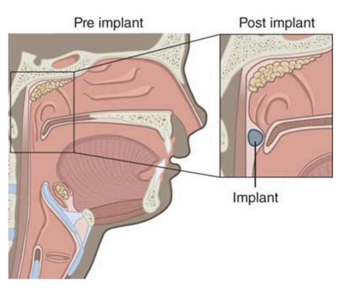
Palatometry
Pseudopalate fitted with electrodes to teach people place of articulation of consonants
CRANIAL NERVES (cont. pg. 25-56)
CRANIAL NERVES- confirmed
CNs that exit medulla
8-12
CNs that exit pons
V, VII
List all CN by name and most basic motor innervation for screening for function
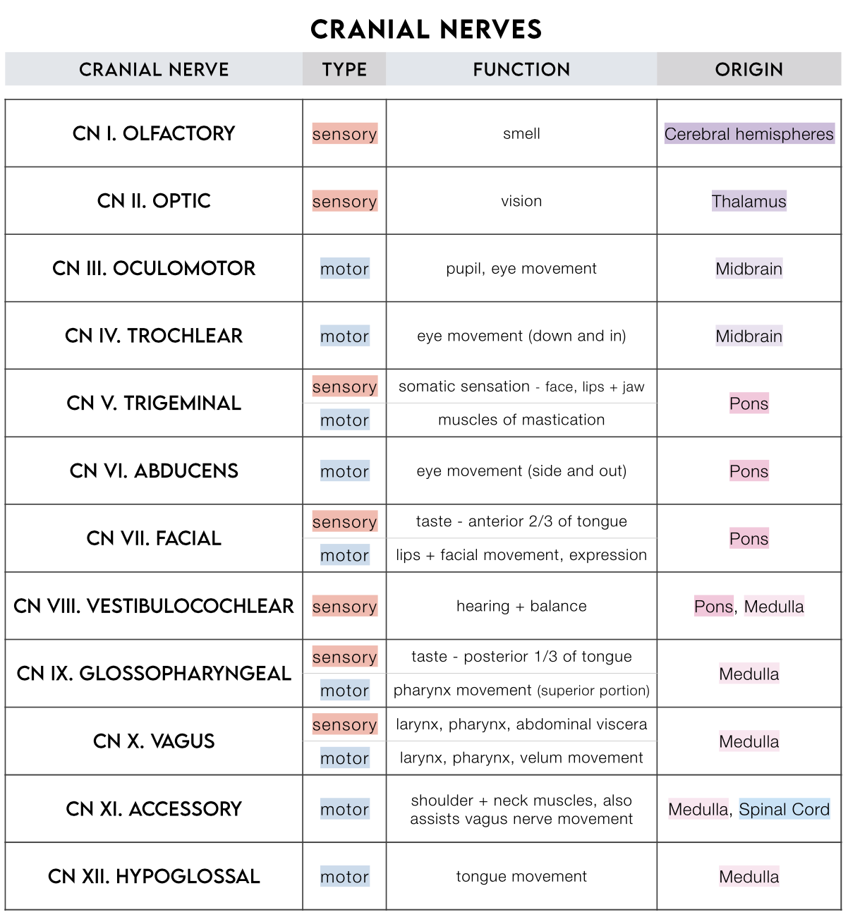
V - name, S (sensory), M (motor)
Trigeminal nerve
S: 3 branches:
-Ophalmic:nose, eyes, forehead
-Maxillary:lips, cheek, upper teeth, palate
-Mandibular:jaw, lower teeth, tongue, part of cheek, part of external ear
M: Jaw muscles including teproalis, lateral and medial pterygoids, master, tensor veli palatine, tensor tympani, mylohyoid, anterior belly of the digastric muscle
Damage: inability to close mouth, difficulty chewing, trigemini neuralgia (sharp pain in face)
VII - name, S, M
Facial nerve
S: ant 2/3 tongue taste
M: Facial expression, speech; Including buccinator, zygomatic, orbicularis iris, obicularis oculists, platysma, stapedius, stylohyoid, frontally, procures, nasalis, depressor labii inferiors, depressor angel iris, auricular muscles, various labial bumbles, posterior belly of the digastric
Damage: Mask-like appearance
VIII - name, S, M
Auditory(Acoustic) / Vestibulocochlear nerve
S: (auditory) hearing, inner hair cells, and (vestibular) balance
M: outer hair cells
Damage: Hearing loss, problems with balance, or both
IX - name, S, M
Glossopharyngeal nerve
S: assists post 1/3 tongue taste, general sensation: tympanic cavity, ear canal, eustachian tube, soft palate, faucial pillars, upper pharynx (gag)
M: Stylopharyngeus, pharyngeal constrictors (clear throat)
Damage: Difficulty swallowing, unilateral loss of gag reflex, loss of taste and sensation from posterior 1/3 of tongue
Parasympathetic: some salivary glands
XI - name, S, M
Spinal Accessory
S: none
M: Trapezius and sternocleidomastoid muscles; head turn, shoulder shrug
Along with vagus: innervate uvula and elevator veli palatine muscles
Damage: Neck weakness, inability to turn head, in ability to shrug shoulders and raise arm above shoulder
X - name, S, M
Vagus nerve
S: digestive system, heart, trachea, pharynx, larynx
M: digestive system, heart, lungs
Splits into SLN and RLN- Innervation to larynx
Pharyngeal Branch: pharyngeal constrictors, muscles of the velum
Damage: difficulty swallowing, paralysis of velum, voice problems if the RLN is damaged
Recurrent laryngeal nerve (RLN)
All motor to remaining intrinsic laryngeal muscles (interarytenoid, posterior cricoarytenoid, thyroarytneoid, and lateral cricoarytenoid muscles)
All sensory to below VFs
RLN can be damaged in thyroid or heart surgery resulting in VF paralysis
Superior laryngeal nerve (SLN)
Internal and external branches
Internal- all sensory info to larynx
External- motor to solely cricothyroid
X and XI-name and function
Pharyngeal plexus
Supplies the upper pharyngeal musculature including salpingopharyngeus, stylopharyngess, superior pharyngeal constrictor, middle pharyngeal constrictor inferior pharyngeal constrict, circopharyngeus, inferior pharyngeal constrictor, thyropharyngeus)
XII - name, S, M
Hypoglossal
S: none
M: all of the intrinsic and the following extrinsic muscles of the tongue: styloglossus, hyoglossus, and genioglossus
Damage: Tongue paralysis, diminished intelligibility, and swallowing problems
Most pharyngeal muscles are innervated by cranial nerves:
X, XI (Vagus, Spinal Accessory)