✅️✅9 - Pelvic Limb Muscles & Joints II
1/116
There's no tags or description
Looks like no tags are added yet.
Name | Mastery | Learn | Test | Matching | Spaced |
|---|
No study sessions yet.
117 Terms
extensors; flexors
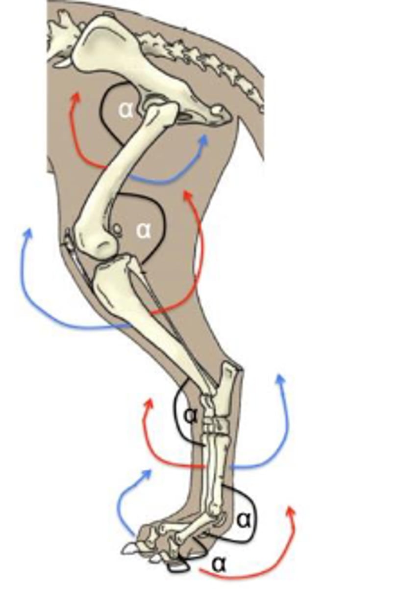
The following muscles are [flexors/extensors] of the hip joint and [flexors/extensors] of the stifle joint:
-Biceps femoris m.
-Semitendinosus m.
-Semimembranosus m.
reviewed
Review
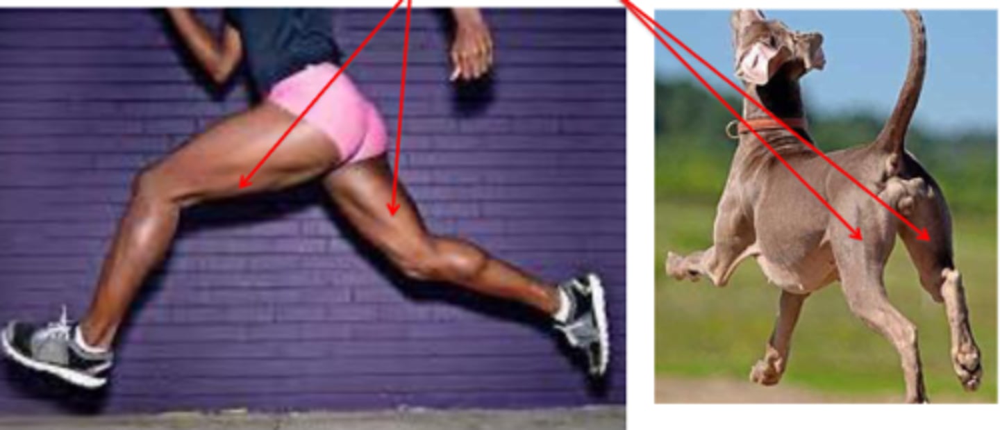
hamstring muscles
What muscle is shown?
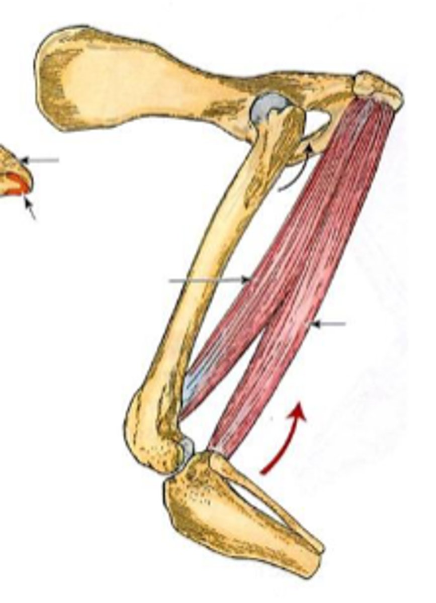
biceps femoris
What muscle is shown?

ischiatic tuberosity and sacrotuberous ligament
Origin of biceps femoris:

-lateral aspect of genual region (viafascia lata)
-tuber calcanei (via crural fascia)
Insertion of biceps femoris:
-extend the coxal joint
-variable action on the genual joint
-extend the tarsocrural joint
Action of biceps femoris:

semitendinosis
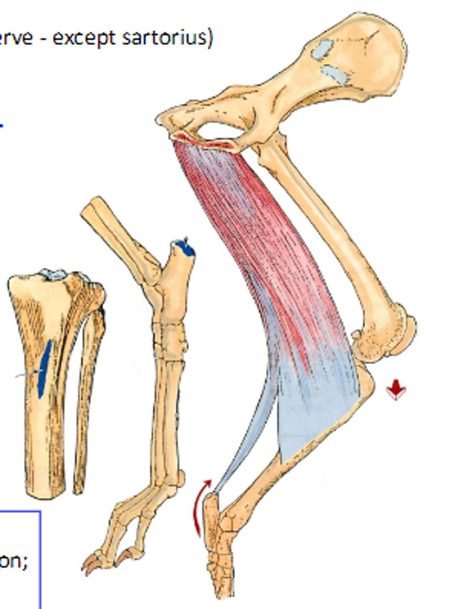
What muscle is shown?

ischiatic tuberosity
Origin of semitendinosus:
-medial aspect of tibia
-tuber calcanei (via crural fascia)
Insertion of semitendinosus:

-extend the coxal joint
-variable action on the genual joint
-extend the tarsocrural joint
Action of semitendinosus:

semimembranosus
What muscle is shown?

ischiatic tuberosity
Origin of semimembranosus:

medial aspect of distal femur andproximal tibia
Insertion of semimembranosus:

-extend coxal joint
-variable action on the genual joint
Action of semimembranosus:
adductors
The following muscles are [abductors/adductors] of the hip joint:
-Gracilis m.
-Pectineus m.
-Adductor m.
-External obturator m.
-Sartorius m.
gracilis
What muscle is shown?

pelvic symphysis
Origin of gracilis:
-medial aspect of the genual region
-tuber calcanei (via crural fascia)
Insertion of gracilis:

-adduct the pelvic limb
-extend the tarsocrural joint
Action of gracilis:

pectineus
What muscle is shown?
iliopubic eminence
Origin of pectineus:

distal body of femur
Insertion of pectineus:

adduct the pelvic limb
Action of pectineus:

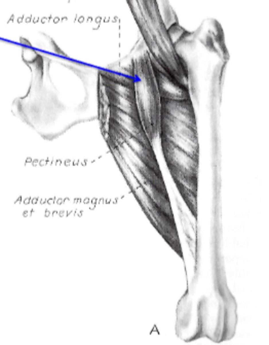
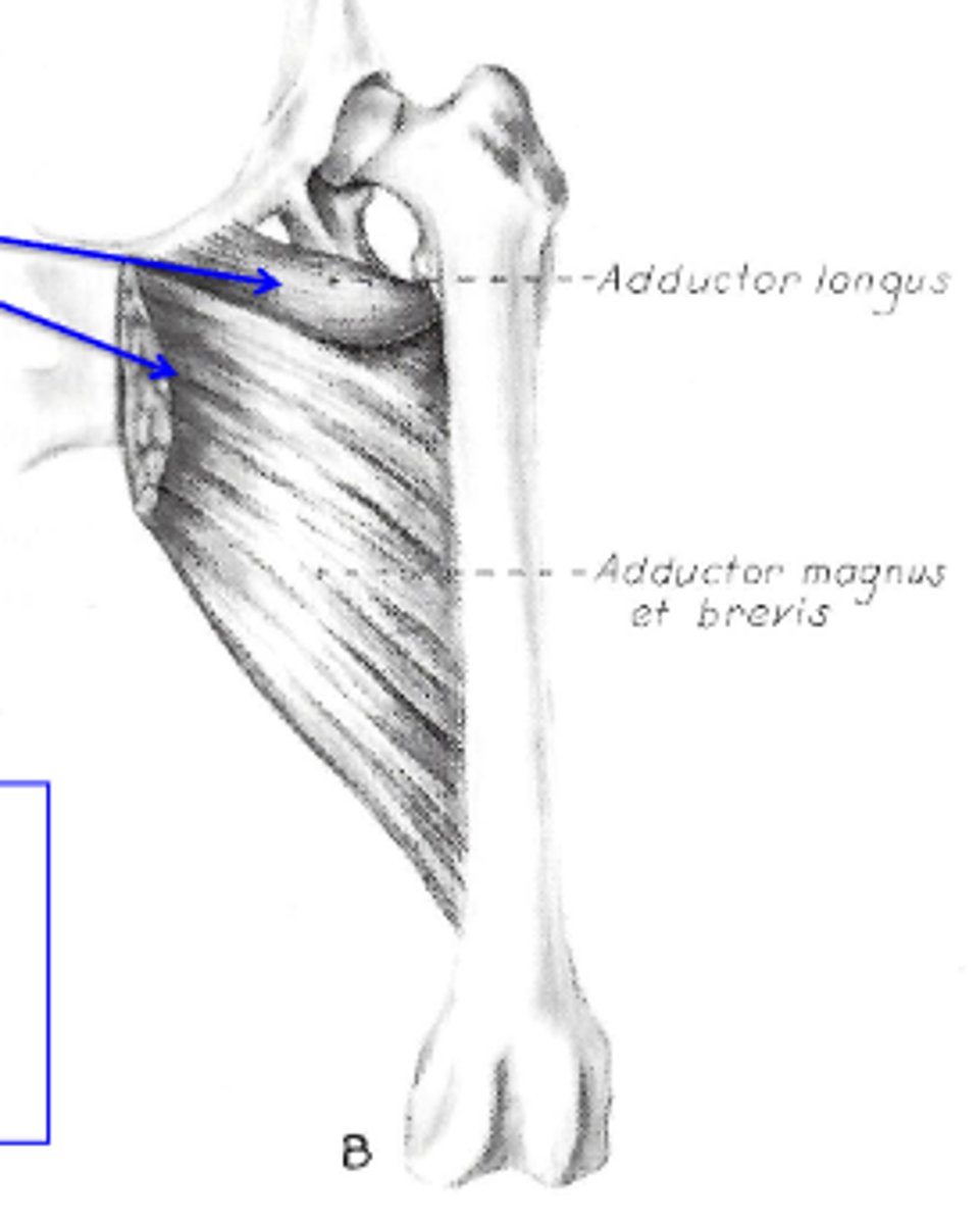
adductor
What muscle is shown?

ventral surface of os coxae
Origin of adductor:

most of the caudal surface of femur
Insertion of adductor:
-adduct the pelvic limb
-extend the coxal joint
Action of adductor:

sartorius
What muscle is shown?

iliac crest
Origin of sartorius:
-Cr: vastus medialis & rectus femoralis
-Cd: medial aspect of the genual region
Insertion of sartorius:

-Cr: extend the genual joint
-Cd: flex the genual joint
Action of sartorius:

quadriceps femoris
What muscle is shown?

-rectus femoris: body of ilium
-vastus: proximal femur
Origin of quadriceps femoris
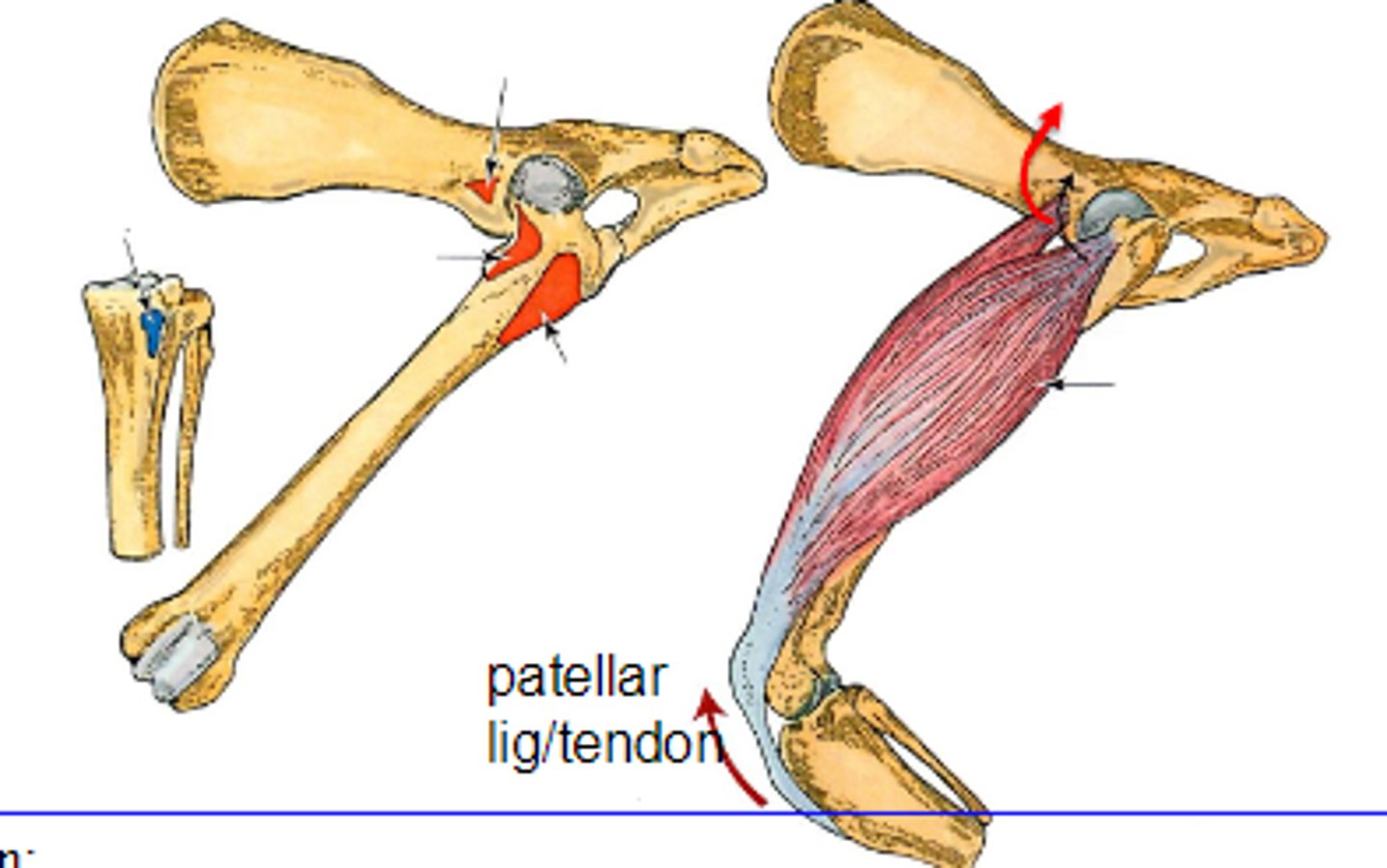
patella and tibial tuberosity via patellar ligament
Insertion of quadriceps femoris

-flex the coxal joint (rectus femoris only)
-extend the genual joint (all 4 heads)
Action of quadriceps femoris

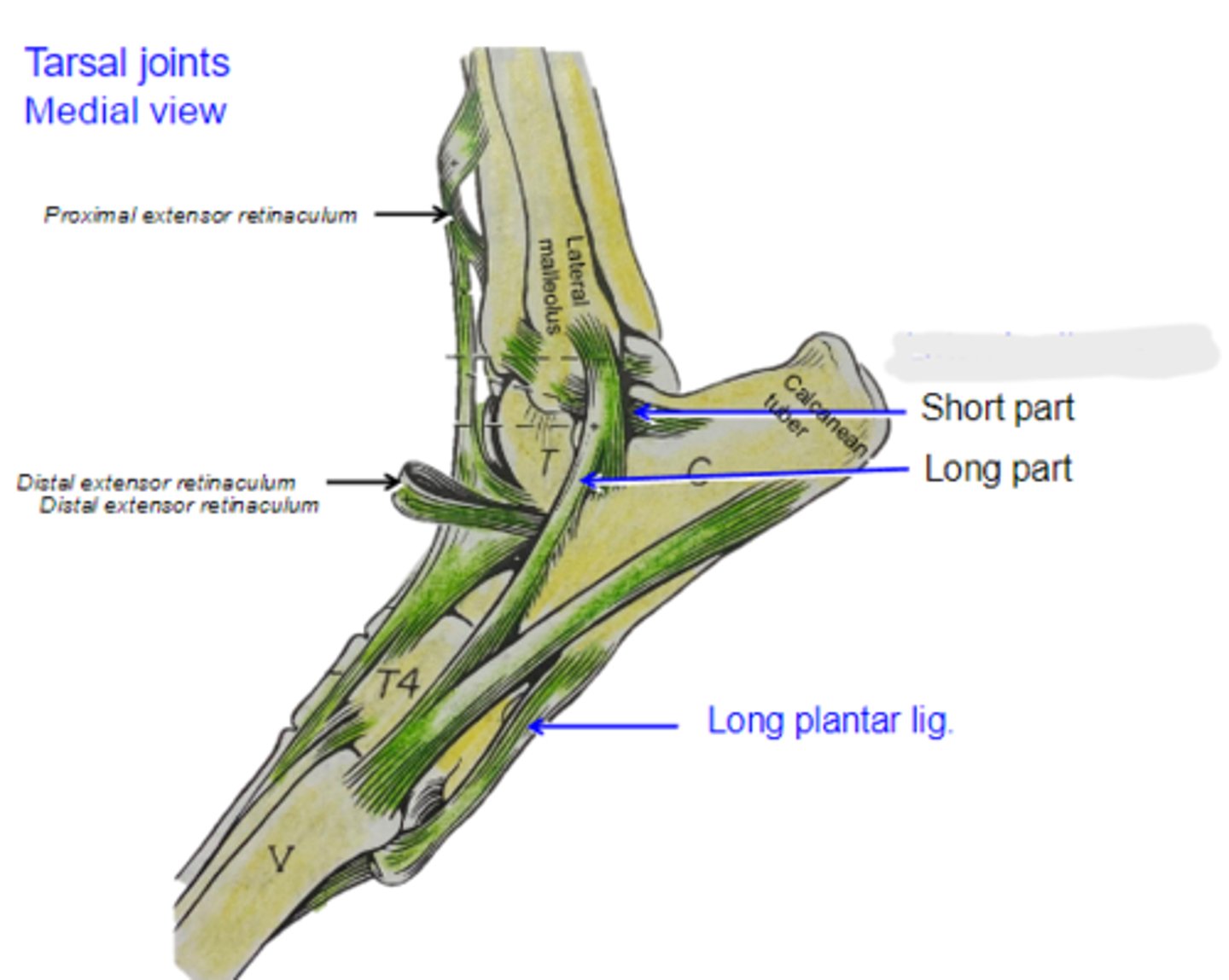
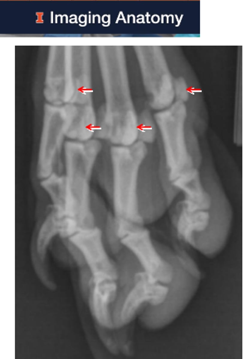
tarsocrural joint
1
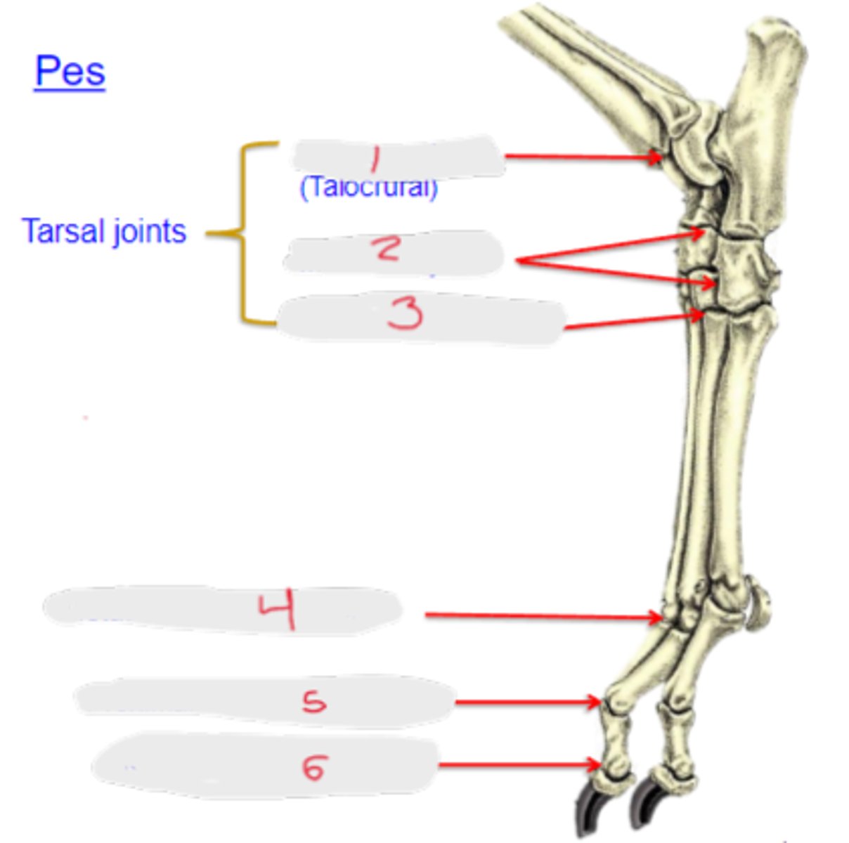
intertarsal joints
2

tarsometatarsal joints
3

metatarsophalangeal joints
4

proximal interphalangeal joint
5

distal interphalangeal joint
6

medial collateral ligament
What is shown?
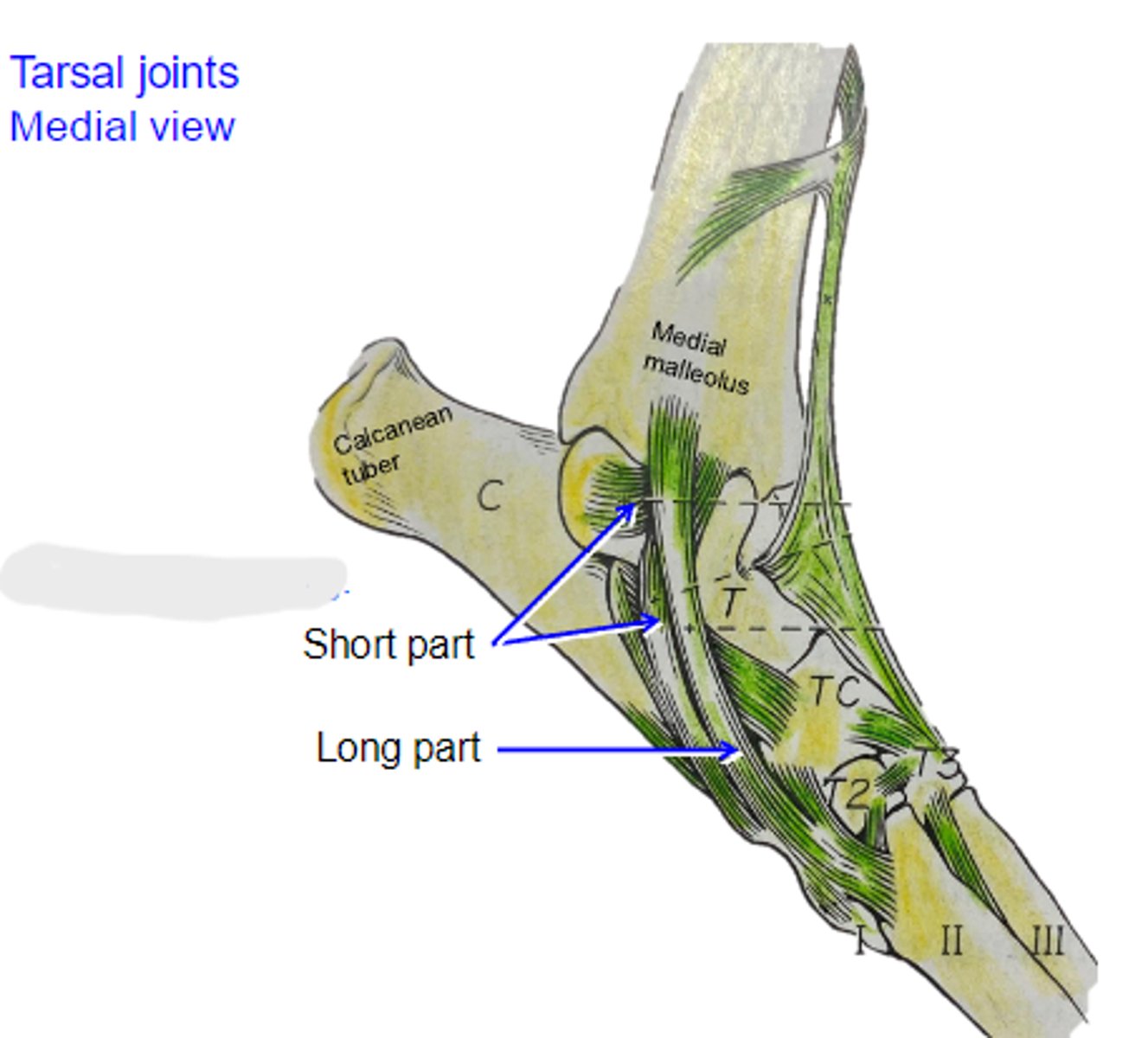
lateral collateral ligament
What is shown?
craniolateral group
Muscles acting primarily on the tarsal and digital joints:
Flexor of tarsal joint & extensors of the digits
caudal group
Muscles acting primarily on the tarsal and digital joints:
Extensors of tarsal joint & flexor of the digits
tarsal joint; digits
The craniolateral muscles serve as the flexors of the _____ joint and extensors of the _____
cranial tibial
What muscle is shown?

lateral aspect of proximal tibia
Origin of cranial tibial
plantar base of 1st and 2nd metatarsal bones
Insertion of cranial tibial

-flex the tarsocrural joint
-rotate pes laterally
Action of cranial tibial

lateral digital extensor
What muscle is shown?
fibula
Origin of lateral digital extensor

extensor process of the distal phalange of fifth digit
Insertion of lateral digital extensor

extend the digital joints of fifth digit
Action of lateral digital extensor

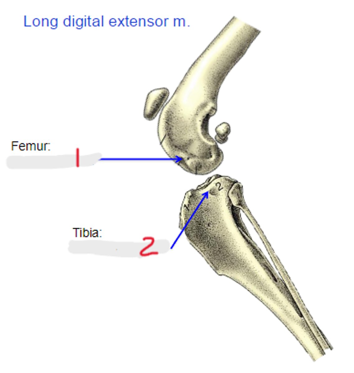
long digital extensor
What muscle is shown?

extensor fossa of femur
Origin of long digital extensor

extensor processes of the distal phalanges
Insertion of long digital extensor

-flex the tarsocrural joint
-extend the digital joints
Action of long digital extensor
extensor fossa
1

extensor groove
2
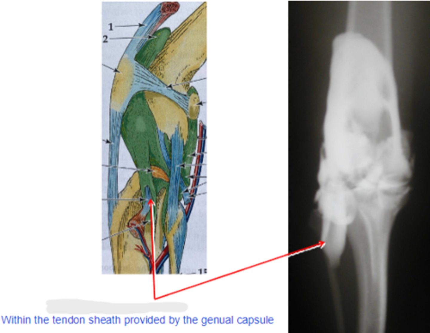
long digital extensor tendon
What is shown?
fibularis longus
What muscle is shown?
on or near lateral collateral ligament of the genual joint
Origin of fibularis longus

4th tarsal bone and plantar base of metatarsal bones
Insertion of fibularis longus

-flex the tarsocrural joint
-rotate the pes medially
Action of fibularis longus

fibularis brevis
What muscle is shown?
distal and lateral surfaces of tibialand fibula
Origin of fibularis brevis

base of metatarsal bone V
Insertion of fibularis brevis

flex the tarsocrural joint
Action of fibularis brevis

extensor tendon sheath
1
extensor retinacula
2

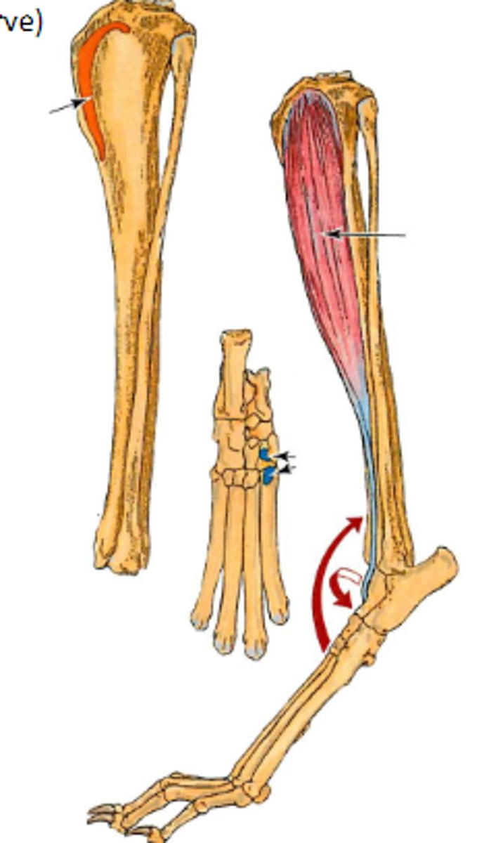
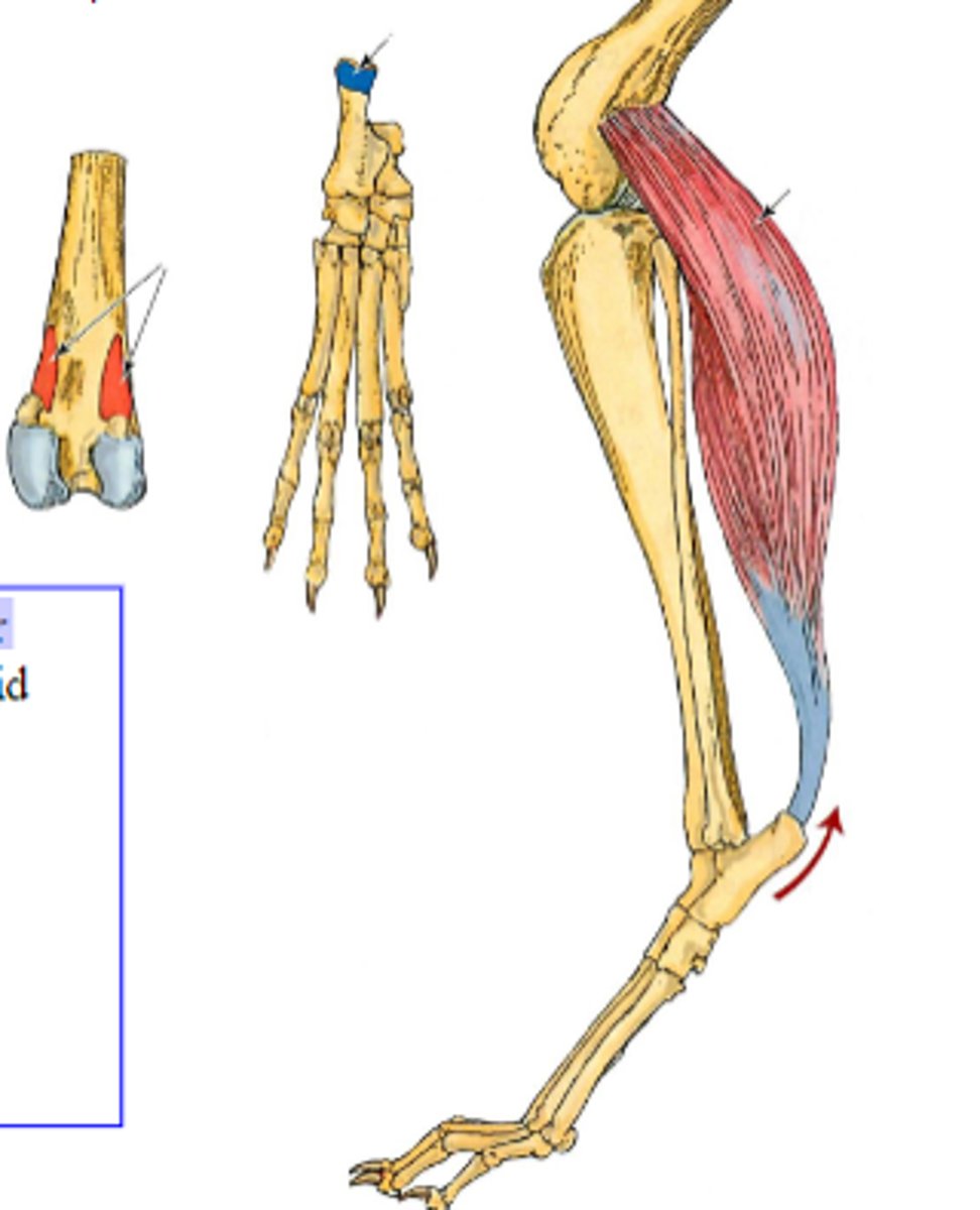
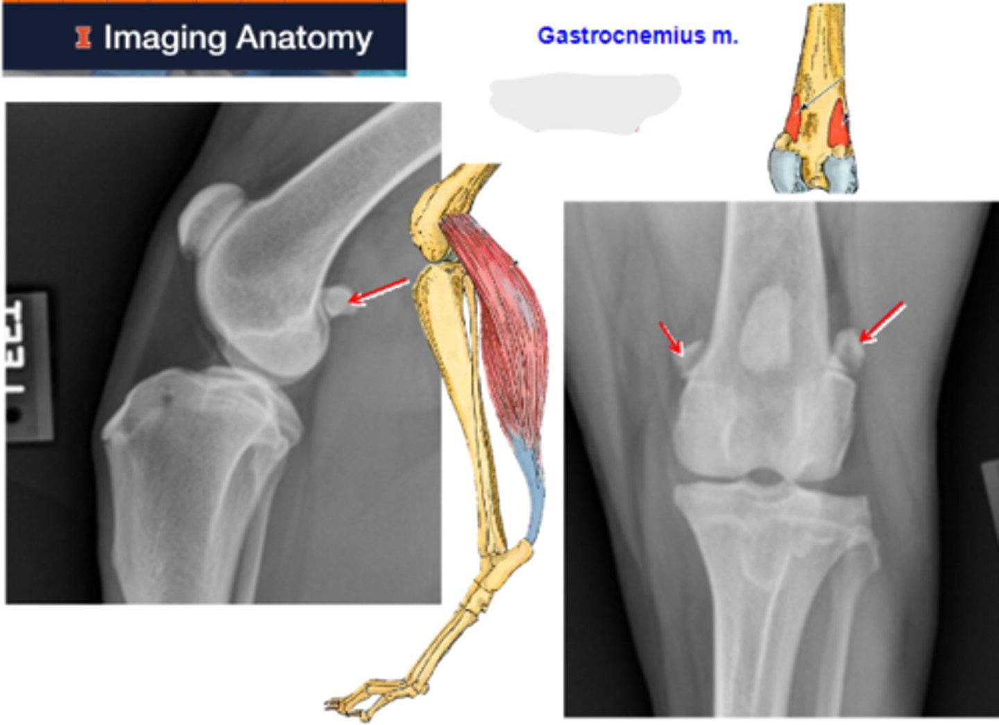
gastrocnemius
What muscle is shown?

medial and lateral supracondylar tuberosities of femur
Origin of gastrocnemius

tuber calcanei
Insertion of gastrocnemius

-flex the genual joint
-extend the tarsocrural joint
Action of gastrocnemius
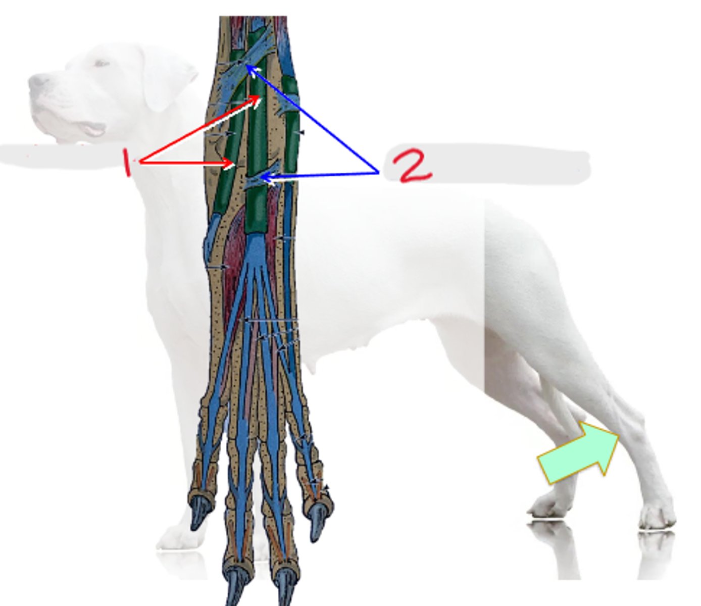
sesamoid bones (fabellae)
What are the arrows pointing to?
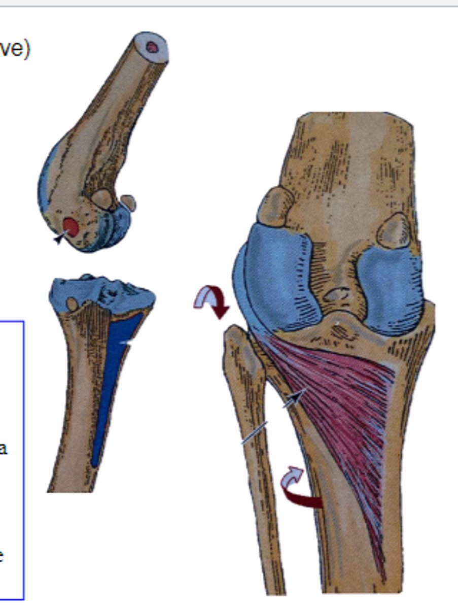
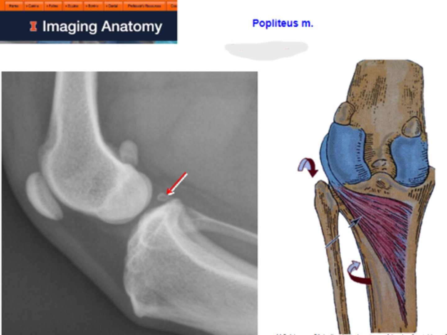
popliteus
What muscle is shown?

lateral condyle of the femur
Origin of popliteus

proximocaudal surface of the tibia
Insertion of popliteus

medial rotation of the genual joint.
Action of popliteus
sesamoid bone
What is the arrow pointing to?
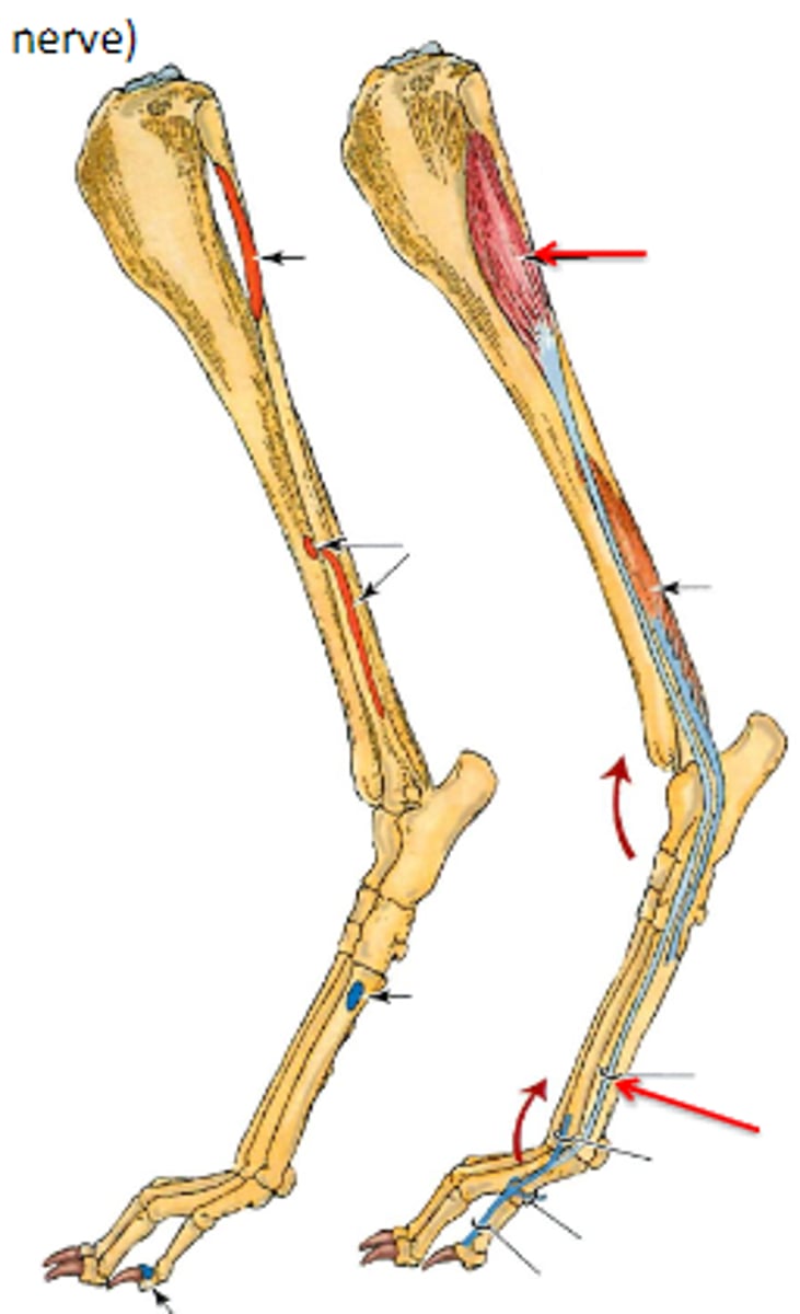
superficial digital flexor
What muscle is shown?

lateral supracondylar tuberosity of femur
Origin of superficial digital flexor

-tuber calcanei
-plantar bases of middle phalanges
Insertion of superficial digital flexor

-flex the genual joint
-extend the tarsocrural joint
-flex the metatarsophalangeal joints
-flex the proximal interphalangeal joints
Action of superficial digital flexor
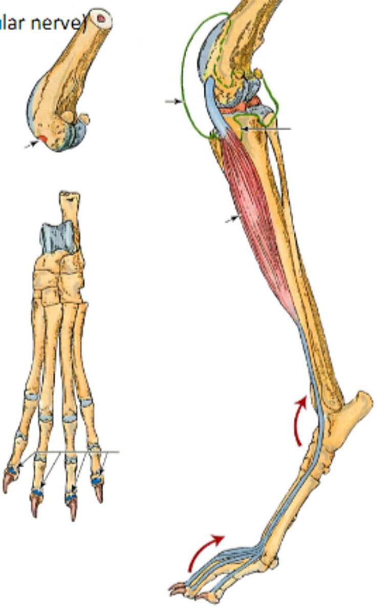
deep digital flexor
What muscle is shown?

proximal caudal aspect of the tibia and fibula
Origin of deep digital flexor

lexor process of distal phalanges
Insertion of deep digital flexor
-extend the tarsocrural joint
-flex the metatarsophalangeal joints
-flex the proximal interphalangeal joints
-flex the distal interphalangeal joints
Action of deep digital flexor

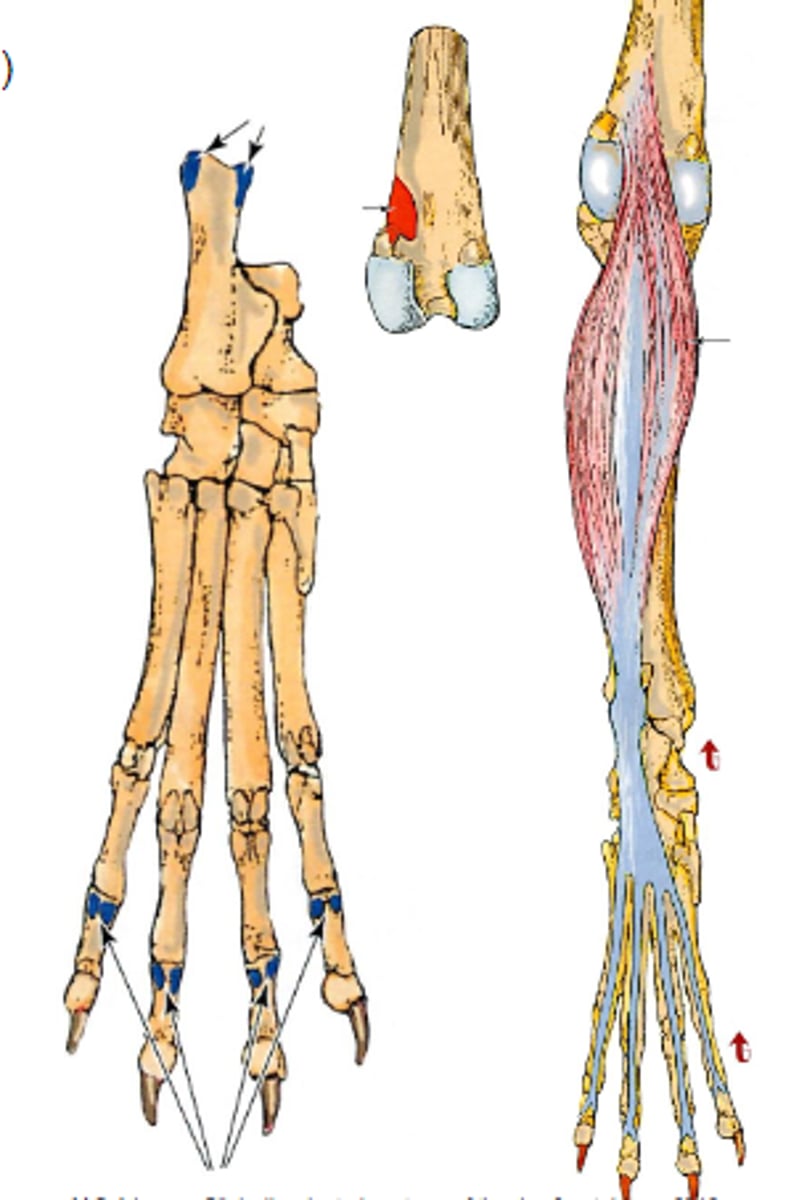
interossei
What muscle is shown?
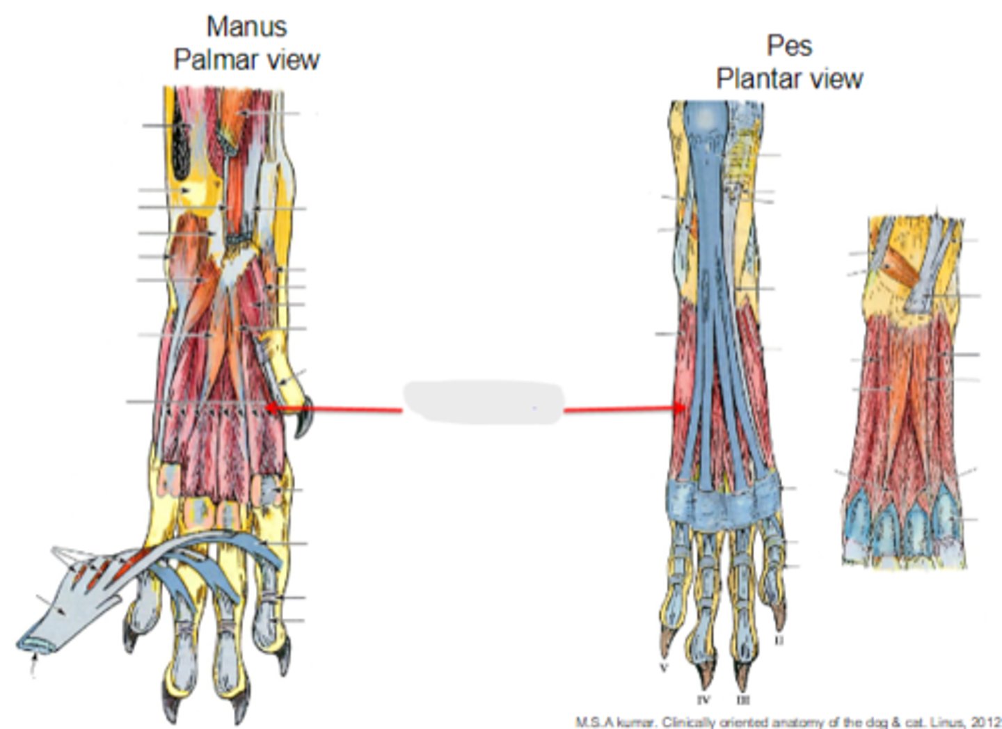
proximal metatarsophalanx sesamoid bones
What is shown?
calcanean bursa
1
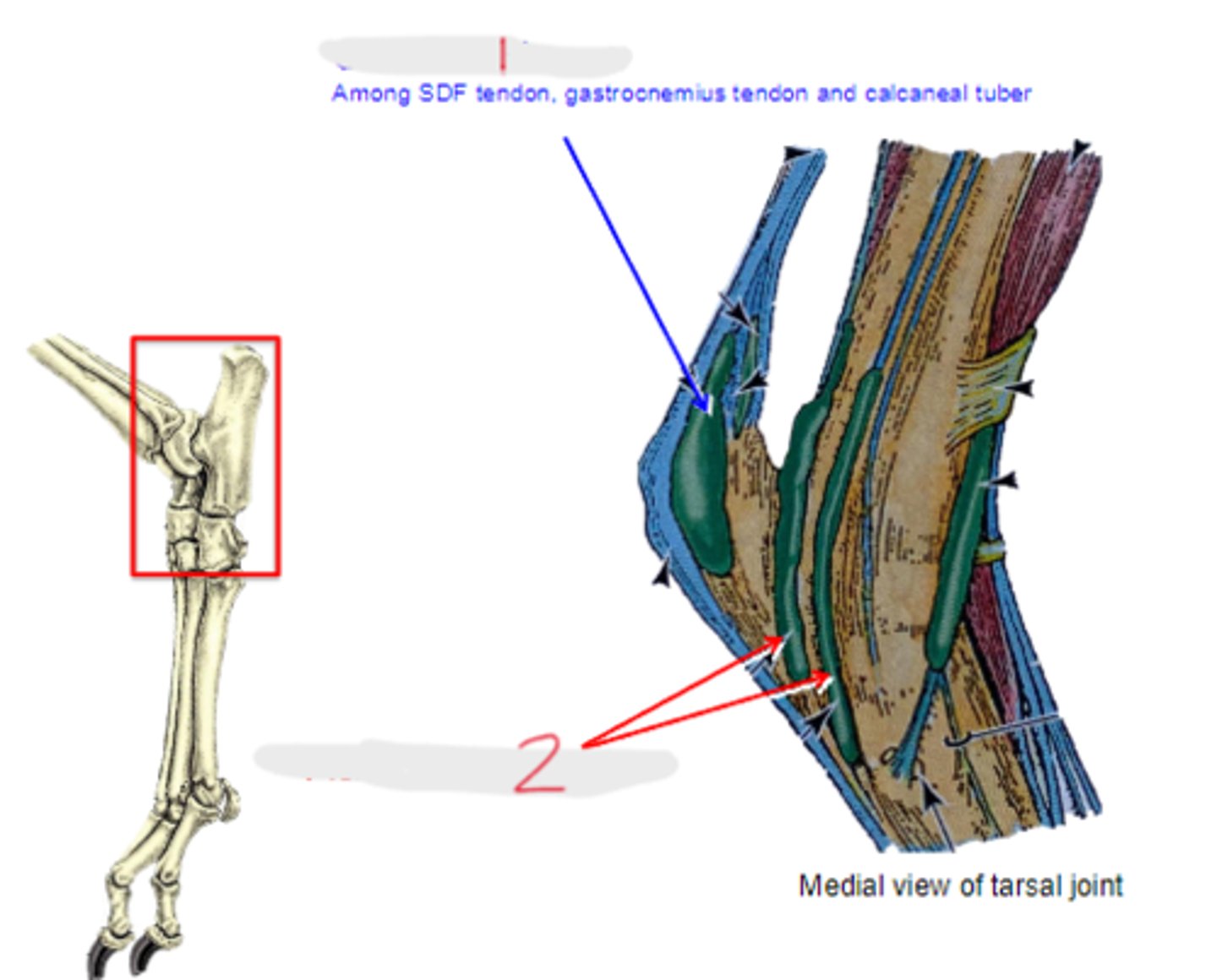
flexor tendon sheath
2

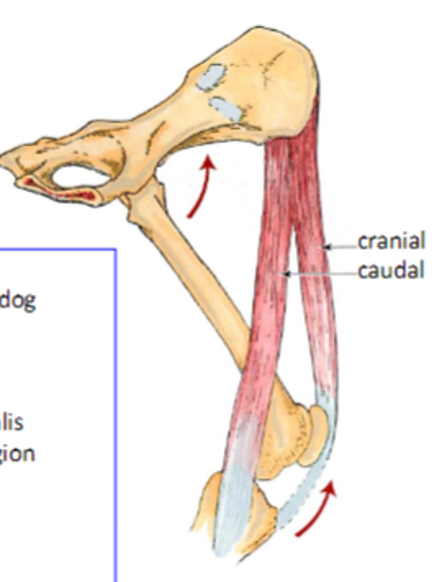
1. biceps femoris
2. semitendinosus
3. semimembranosus
What are the 3 "hamstring" muscles?
extend; flex
The "hamstring" muscles [flex/extend] the hip joint and [flex/extend] the stifle joint
biceps femoris
What muscle of the hamstrings is the most lateral?
semimembranosus
What hamstring muscle is most medial?
ischiatic tuberosity
The biceps femoris, semitendinosus, and semimembranosus all originate where?
semimembranosus
The [semimembranosus/semitendinosus] has 2 heads