Bio test 28.11.24
1/212
There's no tags or description
Looks like no tags are added yet.
Name | Mastery | Learn | Test | Matching | Spaced |
|---|
No study sessions yet.
213 Terms
Contrast Positive and Negative Tropism
Positive tropism: Growth towards the stimulus.
Negative tropism: Growth away from the stimulus.
Example: Stems show positive phototropism (towards light); roots exhibit negative phototropism (away from light).
Example: Roots display positive gravitropism (towards gravity); stems exhibit negative gravitropism (against gravity)
Outline Phototropism and Gravitropism in Roots and Stems
Phototropism
Directional response to light.
Stems grow towards light (positive phototropism).
Roots grow away from light (negative phototropism).
Gravitropism (Geotropism)
Directional response to gravity.
Roots grow downwards into the soil (positive gravitropism).
Stems grow upwards against gravity (negative gravitropism).
Cause of Positive Phototropism
Triggered by uneven light distribution on plant shoot.
Auxins (phytohormones) accumulate on the shaded side of shoot.
Auxins promote cell elongation on shaded side, causing shoot to bend towards light source.
Consequence of Positive Phototropism
Maximises light absorption for photosynthesis.
Enhances plant growth and survival in light-limited environments.
Facilitates optimal energy production by aligning leaves towards light.
Define Phytohormone
Chemical messengers in plants regulating growth, development, and responses to stimuli.
Synthesised in various plant tissues, transported via xylem and phloem.
Examples:
Auxins (e.g., indole-3-acetic acid, IAA).
Cytokinins.
Gibberellins.
Abscisic acid (ABA).
Ethylene.
Role of phytohormone Auxin
Promote cell elongation (tropic movements).
Control apical dominance by inhibiting lateral bud growth.
growth hormones produced in the shoot (apical meristem)
Role of phytohormone cytokinins
synthesised in the roots and pass into the leaves and fruits where they:
Stimulate cell division and differentiation.
Delay ageing in leaves and fruits.
Role of phytohormone Gibberellins
Synthesised in the apical meristems of roots and shoots, young leaves and embryos:
Stimulate stem elongation, seed germination, and fruit maturation.
Break seed dormancy.
Role of phytohormone Abscisic Acid ABA
Inhibits growth, induces seed dormancy.
Promotes leaf abscission under stress conditions.
Role of phytohormone Ethylene
Accelerates fruit ripening.
Induces leaf and flower abscission
a gas and is produced by ageing tissues
Mechanism of auxin movement:
Entry into cells: Auxin enters plant cells passively by diffusion or via membrane proteins called auxin influx carriers.
Dissociation within the cell: Inside the cytoplasm, auxin dissociates into IAA⁻ ions, which cannot exit passively.
Efflux: Auxin efflux carriers, located on specific sides of the cell membrane, actively pump auxin ions out using ATP.
Directionality: The localization of efflux carriers determines the polar (directional) movement of auxin, ensuring its concentration gradient is maintained.
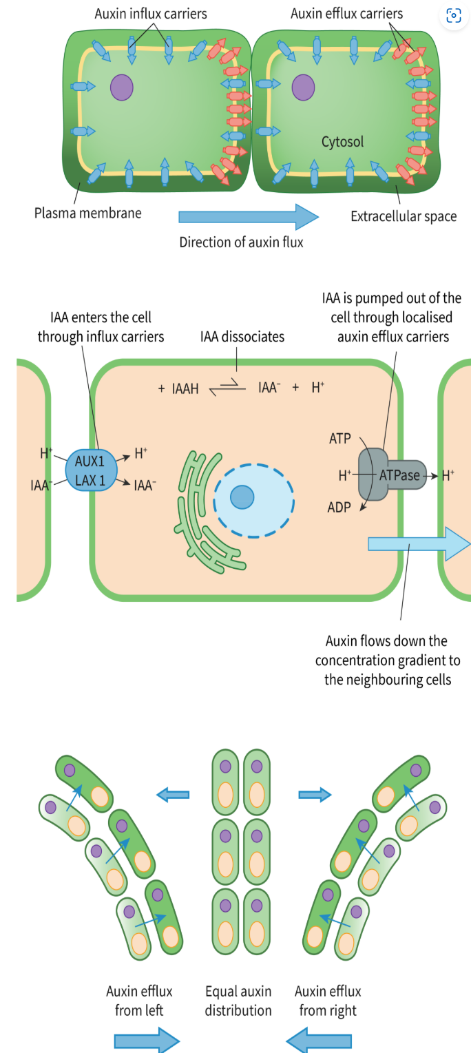
Phototropism and auxin concentration:
In phototropism, auxin redistributes unevenly in response to light.
Auxin accumulates on the shaded side of the plant, causing faster cell elongation there.
This uneven growth results in bending towards the light.
Mechanism of auxin action (Acid Growth Theory):
Auxin binds to receptors on cells on the shaded side.
Activation of H⁺-ATPase pumps leads to proton (H⁺) movement into the cell wall, lowering pH.
Acidic conditions activate expansins, proteins that loosen cellulose cross-links in the cell wall.
Potassium ions enter cells, decreasing water potential and causing water to flow in by osmosis.
Increased turgor pressure stretches the cell wall, elongating cells.
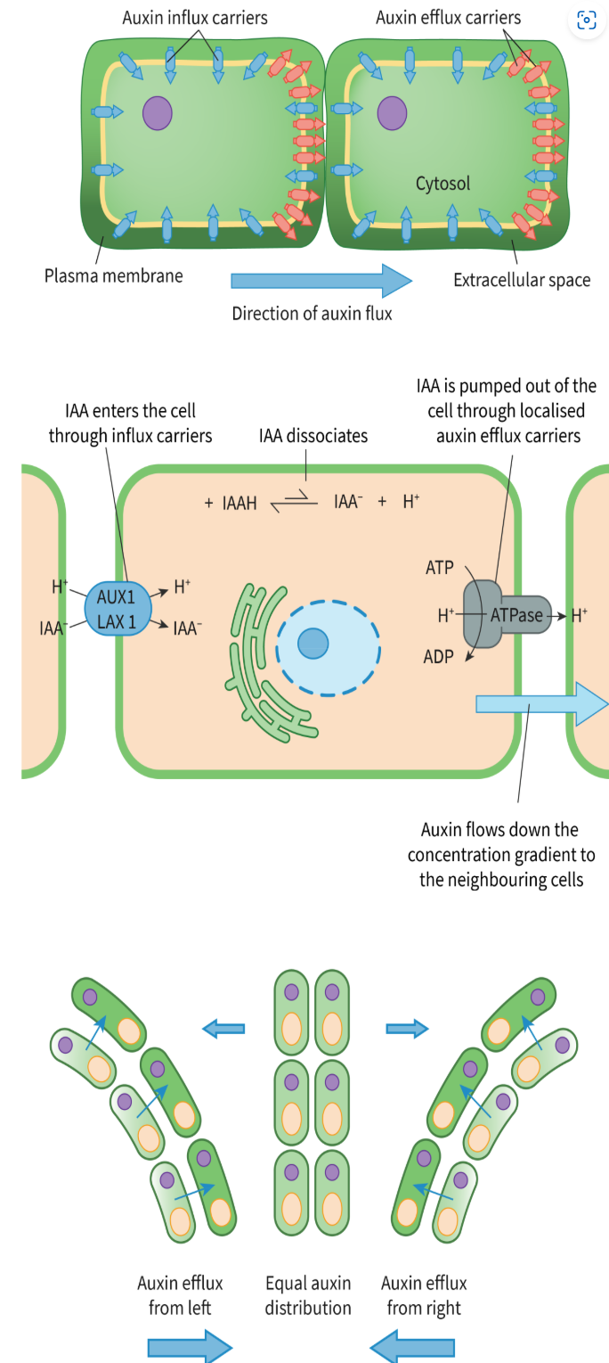
Source and transport of auxin and cytokinin
Auxin is produced in the shoot apical meristem and moves down the plant through vascular tissues.
Cytokinin is produced in the root apical meristem and moves upward.
Regulation of root and shoot growth Auxin and Cytokinin:
High auxin-to-cytokinin ratios promote root development.
High cytokinin-to-auxin ratios stimulate shoot and bud development.
Auxin suppresses lateral bud formation through apical dominance, while cytokinin promotes lateral bud growth.
Together, auxin and cytokinin regulate cell division (auxin promotes) and differentiation (cytokinin promotes).
Function of fruits:
Protect seeds during development.
Facilitate seed dispersal through attraction of animals or environmental mechanisms.
Changes during fruit ripening:
Softening of the fruit due to breakdown of cell walls.
Conversion of starches into sugars, making the fruit sweeter.
Reduction in bitter phenolic compounds.
Color changes as chlorophyll degrades and other pigments are synthesized.
Release of volatile compounds responsible for characteristic aromas.
Positive feedback mechanism of fruit ripening:
Initially, unripe fruit produces small amounts of ethylene gas.
Ethylene stimulates its own synthesis by activating genes responsible for ethylene production.
As ethylene levels increase, ripening processes (e.g., cell wall softening, sugar production) accelerate.
Increased ethylene leads to further ethylene synthesis, creating a self-amplifying loop.
Reasons for rapid and synchronized fruit ripening evolution:
Ensures maximum attractiveness to seed dispersers like animals at the right time.
Promotes efficient seed dispersal when fruits are in optimal condition.
Prevents prolonged exposure to predators or environmental threats, reducing the risk of seed loss.
Synchronization allows a uniform harvest period, beneficial in certain environments
What is the function of brain?
Processing - the brain receives information from multiple receptors via sensory neurones. It processes the information and then and then sends signals via motor neurones to effectors which then make a response.
Memory and learning. The brain exhibits plasticity. This means that new information and experiences can change neural pathways. This happens as new synapses are made, strengthened (due to more use) or removed.
What are the different types of memory?
Explicit: conscious memory that comes from personal experience and require intentional recall
Implicit: this is unconscious/ automatic recall that comes from daily experience and that we don't think about
What do receptor cells do?
Detect changes in the environment and converts the information into electrical signal through transduction (which is the conversion of a physical stimulus into an electrical signal called an action potential that is carried along a neurone.)
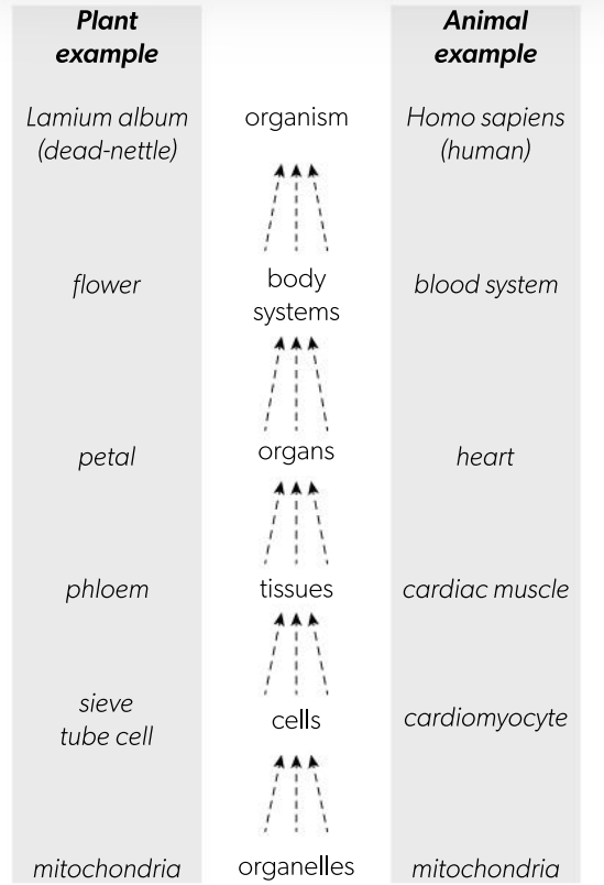
What’s system integration and provide an example.
The cells within a multicellular organisms interact with each other at multiple levels in a hierarchy of organisation, shown in the flow chart.
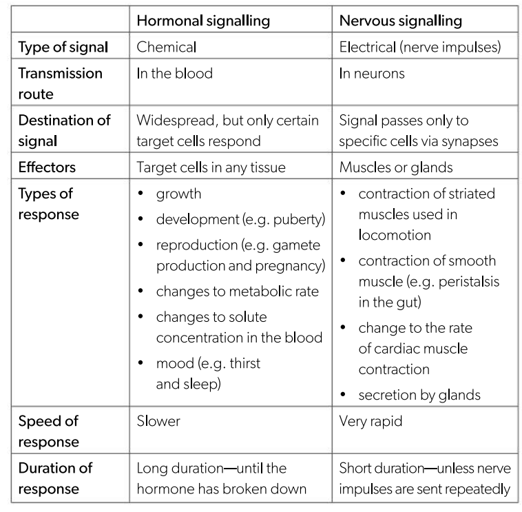
How does integration of organs in an animal work? provide two examples and compare them.
Integration of organs in an animal requires transport of materials and energy, and effective communication.
What’s the role of blood in the transport of material and energy between organs.
Blood circulates through all organs and almost all tissues, transporting:
- energy in the form of substrates of respiration, such as glucose
- oxygen for aerobic respiration
-water and carbon compounds needed for growth or repair
- waste products of metabolism.
Label and state the function of the hypothalamus
link between nervous and endocrine systems.
- monitors temperature, hunger, and thirst
- Sends signals through nerves for quick responses
- Triggers hormone release for longer-term changes
The nervous system enables quick responses by transmitting electrical signals through neurons, allowing for immediate reactions to stimuli.
The endocrine system produces hormones that create long-lasting effects, influencing processes like growth and metabolism over time.
These systems integrate through the hypothalamus, which coordinates their activities to maintain balance and homeostasis in the body.
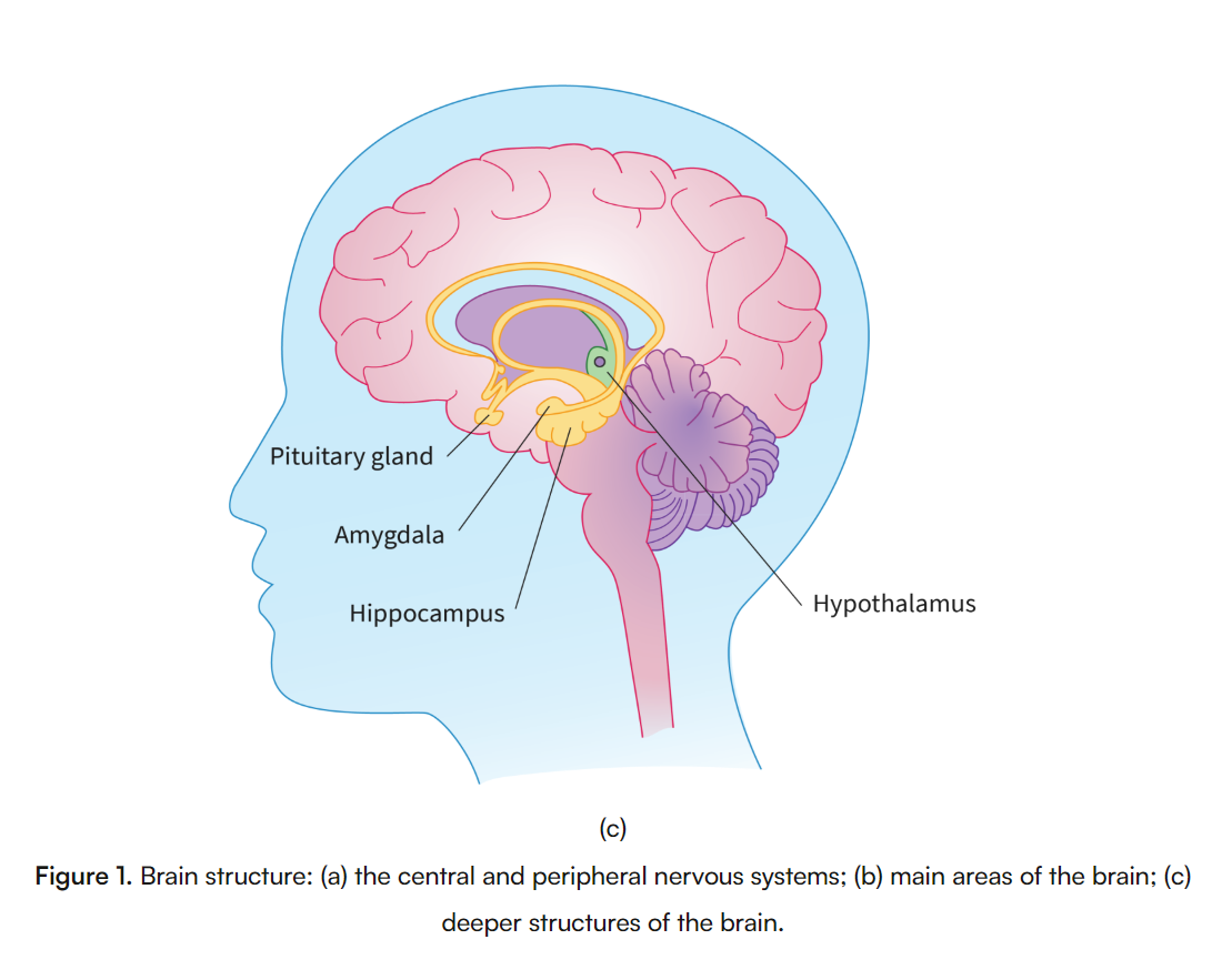
Compare and Contrast Unconscious and Conscious processing
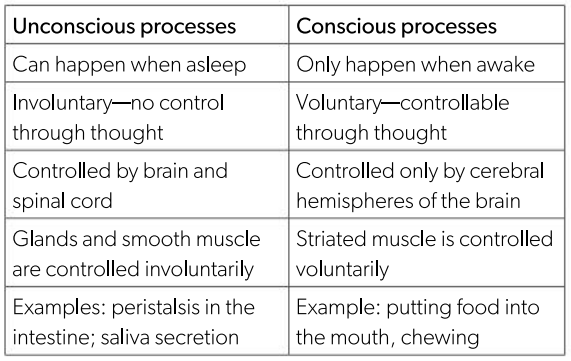
What are the organs of the central nervous system?
Brain and Spinal cord (which can only coordinate unconscious processes).
What is the function of sensory neurons? provide the different types of sensory receptors.
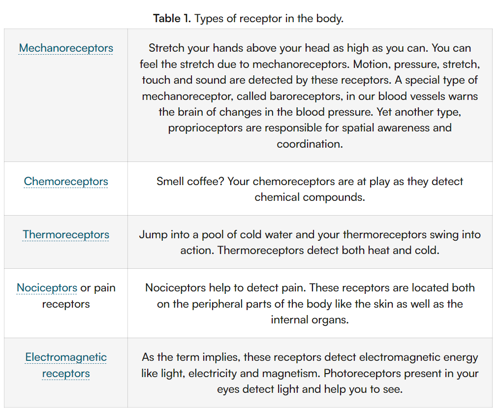
receptor cells located in the skin and sense organs, detect changes in the external environment wheich then pass these impulses to sensory neurons.
Sensory neuron: Nerve cells which transmit sensory input from a sense organ to the central nervous system.
Sensory receptors: An organ having nerve endings (for example in the skin) that respond to stimulation.
Sensory Input: Stimuli that are perceived by our senses like smell, sight, touch, taste and hearing.
What are the different types of motor neurones?
Conveys messages from the CNS to Muscles
The upper motor neurons that carry the impulses from the brain to the spinal cord.
The lower motor neurons that travel from the spinal cord to the muscles.
How does the motor neurone carry its function and where?
Cerebral Hemispheres: Located above the brainstem, the cerebral hemispheres are responsible for higher brain functions such as thought, memory, and sensory processing. They control the opposite side of the body, integrating information and coordinating actions.
Primary Motor Cortex: Situated in the frontal lobe of the cerebral hemispheres (specifically in the precentral gyrus), the primary motor cortex is crucial for planning and executing voluntary movements. It sends signals to motor neurones that activate skeletal muscles.
Skeletal Muscles: Found throughout the body, skeletal muscles are attached to bones and enable voluntary movements like walking and lifting. They contract in response to signals from motor neurones, allowing for precise control of movement.
Connection: The motor neurones act as the communication link between the primary motor cortex and skeletal muscles. When the primary motor cortex sends a signal to a motor neurone, it triggers the corresponding skeletal muscle to contract, resulting in movement.
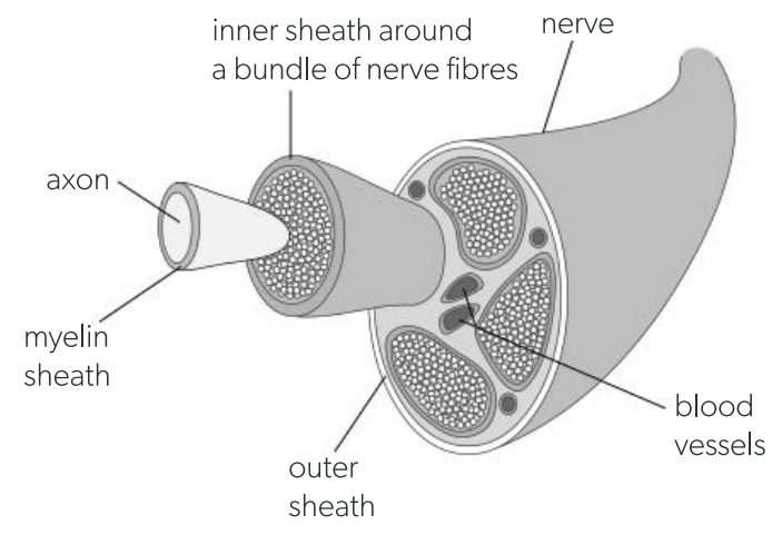
What is a nerve? define and draw
A nerve is a bundle of nerve fibres enclosed in a protective sheath.
Most nerves contain nerve fibres of both sensory and motor neurons. However, some contain only sensory neurons (e.g. the optic nerve) and some contain only motor neurons (e.g. the oculomotor nerve).
Define reflex and reflex arc
A reflex is an automatic, involuntary response to a specific stimulus.
A reflex arc is the neural pathway that mediates a reflex action.
Outline and explain the pain reflex arc
RSIME
Receptor cells perceive the stimulus. Pain and heat are perceived directly by nerve endings of sensory neurons in the skin, so there is no need for a separate receptor cell. This is how the stimulus is perceived when the hand touches a hot object.
Sensory neurons receive signals, either from receptor cells or from their own sensory nerve endings and pass them as nerve impulses to the brain or spinal cord, via long axons. These axons end at synapses with interneurons in the grey matter of the spinal cord or brain
Interneurons are located in the grey matter of the brain and spinal cord. They have many branched fibres called dendrites, along which nerve impulses travel. Interneurons process signals brought by sensory neurons and make decisions about appropriate responses. They do this by combining impulses from multiple inputs and then passing impulses to specific other neurons. The decision-making process in a reflex action is very simple because there may be only one interneuron connecting a specific sensory neuron to the motor neuron that can cause an appropriate response.
Motor neurons receive signals via synapses with interneurons. If a threshold potential is achieved in a motor neuron, an impulse is passed along the axon which leads out of the central nervous system (CNS) to an effector. The axon does not change its position or connections, so the impulse always travels to the same effector cells.
Effectors carry out the response to a stimulus when they receive the signal from a motor neuron. The two types of effector are muscles and glands. When the hand has touched a hot object, the effectors are muscles which flex the arm at the elbow, pulling it away.
What’s the difference between grey and white matter?
Aspect | Grey Matter | White Matter |
Composition | Contains the cell bodies of neurons, dendrites, and unmyelinated axons. | Composed of myelinated axons, with few or no cell bodies. |
Color | Grey in appearance due to the lack of myelin and the presence of cell bodies. | White in appearance due to the high lipid content of myelin. |
Function | Primarily processes information, including integration of sensory input and generation of responses. | Facilitates the transmission of electrical signals between different regions of the nervous system. |
Location in the Brain | Found on the outer regions (cortex) of the cerebrum and cerebellum. | Found in the inner regions beneath the grey matter in the brain. |
Location in the Spinal Cord | Found in the inner regions, forming an H-shaped core. | Found in the outer regions surrounding the grey matter. |
Myelination | Largely unmyelinated. | Heavily myelinated. |
proprioceptors
specialized sensory receptors located in muscles, tendons, and joints that provide the brain with information about body position, movement, and spatial orientation.
What’s myelin Sheath made up of?
It is created by the Schwann cells of the PNS.
Function of cerebellum and identify its location in the brain
Coordination of voluntary muscle movements
Maintenance of balance and posture
Motor learning
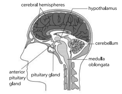
What is the circadian rhythm and how does it work?
Physical, behavioural, and mental changes that occur in the body in a 24-hour cycle.
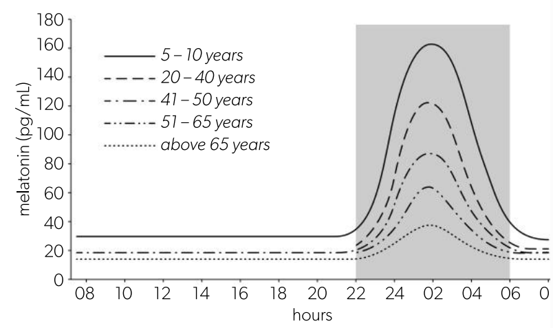
The rhythm is set by the suprachiasmatic nuclei which control the secretion of the hormone melatonin by the pineal gland.
Melatonin secretion increases in the evening and drops to a low level at dawn. The hormone is rapidly removed from the blood by the liver, so blood concentrations rise and fall rapidly in response to rate of secretion. The graph shows melatonin concentrations at different ages, with the period in which it is dark shown in grey.
How does the circadian rhythm work?
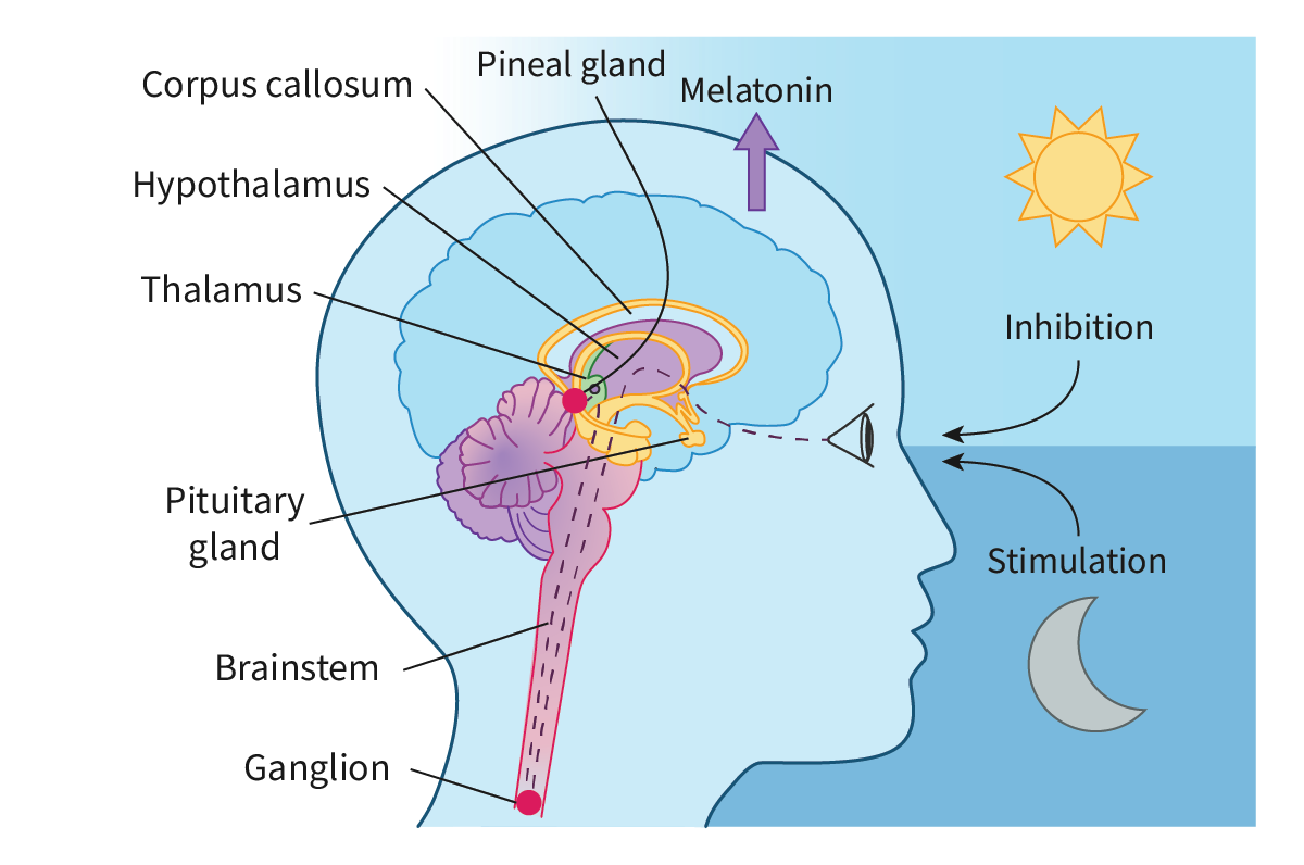
Changes in the levels of light are detected by the photoreceptors of the eye.
This is conveyed to the principal circadian clock of the body, a small structure located in the hypothalamus.
During the day, the circadian clock detects the high levels of light and ‘instructs’ the pineal gland to reduce the secretion of melatonin, whereas at night, the reverse happens.
The rise in melatonin level leads to the body’s preparation for sleep, with a lowering of the core temperature and hence, melatonin is often called the sleep hormone.
What happens during a stressful situation?
The hypothalamus sends impulses through the nerves of the autonomic nervous system.
This activates the adrenal glands.
The glands secrete epinephrine (see section C2.1.10) into the bloodstream
Epinephrine secretion by the adrenal glands:
adrenaline, is secreted by the adrenal medulla, the inner part of the adrenal glands, which are located above the kidneys.
It is released into the bloodstream in response to signals from the hypothalamus during stressful or emergency situations, part of the body’s fight or flight response.
Mechanism of action of epinephrine as a signaling molecule
Epinephrine binds to adrenergic receptors (alpha, beta) on target cell surfaces, activating G-protein-coupled pathways.
Beta-1 receptor activation increases cyclic AMP (cAMP) in cardiac cells, activating protein kinase A (PKA), enhancing calcium influx, increasing heart rate, and muscle contraction.
Beta-2 receptor activation in liver stimulates glycogenolysis, releasing glucose into bloodstream for energy.
Beta-2 receptor activation in smooth muscle relaxes bronchial and vascular smooth muscle, causing bronchodilation and vasodilation.
Effects of epinephrine on body:
Skeletal muscles:
Vasodilation increases blood flow, delivering oxygen and nutrients.
Glycogenolysis releases glucose, providing energy for muscle activity.
Liver:
Epinephrine activates glycogenolysis, raising blood glucose levels for rapid energy supply.
Bronchi and bronchioles:
Bronchodilation improves airflow into lungs, increasing oxygen intake.
Ventilation:
Increases respiratory rate and tidal volume, enhancing oxygen uptake.
Heart rate:
Increases heart rate via beta-1 receptors in sinoatrial node, boosting circulation to vital organs.
Cardiac output:
Increases due to higher heart rate and stroke volume, improving blood flow.
Vessel dilation:
Vasodilation increases blood flow to muscles.
Vasoconstriction reduces blood flow to non-essential areas, prioritizing brain and muscle.
What is the hypothalamus-pituitary complex?
the command centre as it produces hormones that either produce direct responses in target tissues or hormones that regulate the synthesis of other hormones
heart rate adjustment
Heart rate is myogenic, meaning it can be modified by neural and endocrine feedback mechanisms to maintain homeostasis.
Structures and functions regulating heart rate
Medulla oblongata: Contains the cardiovascular centre, which processes sensory input from baroreceptors and chemoreceptors and modulates heart rate.
Sympathetic nerve: Activates in response to low blood pressure or low oxygen levels, increasing heart rate and stroke volume by releasing norepinephrine.
Vagus nerve: Part of the parasympathetic nervous system, decreases heart rate when blood pressure is high or oxygen levels are adequate by releasing acetylcholine.
Baroreceptors: Mechanoreceptors in carotid sinus and aortic arch detect blood pressure. Increased pressure triggers parasympathetic activation, while decreased pressure activates the sympathetic system.
Chemoreceptors: Located in carotid and aortic bodies, they sense changes in blood pH, CO₂, and oxygen levels, sending signals to the medulla oblongata to adjust heart rate.
Factors increasing heart rate
Low blood pressure: Baroreceptors detect pressure drop, activating sympathetic response to increase heart rate.
Low oxygen or high CO₂: Chemoreceptors detect these changes, triggering sympathetic response to increase heart rate and improve oxygen delivery.
Exercise: Increased demand for oxygen and removal of CO₂ stimulates sympathetic activation.
Factors decreasing heart rate
High blood pressure: Baroreceptors detect stretch, triggering parasympathetic response to lower heart rate.
High oxygen levels: Reduced need for oxygen uptake reduces sympathetic drive, leading to a lower heart rate.
Resting state: The parasympathetic system dominates during relaxation, slowing heart rate.
CO₂ production and blood pH relationship
CO₂ dissolves in blood, forming carbonic acid, which dissociates into hydrogen ions (H⁺) and bicarbonate. Increased CO₂ leads to a decrease in pH (more acidic), signaling the need for faster ventilation to expel CO₂
Feedback loop regulating ventilation rate:
Chemoreceptors: Detect elevated CO₂ and lowered pH levels in blood.
Brainstem (medulla oblongata): Processes signals from chemoreceptors and activates ventilation centers.
Diaphragm and intercostal muscles: Increased signal to these muscles causes enhanced contraction and relaxation, increasing breathing rate and depth, expelling excess CO₂.
Hyperventilation response to exercise:
During exercise, increased CO₂ production leads to a drop in blood pH, stimulating chemoreceptors.
The brainstem activates the respiratory muscles to increase ventilation, expelling CO₂ to restore pH balance and maintain oxygen supply.
Role of CNS and ENS in digestion:
Central nervous system (CNS): Voluntary control of swallowing and initial digestive processes.
Enteric nervous system (ENS): Coordinates involuntary processes such as peristalsis (muscle contractions that move food through the gut), enzyme release, and nutrient absorption, from the esophagus to the rectum.
Voluntary and involuntary control of gut movements:
Voluntary control:
Swallowing: Initiated by CNS, voluntary control of food intake.
Egestion (defecation): Typically under CNS control, although can be partially involuntary.
Involuntary control:
Peristalsis: Rhythmic contractions of smooth muscle in the gut controlled by ENS.
Secretion and mixing: Digestive enzymes and bile are secreted under ENS control, ensuring proper digestion.
Absorption: ENS regulates blood flow to the gut for nutrient absorption, ensuring efficiency.
List three major types of cause of disease.
Pathogens (infectious agents like bacteria, viruses, fungi, etc.)
Genetic factors (inherited conditions)
Environmental factors (e.g., exposure to toxins, poor nutrition)
Define pathogen
A microorganism or other agent (such as viruses) that can cause disease in another organism.
A pathogen is an organism or infectious agent that causes disease. It invades a host and disrupts its normal functioning.
List major pathogen types
Bacteria: Unicellular, prokaryotic organisms. Ex: Tuberculosis, cholera.
Viruses: Require living cells to replicate. Ex: COVID-19, influenza.
Fungi: Eukaryotic organisms, some of which cause disease. Examples: Athlete’s foot, ringworm.
Protists: Unicellular, eukaryotic organisms. Ex: Malaria, toxoplasmosis.
Prions: Infectious proteins that cause neurodegenerative diseases. Ex: Bovine spongiform encephalopathy (mad cow disease).
Define “primary defense.”
refers to the body's first line of defense, which prevents pathogens from entering the body, mainly through physical and chemical barriers such as skin and mucous membranes.
The first line of defence by the body is non-specific and prevents the entry of any potential pathogen into the body.
Outline the role of skin in the defence against pathogens.
Skin acts as a physical barrier with layers of dead, keratinized cells in the epidermis.
Constant shedding of outer skin layers removes attached microbes.
Skin secretes sweat and sebum, lowering pH to around 4-5, inhibiting bacterial growth.
Outline the role of sebaceous glands in the defence against pathogens.
Sebaceous glands secrete sebum containing fatty acids and lipids that lower skin pH.
Antimicrobial properties in sebum prevent bacterial and fungal infections.
Sebum contains antimicrobial peptides that directly neutralize harmful microbes.
Outline the role of mucous membranes in the defence against pathogens.
Mucous membranes line internal body cavities such as the respiratory, digestive, and urogenital tracts, secreting mucus that traps pathogens, preventing them from reaching deeper tissues.
Mucus contains the enzyme lysozyme, which breaks down bacterial cell walls, effectively destroying bacteria that come in contact with it.
In the respiratory tract, cilia (tiny hair-like structures) move the mucus, along with trapped pathogens, toward the throat, where it can be expelled by coughing or sneezing, thus physically removing microbes from the body.
State two benefits of blood clotting when skin is cut.
Prevents excessive blood loss.
Blocks the entry of pathogens at the injury site.
Outline two roles of platelets in the blood clotting cascade.
Platelets accumulate at the site of injury and form a plug to seal the wound.
They release clotting factors, triggering the clotting cascade that leads to the formation of a stable clot.
Describe the blood clotting cascade, including the role of platelets, clotting factors, prothrombin, thrombin, fibrinogen and fibrin.
Platelets rush to the injury site, clumping together to form a plug, acting as a physical barrier that immediately reduces bleeding.
Platelets release clotting factors like thromboplastin and calcium ions, initiating the clotting cascade.
Clotting factors activate prothrombin, converting it into thrombin. Thrombin then converts fibrinogen into fibrin.
Fibrin forms a mesh that traps red blood cells and platelets, creating a stable clot that seals the wound and prevents pathogen entry.
Distinguish between innate and adaptive immunity, including the types of cells and timing of response to infection.
FeatureInnate Immune SystemAdaptive Immune System | ||
Timing of Response | Immediate (minutes to hours) | Delayed (days to respond) |
Specificity | Non-specific, same response to all pathogens | Highly specific, targets particular pathogens (e.g., viruses, bacteria) |
Cells Involved | Phagocytes (e.g., macrophages, neutrophils, monocytes) | Lymphocytes (B cells, T cells) |
Memory | No memory, same response each time | Has memory, quicker and stronger response on re-exposure |
Defense Mechanism | Physical barriers (skin, mucous membranes), inflammation, phagocytosis | Antibody production, cytotoxic T cells |
Duration of Response | Short-term, rapid but limited in duration | Long-term, can last years due to immunological memory |
Communication with Other System | Communicates and activates adaptive immune system if unable to eliminate pathogen | Relies on innate system to activate and present pathogens for targeted attack |
Function of phagocytic white blood cells in defence
Phagocytes like neutrophils and macrophages are crucial in the non-specific immune response, targeting a wide range of pathogens.
They recognize pathogens through receptors on their surface, which bind to the pathogen, initiating phagocytosis.
Phagocytes engulf and digest pathogens, eliminating them from the body before they can spread or cause harm.
Process by which a macrophage destroys a pathogen
Recognition and binding: The macrophage identifies the pathogen by binding to it through receptors on its plasma membrane.
Engulfment: The macrophage extends pseudopodia around the pathogen, engulfing it into a vesicle known as a phagosome.
Phagosome maturation: The phagosome fuses with lysosomes, forming a phagolysosome that contains digestive enzymes.
Digestion: The enzymes break down the pathogen’s components, killing it.
Expulsion: The digested materials are either used by the macrophage or expelled from the cell as waste.
What are the professional phagocytes of the body?
macrophage
neutrophils
Monocytes
Structure and Function of Lymphocyte Cells
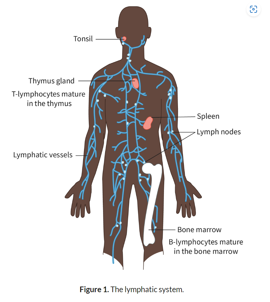
B-Lymphocytes (B-Cells): Develop in the bone marrow. Produce antibodies that bind to specific pathogens. Key role in humoral immunity.
T-Lymphocytes (T-Cells): Mature in the thymus. Two types: Helper T-cells, which activate other immune cells, and Cytotoxic T-cells, which kill infected cells. Involved in cell-mediated immunity.
B-Lymphocytes (B-Cells):
B-lymphocytes are a type of white blood cell that develop in the bone marrow and play a critical role in the adaptive immune response.
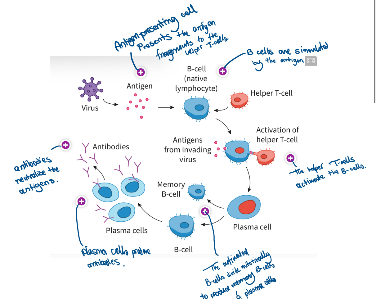
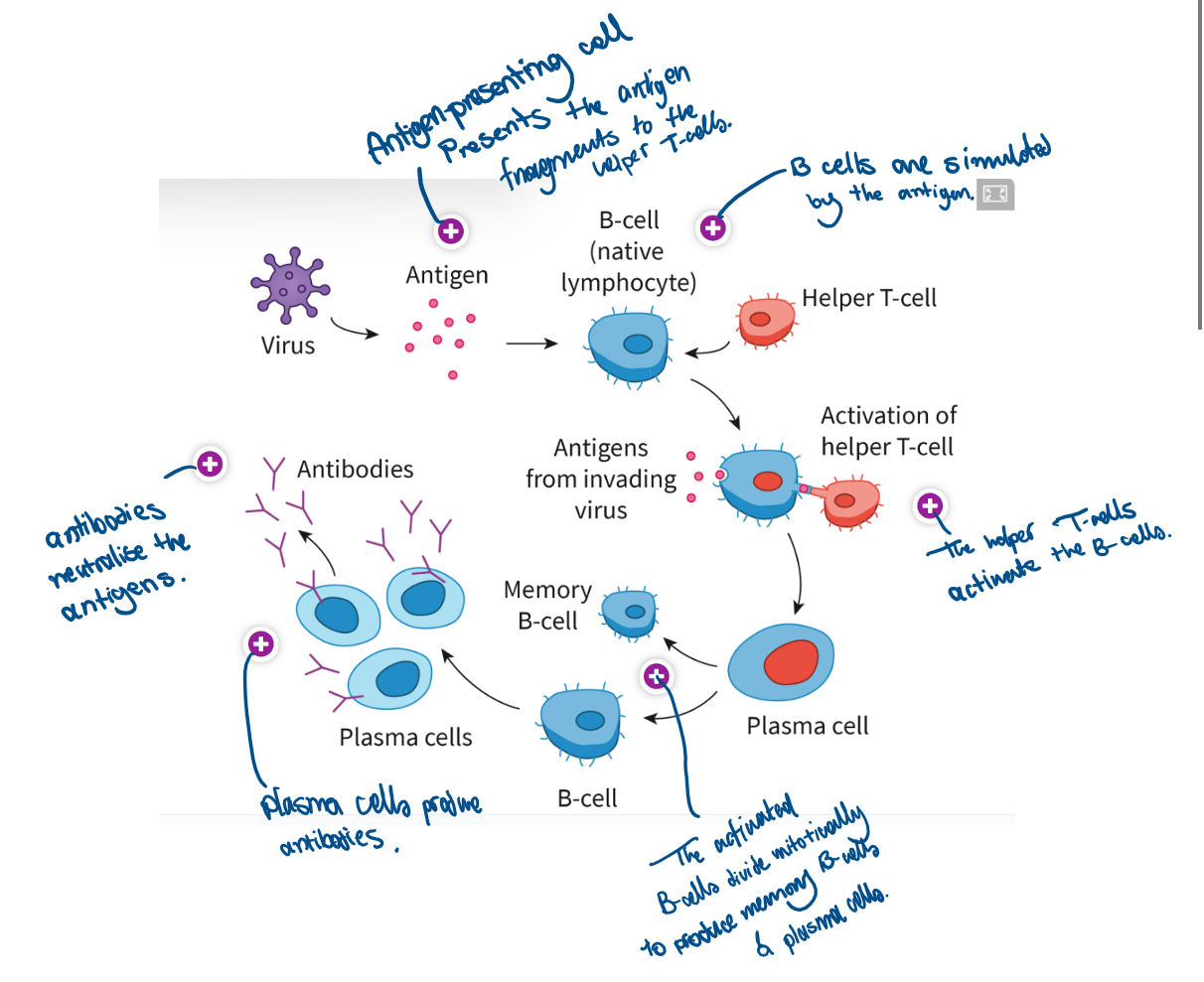
Once activated by a specific antigen, B-cells differentiate into plasma cells, which are specialized to produce large quantities of antibodies. These antibodies are highly specific and bind to antigens on the pathogen, marking it for destruction by other immune cells or neutralizing its harmful effects.
B-cells are responsible for humoral immunity, which refers to the production of antibodies that circulate in the bloodstream and lymph. Humoral immunity is essential for targeting pathogens in the blood and extracellular fluids.
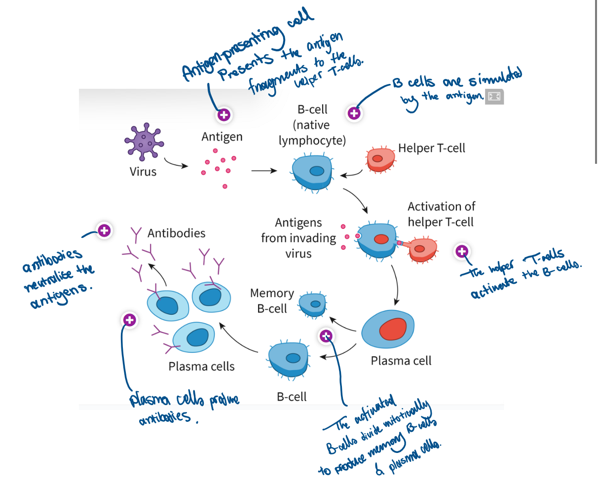
B-cells also generate memory B-cells after the initial infection, allowing for a faster and more efficient immune response upon future exposures to the same pathogen.
T-Lymphocytes (T-Cells):
T-lymphocytes mature in the thymus and are also involved in the adaptive immune system. T-cells are primarily responsible for cell-mediated immunity, which involves the direct destruction of infected or abnormal cells.
There are two main types of T-cells:
Helper T-cells (CD4+ cells): These cells play a central role in orchestrating the immune response. They activate B-cells, cytotoxic T-cells, and macrophages by releasing signaling molecules called cytokines. Helper T-cells are essential for the initiation and coordination of the immune response.
Cytotoxic T-cells (CD8+ cells): These cells directly attack and kill cells that are infected by viruses or have become cancerous. They recognize infected cells through antigens presented on the cell surface and release enzymes that induce cell death (apoptosis).
Like B-cells, T-cells also form memory T-cells, which remain in the body after an infection and respond rapidly if the same antigen is encountered again, providing long-term immunity.
Interaction Between B- and T-Cells:
Helper T-cells activate B-cells by recognizing the same antigen and secreting cytokines that stimulate B-cells to proliferate and differentiate into plasma cells.
This cooperation between B- and T-cells ensures a coordinated and highly specific immune response tailored to eliminating pathogens effectively.
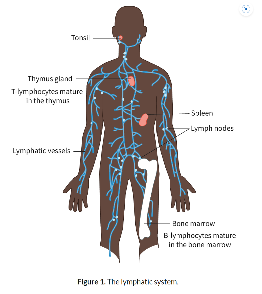
Location of lymphocytes in the body
blood, lymphatic vessels, and secondary lymphoid organs such as lymph nodes, spleen, and tonsils.
Definition of “Specific” in Immune Response:
"Specific" means that lymphocytes recognize and target particular antigens on pathogens, leading to an immune response tailored to that specific pathogen.
Definition of “Antibody”:
Antibodies are Y-shaped proteins produced by B-cells. They bind to specific antigens on pathogens, neutralizing them or marking them for destruction.
Role of Lymphocytes in Producing Antibodies:
B-Cells recognize a specific antigen, become activated, and differentiate into plasma cells, which secrete large amounts of antibodies. These antibodies bind to the antigen and help eliminate the pathogen.
Definition of Antigen:
An antigen is any substance that triggers an immune response by stimulating the production of antibodies. Antigens are usually proteins found on the surface of pathogens.
Antigens are typically proteins, glycoproteins, or lipoproteins found on the surface of pathogens. They are recognized by immune cells due to their unique molecular structure.
Cause and Consequence of Antibody Binding to an Antigen:
Cause: Antibodies bind to antigens due to complementary shapes, like a lock and key, targeting specific pathogens.
Consequence 1 – Neutralization: Antibodies block pathogen entry into host cells by binding to key sites, preventing infection.
Consequence 2 – Opsonization: Antibodies mark pathogens for destruction by phagocytes, making them easier to recognize and engulf.
Consequence 3 – Complement Activation: Antigen-antibody complexes trigger complement system, which punches holes in the pathogen's membrane, leading to membrane attack complexes that destroy pathogens.
Difference Between ABO Blood Antigens:
Blood groups are determined by the presence of specific antigens on the surface of red blood cells:
Group A: Has A antigens.
Group B: Has B antigens.
Group AB: Has both A and B antigens.
Group O: Has no A or B antigens.
Consequence of Mismatched Blood Transfusions:
When incompatible blood is transfused, the recipient’s antibodies bind to the donor’s RBC antigens, leading to agglutination (clumping of RBCs) - clumping blocks blood vessels and disrupts normal blood flow.
and hemolysis (rupturing of RBCs) releasing hemoglobin into the bloodstream. Free hemoglobin can damage organs, particularly the kidneys, leading to kidney failure, shock, and in severe cases, death.
Describe activation of helper T lymphocytes by a macrophage
Phagocytes that digest a pathogen retain fragments of it. Antigen-presenting cells present the fragments to specific helper T-cells, thereby activating the latter.
What happens once a T-cell is activated?
results in an increase in the number of helper T-cells that recognise the same antigen
activates the B-cells specific to the same antigen
activates cytotoxic T-cells specific to the same antigen.
What does the activation of B-cells depend on?
-when the receptor present on the surface of a B-cell recognises and binds to a specific antigen.
-the stimulation by an activated helper T-cell that recognises the same antigen.
Outline the role of the helper T lymphocytes in the activation of B lymphocytes
Activated helper T-cells release cytokines that stimulate specific B-cells. Helper T-cells also interact with B-cells directly by binding to the same antigen, ensuring activation.
B-cells secrete antibodies only after activation by helper T-cells through antigen recognition and cytokine signaling.
Describe clonal selection of plasma B cells
Only the B-cells with receptors specific to the antigen are selected to multiply. These cells undergo repeated mitosis to form identical clones.
Plasma B-cells produce antibodies only after maturation and differentiation to enhance their capacity for large-scale protein synthesis.
Define immunity
Immunity is the body’s ability to resist infection due to the presence of memory cells, which enable a rapid and specific immune response to previously encountered pathogens.
Outline the role of memory B cells in maintaining immunity
Memory B-cells persist in the bloodstream and lymph nodes after an initial infection. They "remember" the antigen and quickly differentiate into plasma cells upon reinfection, producing antibodies more efficiently.
Compare the primary and secondary immune responses to specific pathogens.
Feature | Primary Immune Response | Secondary Immune Response |
Magnitude | Smaller response; fewer antibodies produced. | Larger response; significantly more antibodies produced. |
Speed | Slower; takes days to weeks to develop. | Faster; begins within hours due to memory cells. |
Duration | Shorter-lasting immunity. | Longer-lasting immunity; sustained by memory cells. 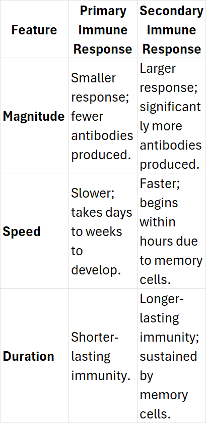 |
What are plasma cells?
B-cells that produce antibodies
Mechanisms of HIV transmissions
Sexual intercourse with an infected person (via vaginal fluids or semen).
Transfusion of infected blood.
Sharing needles or syringes.
From mother to child during childbirth or breastfeeding.
Describe the consequences of HIV on the immune system
HIV targets helper T-cells (CD4+ T-cells), critical for activating B-cells and cytotoxic T-cells.
Viral replication in T-cells leads to cell lysis, reducing helper T-cell count.
A weakened immune system makes the individual vulnerable to opportunistic infections.
The immune response is progressively impaired, culminating in AIDS.
Outline the relationship between HIV and AIDS
HIV is the causative agent of AIDS (Acquired Immunodeficiency Syndrome).
HIV attacks the immune system, specifically the helper T-cells, which weakens the body’s ability to fight infections.
Over time, as the number of helper T-cells decreases, the individual becomes vulnerable to severe infections and certain cancers, a condition known as AIDS.
The transition from HIV to AIDS occurs when the immune system is significantly impaired, and the individual becomes highly susceptible to opportunistic infection
The different stages of HIV
Stage 1
Acute HIV infection is the initial stage where the HIV multiplies and destroys the helper T-cells. During this period, the level of HIV in the blood is high, which increases the risk of transmission.
Stage 2
Chronic HIV infection is the stage where the HIV multiplication drops to low levels. Individuals in this stage may not have obvious symptoms. This stage could last for several years.
Stage 3
At the final stage, the body cannot fight opportunistic infections. The individuals suffer from a range of conditions affecting different organ systems due to the progressive weakening of the immune system, leading to the term, ‘acquired immuno-deficiency syndrome’ or AIDS.
What’s a retrovirus and example of retrovirus
A retrovirus is a virus that has RNA as its genetic material. On infection, reverse transcription occurs and the RNA is converted to DNA.
Outline the natural function of antibiotics when secreted from saprotrophic fungi
Antibiotics are naturally produced by saprotrophic fungi to compete for resources by inhibiting or killing other microorganisms that could be harmful or take up nutrients. This gives the fungi a competitive advantage in their environment.
Antibiotic: A substance that can kill or inhibit the growth of certain bacteria.
Outline the function of antibiotics when used in medical treatment.
Antibiotics are used in medicine to treat bacterial infections by either inhibiting bacterial growth (bacteriostatic) or killing bacteria directly (bactericidal). They target specific bacterial structures, such as cell walls or protein synthesis machinery, which are not present in human cells.
State why antibiotics fail to control viral infections.
Antibiotics are ineffective against viral infections because viruses do not have cellular structures that antibiotics can target. Viruses hijack host cells for replication and lack the independent metabolic processes that antibiotics typically disrupt in bacteria.
Describe how natural selection leads to the development of antibiotic resistance in bacteria.
When bacteria are exposed to antibiotics, some bacteria may have genetic mutations that give them resistance, such as the ability to pump out the drug or break it down. These bacteria survive the treatment and reproduce, passing on the resistant traits to offspring, gradually increasing the population of resistant bacteria.
Discuss the medical cause and consequences of evolution of antibiotic resistance
The overuse and misuse of antibiotics (e.g., incorrect prescriptions or incomplete courses of treatment) accelerate the development of resistance. This makes infections harder to treat, resulting in longer hospital stays, higher healthcare costs, and increasing mortality rates due to multidrug-resistant (MDR) bacteria
Outline the major routes of pathogen transmission.
Pathogens can be transmitted from animals to humans through direct contact (e.g., bites, scratches), indirect contact (e.g., contaminated food or water), or vector organisms (e.g., mosquitoes transmitting viruses like Japanese encephalitis).
Outline the reason why most infectious agents are species-specific.
Most infectious agents are species-specific due to differences in host cell receptors, immune systems, and cellular environments.
Pathogens are adapted to infect particular species where they can effectively replicate and thrive.
Define zoonosis.
A zoonosis is an infectious disease that can be transmitted from non-human animals to humans, typically involving pathogens like viruses, bacteria, or parasites that have reservoirs in animal species.
Mycobacterium tuberculosis, the causative agent of tuberculosis, can be transmitted from cattle to humans through contaminated dairy products
List two reasons that will lead to an increase in the appearance of zoonotic diseases in humans.
Increased human-wildlife interaction due to habitat encroachment
global travel and trade, which facilitate the spread of zoonotic diseases across regions and species.