Exam 2: Visual Fields
1/23
There's no tags or description
Looks like no tags are added yet.
Name | Mastery | Learn | Test | Matching | Spaced |
|---|
No study sessions yet.
24 Terms
How are visual fields’ left and right hemifields defined? What do you see with each eye?
How are they defined? Hemifields are defined as the left and right hemifields by drawing a line starting at your nose
What do you see with each eye? (largely binocular vision, but monocular in the periphery of the visual field) Each eye gets information from BOTH hemifields (binocular), however, vision is monocular in the periphery (you can see it only with one eye)
Describe the cross-over system and how it’s accomplished
The brain uses a cross-over system so that the left half of the brain perceives (and controls, in this case of the motor system) the right side of the body. Because both eyes see both hemifields, this needs to be sorted out by the selective crossover of fibers (ex. Vision in the periphery of the visual field is monocular on that same side, i.e. you can see the right peripheral part of the visual field only with your right eye, therefore, it HAS to cross over to the other side of the brain). The crossing takes place at the optic chiasm. Each retina has its temporal side (towards the temporal bone) and its nasal side (towards the nose).
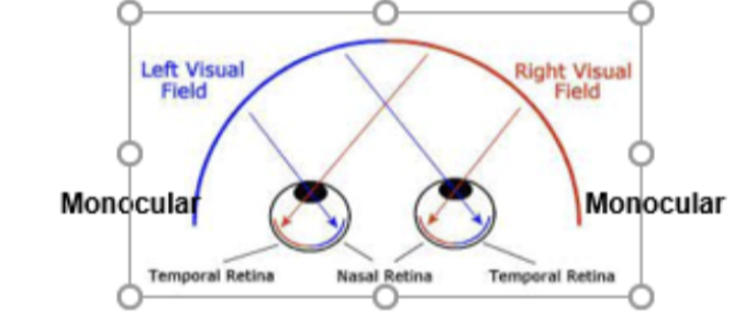
What part of the retina in each eye is activated by light rays from the left or right visual hemifields?
Monocular vision (ie the most peripheral part of the right hemifield is seen only by the right eye) is mediated by the most medial portion of the nasal retina
Which fibers cross at the optic chiasm? Provide an example.
Fibers from the nasal retinas cross to reach the opposite hemisphere. The temporal fibers do not cross since they already carry information from the opposite visual field. Therefore, they project to the same side of the hemisphere. The reason is so that each hemisphere can get complete input from the contralateral visual hemifield.
Ex. The light rays from the right hemifield hit the left temporal retina, and those fibers continue to the left V1 (primary visual cortex). At the same time, information from the right hemifield will hit the right nasal retina and cross over to the left V1. (You can figure this out by using your finger and following the oath of light rays entering your eye(s))
Where do retinal fibers end up?
90% relay in the lateral geniculate nucleus (LGN) of the thalamus on their way to V1 (the primary visual cortex), which has retinotopic maps of the contralateral hemifield.
However, the remaining 10% go to the superior colliculus (eye movements), midbrain (pupillary reflex), and the suprachiasmatic nucleus (hypothalamus) - for the light/dark cycle (circadian rhythm)
What is the retino-geniculo-calcarine pathway? Where does it end up?
The pathway from the LGN of the thalamus to the V1 (the primary visual cortex), which has retinotopic maps of the contralateral hemifield.
The main visual projection pathway ends in V1, which subserves conscious visual analysis
Describe the primary visual pathway.
retina → optic nerve → optic chiasm → thalamus (LGN) → occipital lobe (V1, BA17)
What is V1? What is represented there?
The primary visual cortex; contains a 2D retinotopic map (a topographic map of the image on the retina); contains cells sensitive to basic visual properties (ie a particular cell fires only to horizontal lines, etc)
Each V1 represents the whole contralateral hemifield in a retinotopic manner (note that the brain is organized so that the left half of the brain controls the right side of the body and vice versa)
What are saccades?
Fast eye movements that allow you to foveate; Necessary because images don’t represent the visual field faithfully. It’s distorted because the foveal areas get a much larger representation in V1. Visual acuity (sharpness) is poor for peripheral retinal images. Saccades keep any object of interest in the fovea.
Describe cortical magnification.
You automatically look directly at anything that grabs your attention, which means that those visual objects will be processed with high precision (they will hit your fovea and will be automatically “magnified.”
V1: What are the following areas, where are they located, and what are their respective functions?
The striate visual cortex surrounded by other cortical visual areas in a ring-like manner.
Each subsequent visual area contains cells sensitive to increasingly more complex visual features
V2: What are the following areas, where are they located, and what are their respective functions?
The prestriate (secondary) visual cortex in the occipital lobe.
It is the first visual association area that is sensitive to combinations such as junctions, texture, etc.
It has powerful feedback connections to V1.
V3: What are the following areas, where are they located, and what are their respective functions?
located in the occipital lobe
dorsal subdivision is sensitive to global motion, and ventral subdivision is sensitive to orientation, depth, and form
V4: What are the following areas, where are they located, and what are their respective functions?
located in the ventral stream
is tuned for processing color and orientation
V5 (MT): What are the following areas, where are they located, and what are their respective functions?
aka middle temporal
sensitive to motion and involved in the guidance of eye movements
What stream does MT belong to?
Belongs to the dorsal stream
What is the relation between V1, V2, V3, V4, and V5 (MT)?
There are very strong feedforward and feedback connections between these areas
What are the 2 main visual pathways?
dorsal
ventral
Dorsal pathway: location? function?
the “where,” “how to,” and “action” streams spread dorsally from V1 via V3, MT
It’s sensitive to the location, distance, and relative position of visual objects, as well as to motion relevant to guiding action, which is essential for tasks such as reaching or grasping. It feeds into the motor cortex.
Ventral pathway: location? function?
the “what” or perceptual stream (spreads along the ventral temporal cortex from V1 and extrastriate areas, including V3 and V4) contains neurons sensitive to size, shape, objects, faces, text, etc
This stream is essential for visual recognition, memory, etc, which helps us to identify/recognize visual objects, including faces, text, etc.
How is the visual cortex organized?
Most connections are reciprocal (they go in both directions). Also, there are numerous connections with the areas outside the visual system
What are bottom-up and what are top-down inputs?
Bottom-up processing proceeds along the visual pathway from LGN → V1 → V2 → etc in a feedforward manner. This pathway is driven by sensory input from the physical world.
Expectations, attention, beliefs, etc drive the top-down processing, which selects and interprets sensory information based on internally driven expectations, memories, etc,
How does bottom-up and top-down inputs affect the experience of perception.
highly susceptible to subjective/top-down influence
Our perception is the result of an interaction between bottom-up and top-down processes. Both streams are active simultaneously and interactively as we look at visual objects around us. The bottom-up processing concerns the actual sensory input and how it proceeds towards higher areas. However, top-down influence is present at all times and influences how we perceive things around us.
Our visual perception is relative, reconstructive, and creative (powerful top-down influences), which helps us construct a coherent perception of the world.
Does the visual perception result in absolute brightness, color, and size judgements?
No, the visual system does not make absolute judgments but instead relies on comparisons. This is especially true for brightness, color, and size. They are all interpreted relative to the brightness/color/size of the objects around them. For example, size is interpreted in the context of perspective cues for distance.