Sensory System (3A)
1/78
There's no tags or description
Looks like no tags are added yet.
Name | Mastery | Learn | Test | Matching | Spaced |
|---|
No study sessions yet.
79 Terms
what stimulus processing is usually processed both subconsciously and consciously
proprioception (position of body in space)
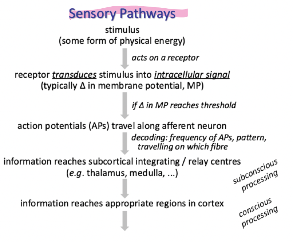
what is the general sensory pathway for unconscious and conscious processing
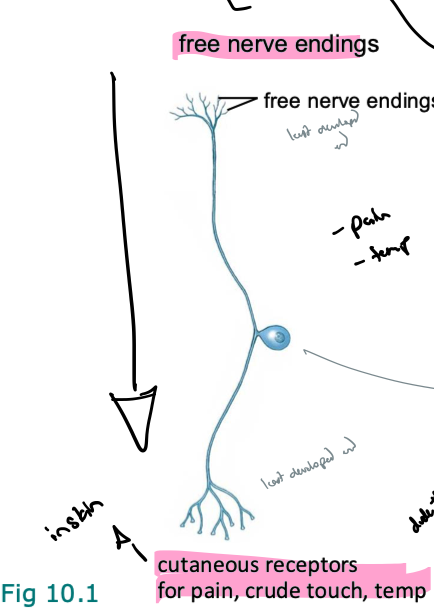
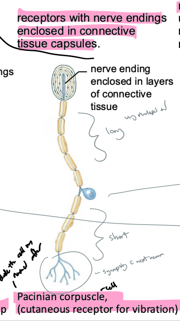
what are free nerve endings, receptors w nerve endings enclosed in connective tissue capsules, and receptors for special senses examples of
Dorsal root ganglion cells
what are neurons with free nerve endings classified as and what do they sense
cutaneous receptors
sense pain, crude touch, temp
what are neurons with nerve endings enclosed in connective tissue capsules classified as and what do they sense
pacinian corpuscles (pacinian = dude the cells are named after, corpuscles = cell)
sense vibration
what are neurons with specialized receptor cells classified as and what do they sense
mechanoreceptors
the receptors for special senses are usually cells that release neurotransmittor onto sensory neurons (neurons are connected to the specialized receptor cells → eg. hair cells in the inner ear)
what type of receptors respond to these types of stimuli…
oxygen, pH, various organic molecules (eg. glucose)
chemoreceptors
what type of receptors respond to these types of stimuli…
pressure (baroreceptors), cell stretch, (osmoreceptors) vibration, accelleration, sound
how does that happen?
mechanoreceptors → deformations of membrane open ion channels
what type of receptors respond to these types of stimuli…
photons
photoreceptors
what type of receptors respond to these types of stimuli…
varying degrees of heat
how does that happen?
thermoreceptors → respond best to inc temps
how does signal transduction within sensory receptors turn into graded potentials
each sensory receptor has an adequate stimulus (type of molecule/energy it responds best to)
stimulus opens or closes ion channels in receptor cell membranes (directly or via second messenger systems)
mostly open cation channels → influx Na or Ca → depolarization
sometimes efflux of K → hyperpolarization
change in mem pot (through graded pot produced) = receptor potential
explain what is meant by sensory neurons’ “receptive fields.“ explain how they work
Somatosensory neurons and visual neurons are activated by stimuli that fall w in a certain physical area
eg. cutaneous receptors → patch of skin, photoreceptors → light falling on area of retina
work bc…
pathway has at least 2 afferent neurons on way to brain
first order (primary) sensory neurons
directly associated w the stimuli
second order (secondary) neuron
relays info from the first neuron to brain
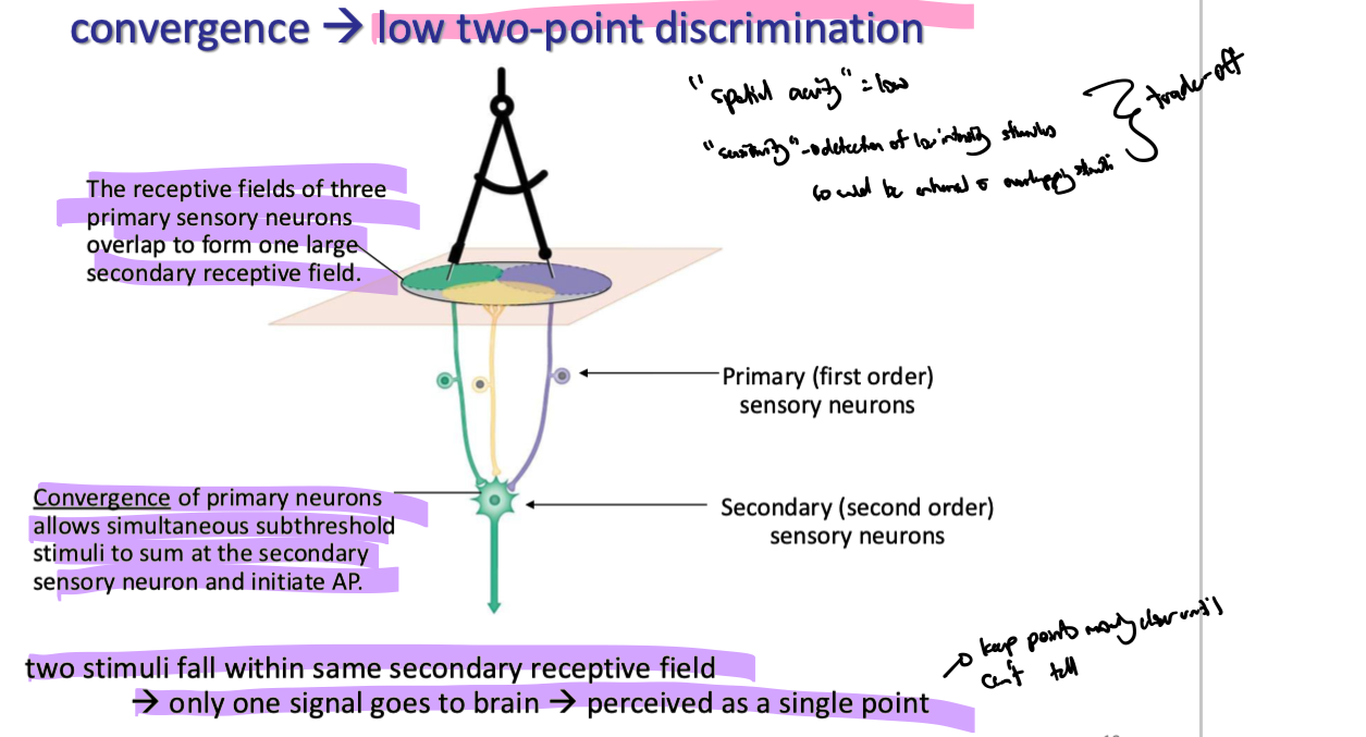
the receptive field is often defined by neurons further up the pathway (ie. primary neurons converge onto one secondary neuron :. sensory input can be gathered from more than one primary sensory neuron in an area
what is 2 point discrimination
bc all primary sensory neurons w overlapping receptive fields converge on one secondary neuron brain cant tell diff btwn primary receptive fields since both signals are being brought to brain from the same pathway
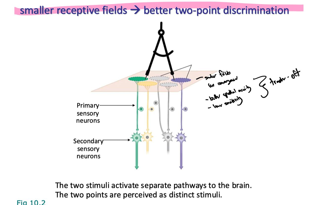
2 point discrimination is the ability to distinguish btwn 2 points of contact on the skin when touched simultaneously instead of feeling a single point → means primary sensory neurons innervating those 2 areas converge onto diff secondary sensory neurons
does convergence of primary sensory neurons make larger or smaller receptive fields
larger (covers larger area)
explain the diff btwn spatial acuity and sensitivity
spatial acuity: ability to distinguish btwn 2 stimuli that are close tg in space
sensitivity: ability to detect low intensity stimuli
explain the relationship btwn spatial acuity and sensitivity
often a trade-off (inversely related)
what is meant by “low” 2 point discrimination? elaborate in regards to spacial acuity and sensitivity
spatial acuity = low → inability to distinguish sensations btwn two objects that are close in space
sensitivity - high
how does the size of a receptive field impact two-point discrimination
smaller fields = less convergence
better spatial acuity
lower sensitivity (harder for threshold to be met for secondary sensory neuron)
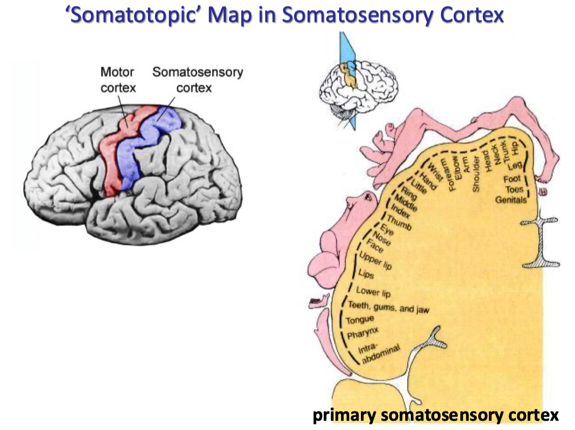
where do all bodily sensations from somatically mapped areas go in the brain
the primary somatosensory cortex
what is a somatotopic map
usually used in the somatosensory cortex
precise mapping of the body's sensory and motor areas onto specific regions in the brain
explain how olfactory sensory info is typically integrated in the CNS (an overview)
olfactory pathways from nose project through olfactory bulb to cortex
explain how most sensory info is typically integrated in the CNS
most sensory pathways project to thalamus which modifies and relays info to cortical centers
eg hearing, taste, vision
thalamus → important relay center for sensory info → signals (most special senses) go here b4 diff regions of brain w some exceptions (ie. olfactory and equilibrium)
explain how equilibrium sensory info is typically integrated in the CNS (an overview)
equilibrium pathways project primarily to cerebellum
MINOR input to thalamus
explain how visceral sensory info is typically integrated in the CNS (an overview)
mostly integrated in brainstem and spinal cord
does not usually reach conscious perception
usually completely subconscious → blood pressure
some can reach consciousness → eg. fullness (through pressure or pain)
how are ALL sensations decoded, processed, and distinguished in the CNS
all stimuli are converted to graded pots then to action potentials (APs)
all APS are identical → if one isnt activated in tract, signal will stop being sent
to distinguish, the CNS must be able to decode…
type of stimulus → modality
location of signal
intensity of signal (how many)
duration of signal (how long)
how does the brain distinguish btwn sensory modalities
depends on receptor type
if there’s adequate stimulus for that receptor type they will activate associated neurons
brain associated info from that receptor type w that modality
eg touch receptors → perceived as touch, photoreceptors perceived as light, etc
how is location of stimuli across the body coded for in the brain? state an example of an exception
most senses are coded according to which receptive fields are activated
eg touch receptors from particular part of body project to specific location in somatosensory cortex
special senses are “special”
eg hair cells in ear respond to diff frequencies but no receptive fields relating to location of sound source
brain uses various strategies to localize origin of sounds, including diff in time of arrival and level in each ear
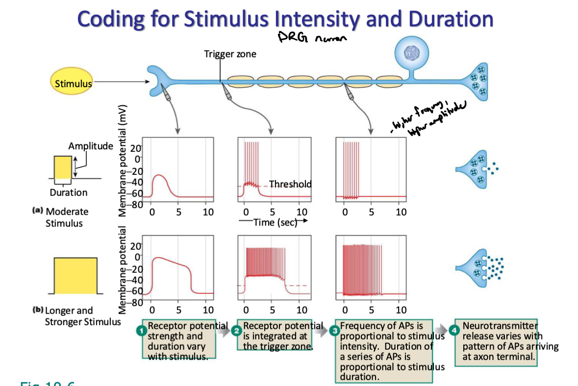
what are the two main ways stimulus intensity is coded? explain each
number of receptors activated (population coding)
diff thresholds for stimulation among group of receptors
low intensity stimulation, most sensitive (lowest threshold) receptors recruited first
as stimulus intensifies, more receptors activated
frequency of APs coming from individual receptor cells
frequency of APs inc with stimulus intensity up to a maximum frequency the axon can transmit
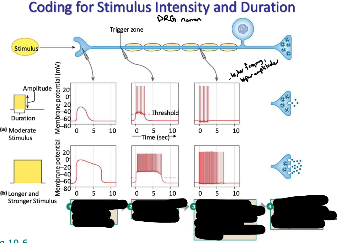
use the diagram to explain how coding for stimulus intensity and duration occurs as signals propagate through DRG neurons
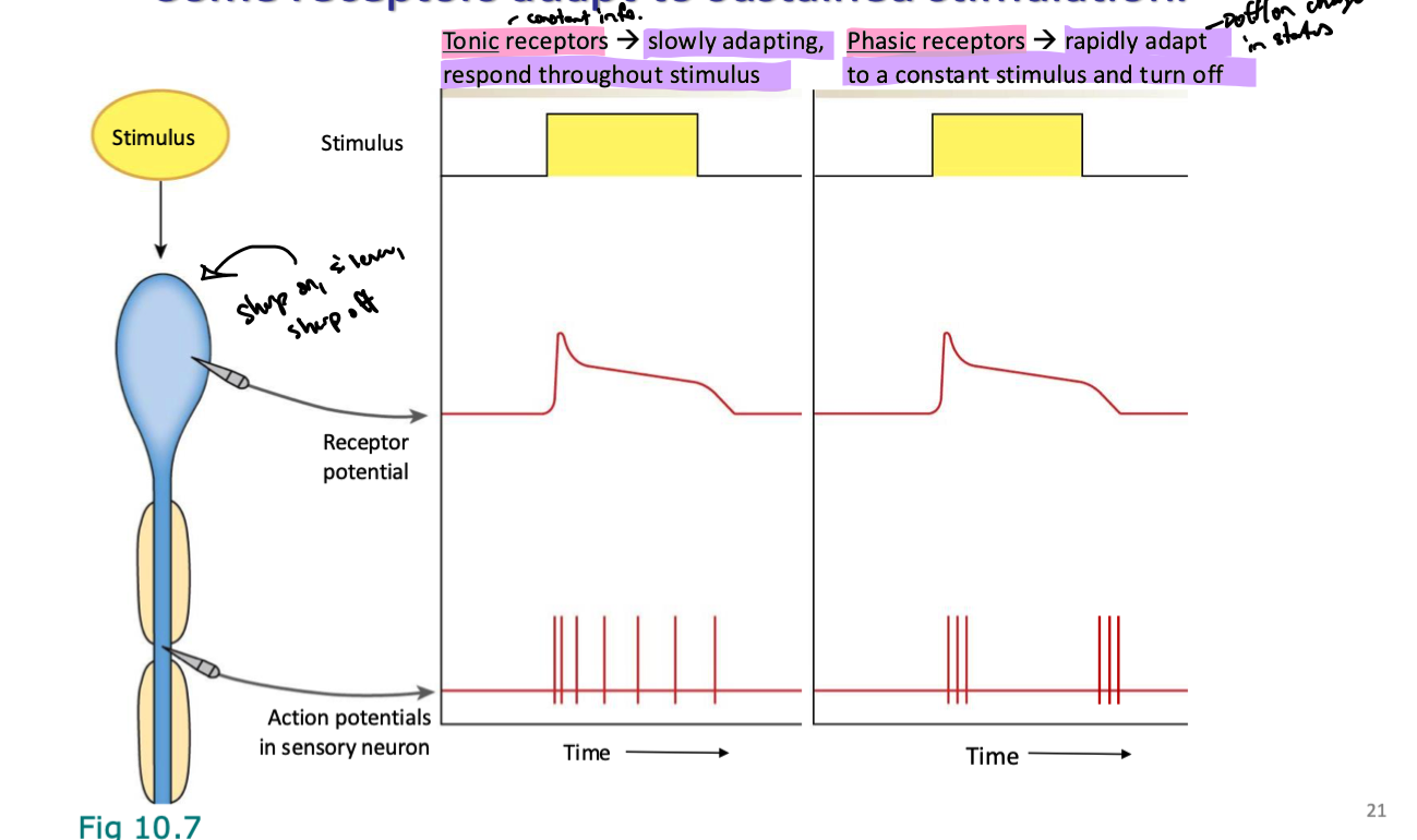
what are tonic receptors? how do they react to sustained stimuli
slowly adapting, respond throughout stimulus
don’t adapt to stimulus touching them so they constantly are active while stimuli are making contact
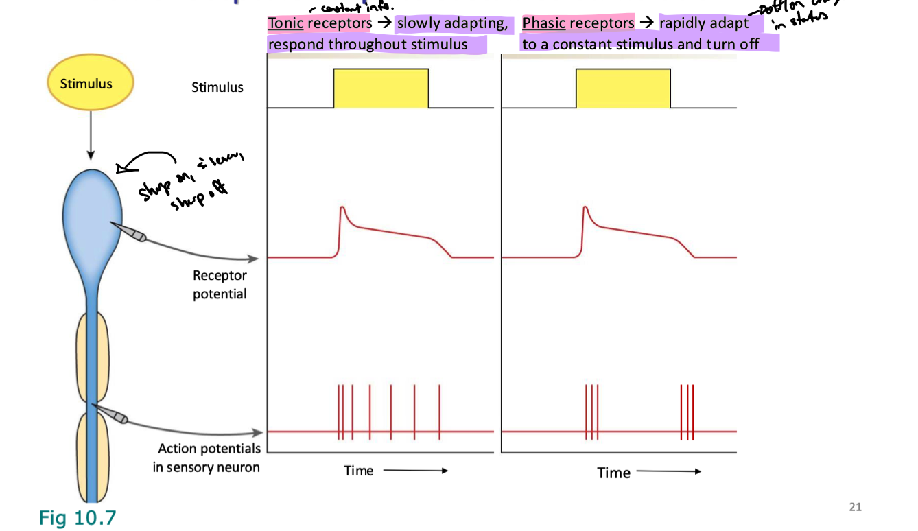
what are phasic receptors? how do they react to sustained stimuli
rapidly adapt to a constant stimulus and turn off (off/on change in status)
send signal initially, then acclimate and stop, then shoot out signals again when stimulus is taken away before quickly adapting again
what is nociception
pain/itching
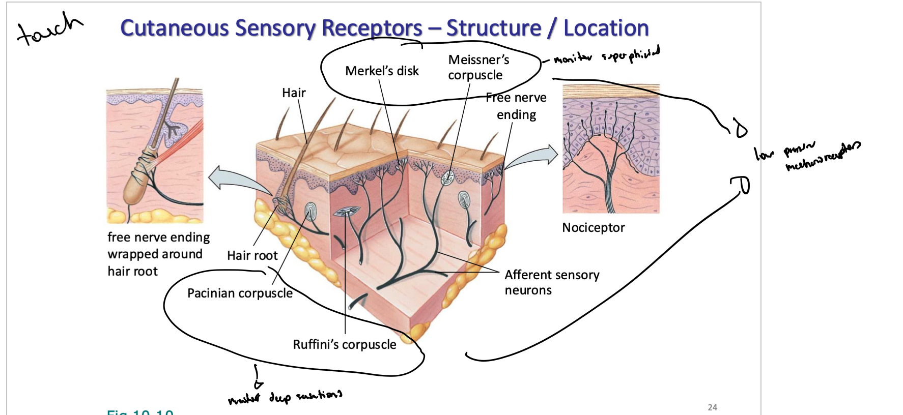
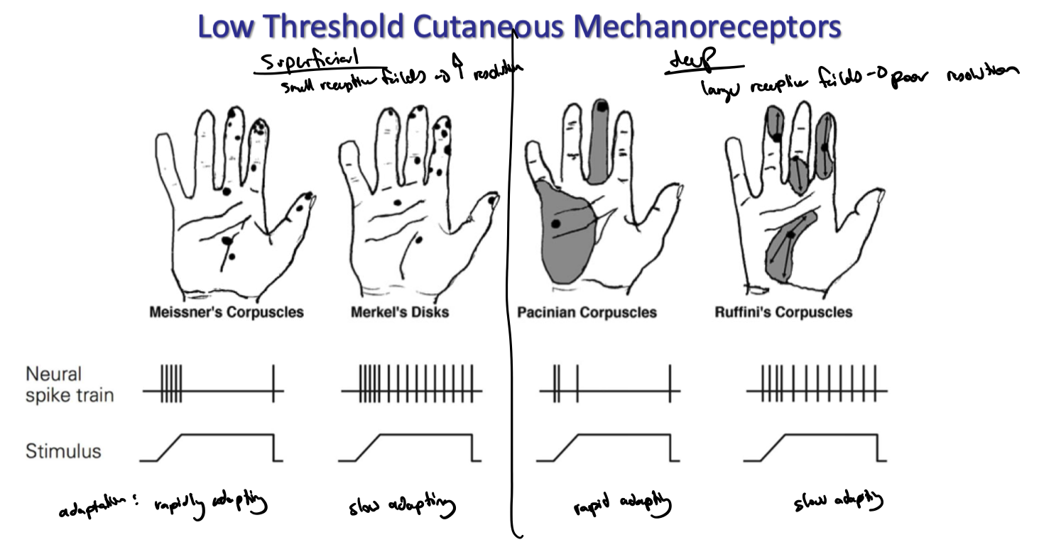
what are the 4 types of cutaneous mechanoreceptors
meissner’s corpuscles
merkel’s disks
pacinian corpuscles
ruffini’s corpuscles
where are merkel’s dicks, meissner’s corpuscles, pacinian corpuscles, and ruffini’s corpuscles located (superficial vs deep)
merkel’s and meissners → monitor superficial skin
pacinian corpuscles and ruffini’s corpuscles → deep sensations
all low pressure mechanoreceptors
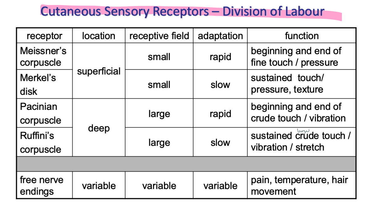
explain the 4 cutaneous mechanoreceptors in terms of deepness in the skin, receptive fields, and adaptation (rapid vs slow)
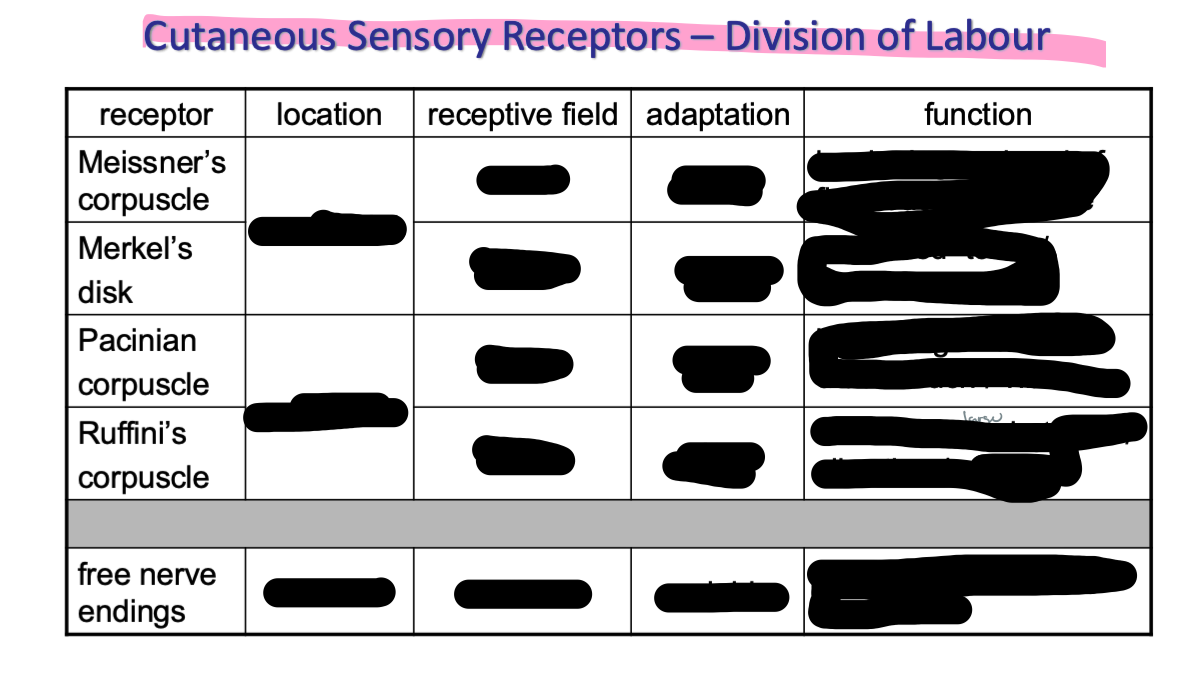
fill out the following
why aren’t nociceptors called “pain receptors”
pain is sensation processed/perceived in brain not stimulus
t/f: all free nerve endings are nociceptors
false → not all free nerve endings are nociceptors but all nociceptors are free nerve endings (found in skin, muscle, joints, etc)
what is nociception mediated by?
what is pain mediated by?
what is the sensation of pain mediated by
nociception is mediated by…
free nerve endings expressing ion channels that respond to variety of strong stimuli (called nociceptors)
chemical/mechanical/thermal
pain is mediated…
via release of local chems (a lot of things - eg K+, histamine, prostaglandins, serotonin, substance P, etc)
can either directly activate nociceptors or sensitize them (invoke inflammatory pain)
which types of channels are found on nociceptors
Transient Receptor Potential (TRP) channels
what are Transient Receptor Potential (TRP) channels
28 diff channels across animal kingdom
expressed on membranes of many diff cell types
mediate a variety of sensations
pain
heat/warmth
cold
some tastes
pressure
vision etc
relatively non-selective ion channels
what are TRPV (vanilloid) receptors
type of TRP (Transient Receptor Potential (TRP)) channel
6 types of TRPVs in humans
respond to…
temp (subtypes have diff ranges)
peper, oregano, wasabi
methanol, pepermint
explain somatic pain
info from nociceptors follow several pathways
spinal reflexes
for somatic pain…
ascending pathways in spinal cord to cerebral cortex
info also sent to limbic system and hypothalamus
can induce emotional reaction
can induce autonomic responses (eg. nausea, vomiting, sweating)
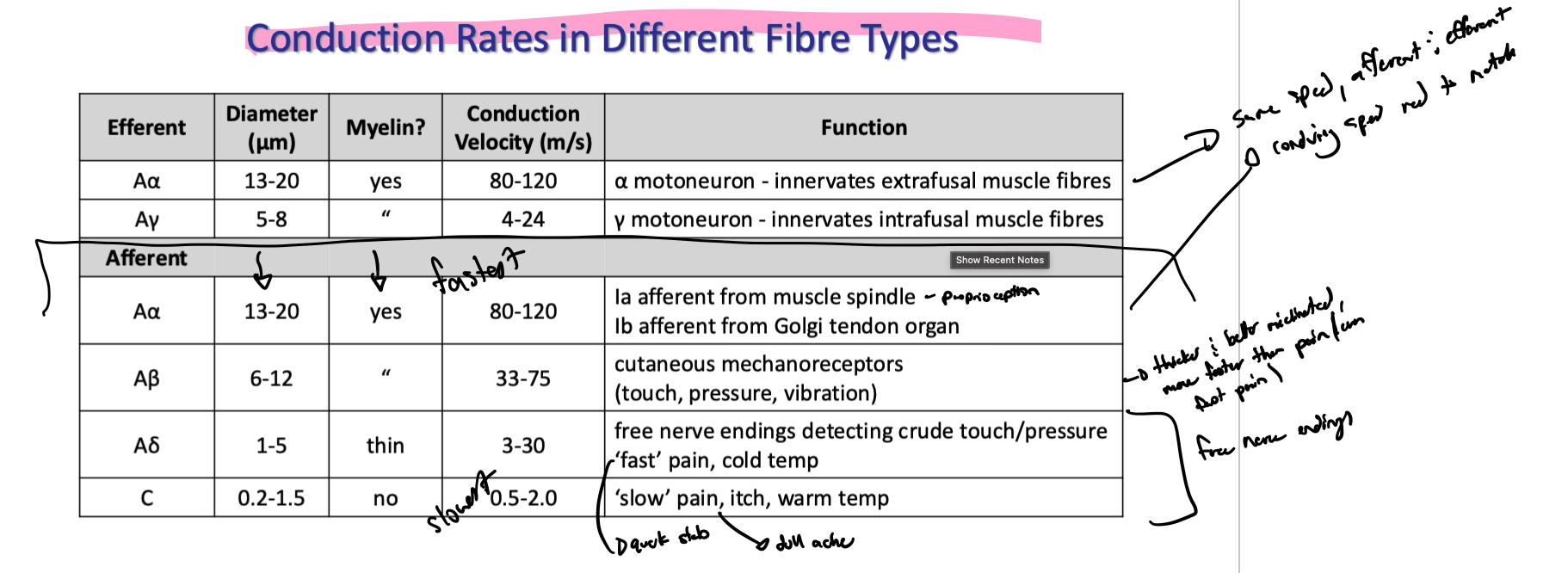
diff types of pain travel on diff fibre types
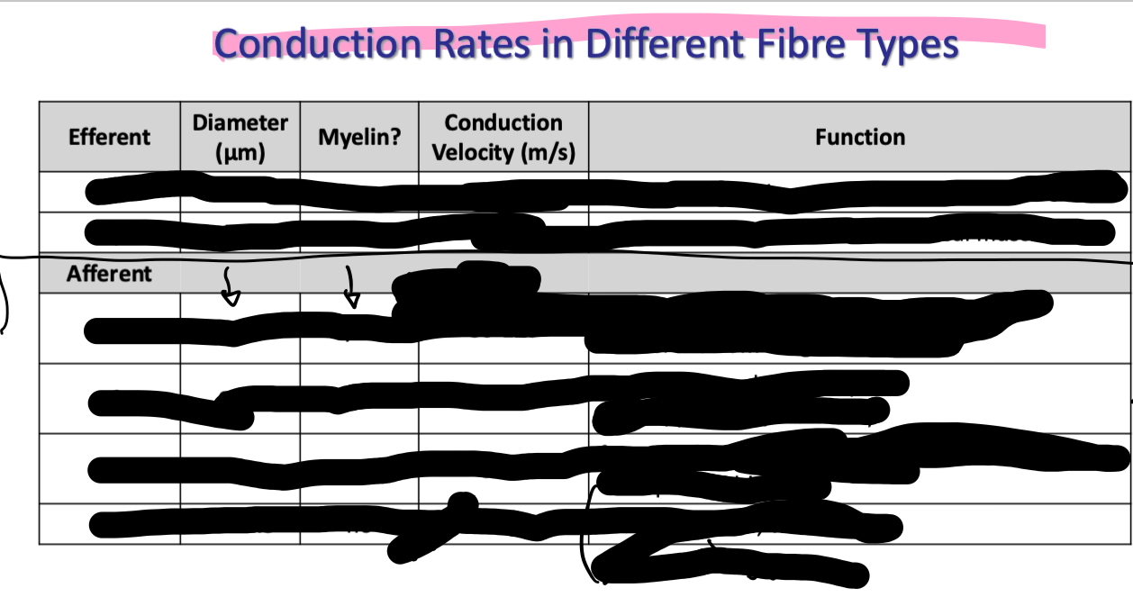
complete the following chart
make q cards for this section if needed
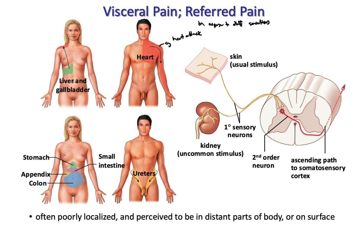
what is referred pain
the experience of pain in a location different from where the injury or disease actually exists
how is visceral pain felt
uncommon sensory stimuli from visceral organs often propagates down primary neurons and converges onto second order sensory neurons in the spinal cord
these second order sensory neurons also typically obtain info from a usual stimulus (eg. pain on skin)
signal goes up to somatosensory cortex and induces same feeling the usual stimulus would
hence why visceral pain is often poorly localized and perceived to be in distant parts of body or on surface
what is muscle tension
Muscle tension -> somatic motor neurons are activated that activate muscle fibers to contract

how does muscle relaxation occur
Muscle relaxation -> sensory input activated inhibitory neurons in the CNS that INHIBIT activity in somatic motor neurons controlling the muscle
Note: no inhibitory neuron that synapses on skeletal muscles to cause them to relax -> relaxation results from the absence of excitatory input by somatic motor neurons
what is muscle tone
Muscle tone: resistance to stretch even when muscle is relaxed and at rest -> result of continuous (tonic) output by alpha motor neurons onto extrafusal muscle fibers
how do proprioceptors work
receptors in joint movements, muscle length, and muscle tension → send info to CNS
depending on appropriate response to body position, CNS activates motor neurons to make motor units contract or activate inhibitory interneurons to inhibit motor neurons → muscles relax
what are the 3 main types of priprioceptors
muscle spindles - monitor muscle strength (length)
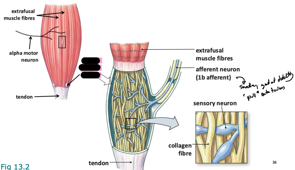
golgi tendon organs - monitor muscle tension
joint receptors - monitor mechanical distortion as bones are repositioned
what are muscle spindles arranged in relation to
parallel to extrafusal muscle fibers (deep to them, and monitor muscle length)
note: muscle spindles are intrafusal muscle fibers
what are golgi tendon organs arranged in relation to
in series with extrafusal muscle fibers (go same way)
they are connected end-to-end, forming a single path, and share the same current
explain 1A vs 1B neurons
1a afferents innervate muscle spindles and provide information about both the rate of change in muscle length (velocity) and the muscle's static length (position), according to the University of Texas Health Science Center at Houston. 1b afferents innervate Golgi tendon organs (GTOs) and provide information about muscle tension (force).
Here's a more detailed breakdown:
1a Afferent Neurons:
Innervation:
Innervate the primary endings of muscle spindles, which are sensory receptors located within muscles that detect changes in muscle length and velocity.
Function:
Provide the central nervous system with information about:
Rate of change in muscle length (velocity): 1a afferents are very sensitive to rapid changes in muscle length and fire at a higher rate during movement than during steady-state conditions.
Muscle length (position): They also transmit information about the static length of the muscle, allowing the brain to know where the muscle is in space.
Response:
1a afferents have a high firing rate during muscle stretch and adapt rapidly to the new length once the stretch stops.
1b Afferent Neurons:
Innervation:
Innervate Golgi tendon organs (GTOs), which are sensory receptors located at the junction between muscle and tendon.
Function:
Provide the central nervous system with information about:
Muscle tension (force): GTOs sense the tension generated by a muscle when it contracts against resistance, according to Physiopedia.
Response:
1b afferents have a low firing rate during passive stretch and their activity increases significantly during active contraction, according to the University of Texas Health Science Center at Houston.
Key Differences:
Location: 1a innervate muscle spindles, while 1b innervate Golgi tendon organs.
Information Transmitted: 1a provide information about muscle length and velocity, while 1b provide information about muscle tension.
Responsiveness to Passive Stretch vs. Active Contraction: 1a have a high firing rate during passive stretch, while 1b have a low firing rate during passive stretch and a high firing rate during active contraction, according to the University of Texas Health Science Center at Houston.
Motor Units and Muscle Receptors (Section 3, Chapter 1 ...
Because they innervate all 3 types of intrafusal fibers, Group Ia afferents provide information about both length and velocity. ..
Department of Neurobiology & Anatomy
Muscle Afferent - an overview | ScienceDirect Topics
Group Ia fibers are termed primary muscle spindle afferents; group II fibers are called secondary muscle spindle afferents. Group ...
The Physiology of the Senses
1a afferents originate from both bag and chain fibers and are therefore sensitive to velocity, the rate of change in length (phasi...
Show all
what are these
golgi tendon organs
central region lacks miofibrils
gamma motor neurons from CNS
alpha motor neyrons
extrafusal muscle fibers
tonically active sensory neurons
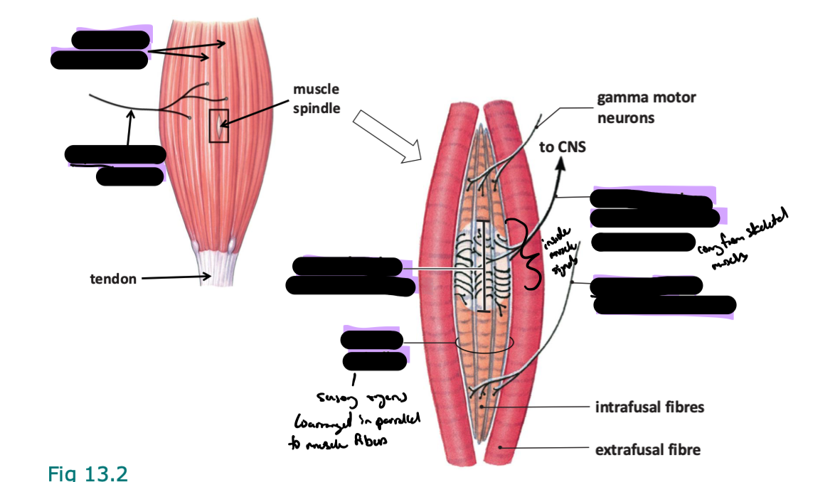
muscle spindles
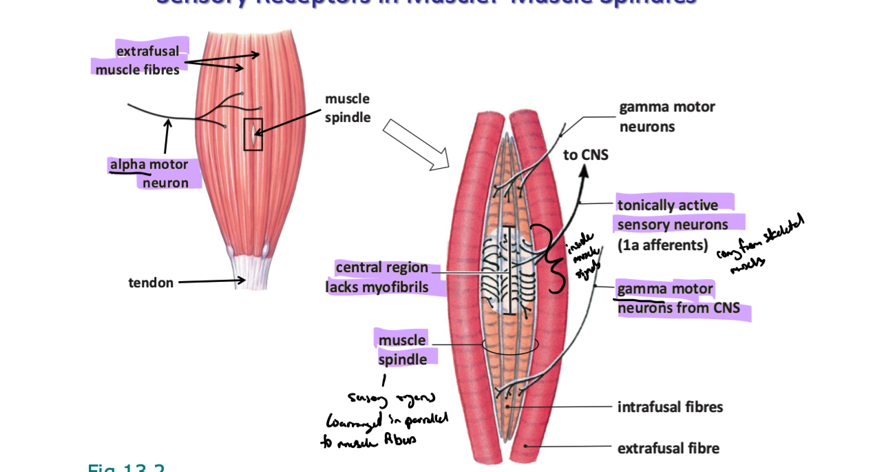
what are intrafusal muscle fibers
A small group of muscle fibers enclosed by a connective tissue capsule
Anchored parallel to extrafusal muscle fibers
Ends of fibers are modified to be contractile, central region is wrapped by sensory nerve endings that are stimulated by stretch
how do muscle spindles work to maintain muscle tone
(hint: reference alpha-gamma coactivation)
each spindle consists of 3-12 intrafusal muscle fibres
arranged in parallel to extrafusal fibres
(almost) every muscle in body has spindles
sensitive to muscle stretch → inc muscle length
tonically active →
mediates stretch reflexes
induces contraction when muscles are stretched
tends to maintain muscle at a constant length
Lie inside skeletal muscle
Contractile ends have their own innervation from gamma motor neurons
When muscles are at rest, each spindle is stretched enough to activate the sensory fibers in the muscle → hence the sensory neurons in spindles are tonically active (send steady stream of action pots to spinal cord even at resting length)
CNS is informed about muscle ‘tone’
These sensory neurons synapse directly onto alpha motor neurons (innervate muscles where spindles are) creating a monosynaptic reflex → :. Tonically active sensory neurons = tonically active alpha motor neurons triggering muscle contraction (muscle tone is the amount of tension that created)
referred to as alpha-gamma coactivation
arranged parallel to muscle fibers (wrap around them)
when muscle fibers stretch, spindles are also stretched and send afferent signal
explain the diff between isotonic and isometric muscle contraction
Isometric exercises involve muscle contraction without a change in muscle length or joint movement, while isotonic exercises involve muscle contraction with a change in muscle length and joint movement
explain what golgi tendon organs (GTOs) do
between muscle fibers and tendons
contraction of muscle causes tendon (and GTO) to stretch
MOST sensitive to isometric contraction (even though works for both)
relatively insensitive to muscle stretch (changes in length)
monitors tension → force of contraction NOT length
info from GTOs combine w info from spindles and joint receptors in CNS integrating centers to monitor/control posture and movement
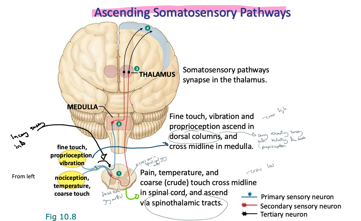
explain the ascending somatosensory pathways in relation to…
fine touch, proprioception, vibration
nociception, temp, coarse touch
make reference to crossing high/low
note:
fine touch, proprioception, vibration…
synapse onto dorsal collumns, ascend in dorsal columns
synapse in medulla, cross high (cross midline in medulla)
nociception, temp, coarse touch
cross midline in spinal cord, synapse onto spinothalamic tracts there (cross low)
ascend via spinothalamic tracts
both somatosensory pathways synapse in thalamus (both are 3 neuron pathways)
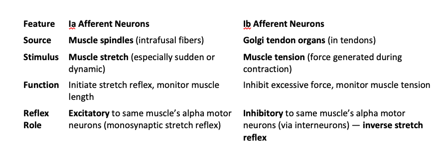
explain the diff btwn 1a afferent neurons and 1b afferent neurons in terms of…
source
stimulus
function
reflex
role
1a afferents innervate muscle spindles and provide information about both the rate of change in muscle length (velocity) and the muscle's static length (position)
1b afferents innervate Golgi tendon organs (GTOs) and provide information about muscle tension (force)
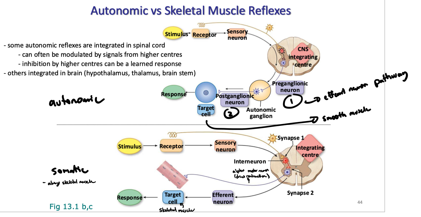
what are 3 ways neural reflexes are classified
according to effector
eg. skeletal muscle
controlled by somatic motor neurons
eg smooth and cardiac muscle, glands, adipose tissue
controlled by autonomic neurons
according to integrating centres
spinal reflexes, “cranial” reflexes
number of neurons in pathway
monosynaptic (only afferent and efferent neurons)
polysynaptic
which types of reflexes are typically mono vs polysynaptic
monosynaptic (only afferent and efferent neurons)
somatic motor reflexes only
polysynaptic
eg autonomic reflexes
reflexes involving interneurons
explain how skeletal muscle reflexes occur in regards to proprioception
(what do/is…
they monitor,
the integrating center
efferent pathway
effectors
monitor:
position of limbs in space, relative pos of body parts
effort exerted in lifting/holding objects
muscle stretch/tone
integrating center: CNS
via networks of excitatory and inhibitory neurons
integrated w spine and/or higher brain regions
efferent pathway:
somatic motor neurons (‘alpha’ motor neurons)
effectors:
contractile skeletal muscle fibers (extrafusal muscle fibers)
explain how autonomic vs skeletal muscle reflexes propagate
explain alpha-gamma coactivation
Movements that decrease muscle length (eg. Contraction):
Gamma motor neurons in normal muscles keep muscle spindles active no matter the muscle length (keep the constant influx of sensory signals)
When alpha motor neurons fire, muscles shorten and release tension on the muscle spindle capsule (in intrafusal muscle fibers) -> gamma motor neurons innervating the contractile ends of the muscle spindle also fire at the same time -> cause muscle to contract and shorten (pulls on central region of spindles and maintains stretch on the sensory nerve endings
Process of excitation of gamma and alpha motor neurons at the same time is called alpha-gamma coactivation → keeps spindles stretched and maintains spindle function when muscle contracts
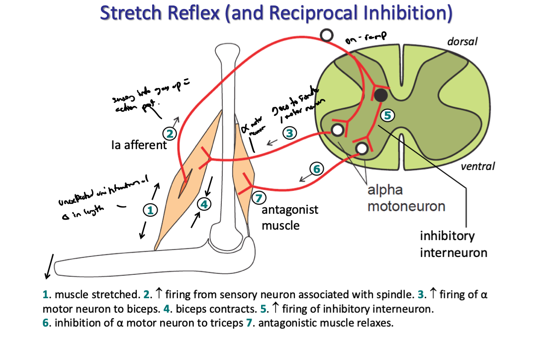
explain reciprocal inhibition
Movements that Increase Muscle length (eg. Stretch):
Any movement that increases muscle length also stretches muscle spindles and causes their sensory fibers to fire more rapidly
Spindle and muscle stretch create a reflex contraction of the muscle to prevent damage from overstretching -> the process of muscle stretch initiating a contraction response is known as a stretch reflex
explain the stretch reflex (and reciprocal inhibition)
(note: bicep is used as an example)
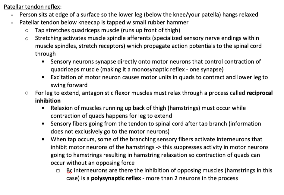
explain the patellar (knee jerk) reflex
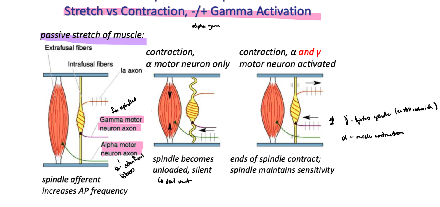
why is the passive stretch of skeletal muscles necessary? how does it work
constant stretch of muscle means spindle afferents are constantly firing
stretch of muscle inc frequency
if only alpha motor neurons contracted (shortened muscle), spindles would become unloaded and silent → wouldnt be able to feel any changes in muscle length → wouldnt know if anything was wrong
gamma motor neurons fire at same time as alpha motor neurons → gamma motor neurons innervate intrafusal muscle fibers (spindles) and keep them taught
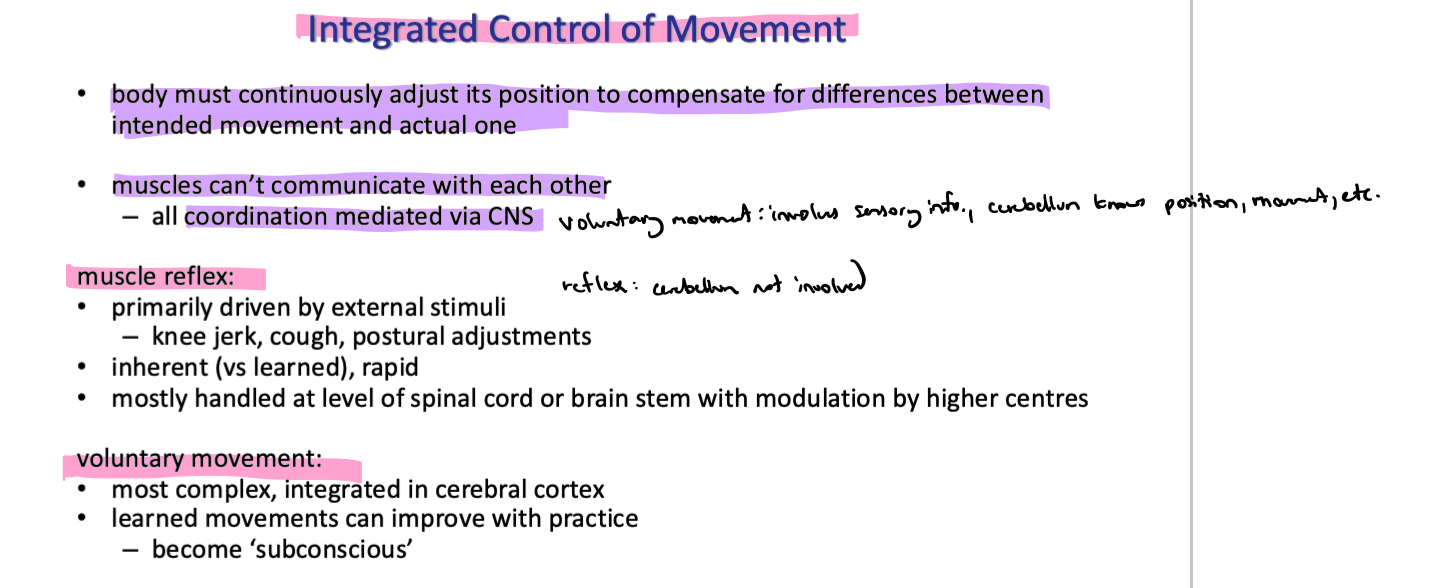
explain the diff between muscle reflexes and voluntary movements
explain the withdrawl reflex
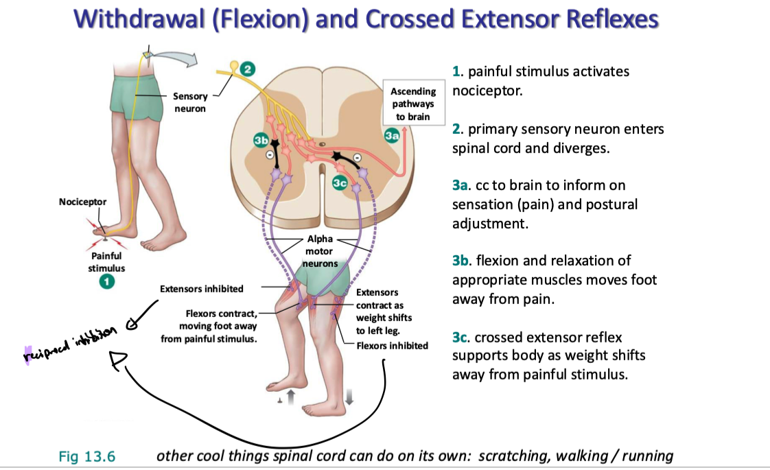
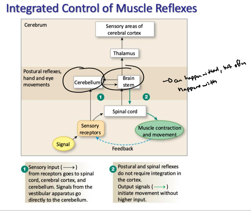
explain the integrated control of muscle reflexes
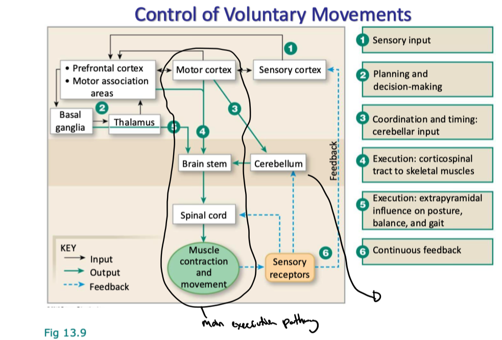
explain the phases of voluntary movement (planning, initiating, executing)
explain the basic mechanism for voluntary movement
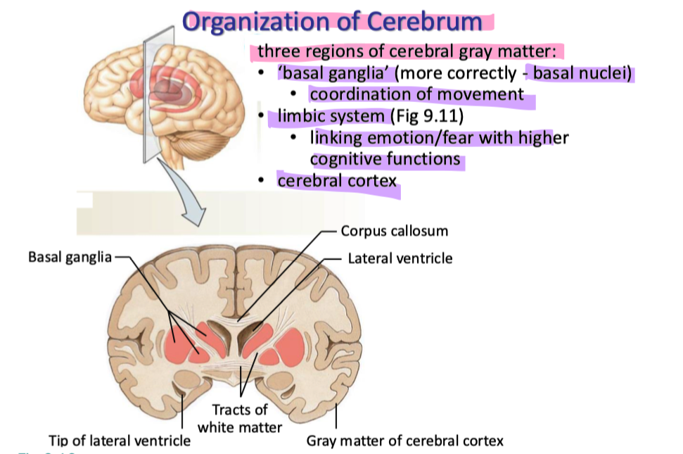
explain the organization of the cerebrum
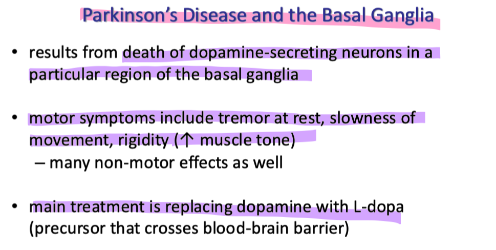
explain parkinson’s disease