A&P I lecture exam 4
1/175
There's no tags or description
Looks like no tags are added yet.
Name | Mastery | Learn | Test | Matching | Spaced | Call with Kai |
|---|
No analytics yet
Send a link to your students to track their progress
176 Terms
Nervous system functions
1- conduction of signals from sensory receptors to processing center
2- processing of brain and spinal cord- interprets these sensory signals and forms a response
3- conducts a signal to an effector
Central nervous system
brain + spinal cord
peripheral nervous system
cranial and spinal nerves
somatic nervous system
voluntary = skeletal muscle
autonomic nervous system
involuntary = smooth and cardiac muscle, glands
nervous system cell type 1 - neurons
conduct electrical signals or action potentials/impulses
nervous system cell type 2- neuroglia
makes up 90% of nervous system cells
support cells
framework for nervous tissue
protection
control composition of extracellular fluid = cerebrospinal fluid in CNS
ependymal cells
assist with producing, monitoring and circulation of CSF
microglial cell
engulf cellular debris, waste and pathogens
astrocyte
many functions:
-maintenance of blood/brain barrier
-structural support
oligodendrocyte
-myelinate CNS axons
-structural support
General Features of a Neuron- Properties
1. Generally amitotic- cannot be replaced
2. Extreme longevity
3. High metabolic rate
Parts of a neuron- cell body(soma)
nucleus, golgi, mito, RER
lack centrioles
abundant cytoskeletal components- neurofibrils
Perikaryon- cytoplasm surrounding nucleus
parts of neuron- axon hillock
initial segment
parts of neuron- axon
carries signals (action potentials) away from cell body
two types of axons
naked- unmyelinated
myelinated- covered with myelin sheath
parts of neuron- dendrites
receives signal
parts of neuron- telodendria
contains axon terminals= synaptic knobs
Formation of the Myelin Sheath
formed by neuroglia
the two cells that form the myelin sheath
1. oligodendrocytes (CNS)
2. Schwann Cells (PNS)- these cells form myelin sheath by wrapping their cytoplasm and membranes around the axon
Nodes of Ranvier(naked axon)
action potentials are conducted from node to node = saltatory conduction(very fast and efficient way to carry an action potential down an axon
functions of myelin sheath
1. Protection
2. Increases speed of action potential conduction b/c myelin is an insulating material
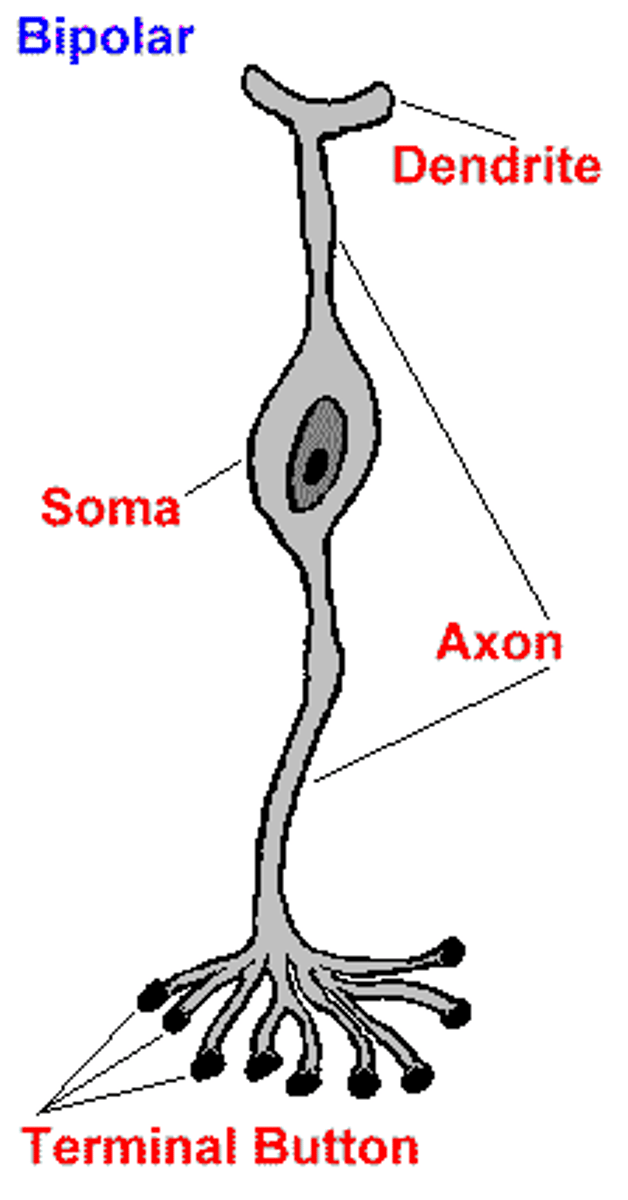
bipolar classification of neurons
Rare: generally associated with special senses
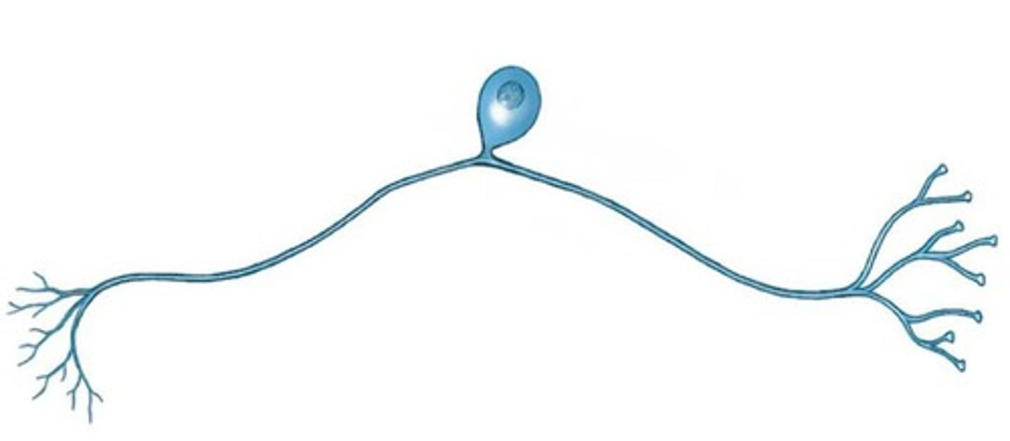
unipolar classification of neuron
structure common to most sensory neurons
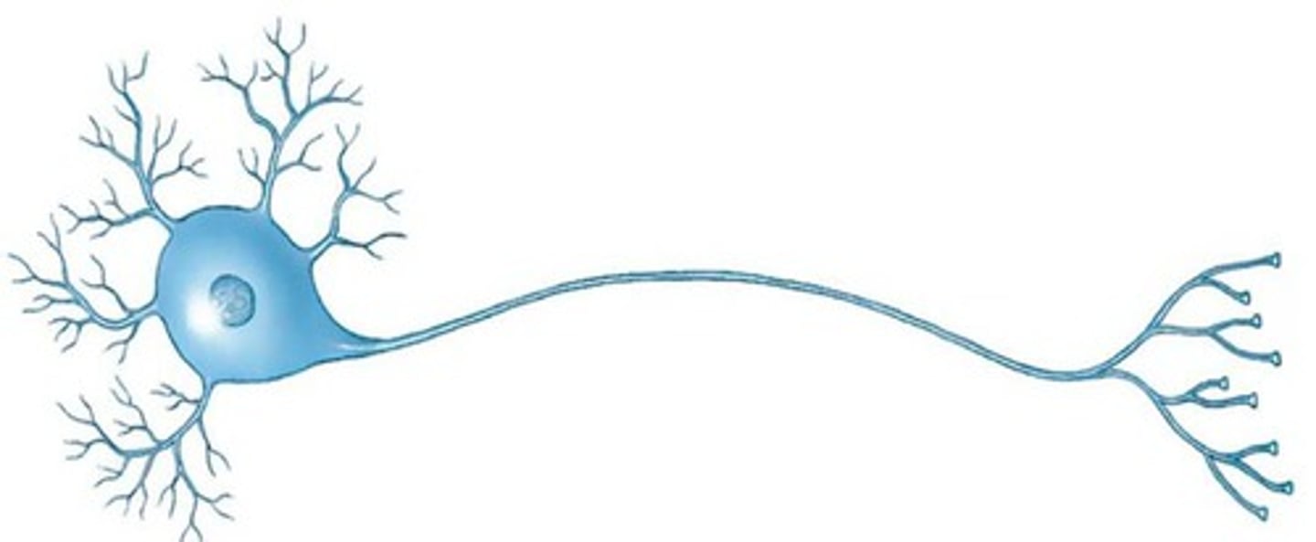
multipolar classification of neuron
structure of most motor neurons and interneurons
sensory neurons (afferent)
carry signals from periphery to CNS
motor neurons (efferent)
carry signals from CNS to muscles and glands
interneurons (association)
most abundant
carry signals b/t sensory and motor neurons
major brain regions
cerebrum- convoluted
major brain regions
diencephalon- cannot be seen externally
a. thalamus- superior portion
b. hypothalmus- inferior portion
major brain regions
brainstem
1. midbrain or mesencephalon
2. pons varolii
3. medulla oblongata
major brain regions
cerebellum
major brain regions
spinal cord
Development of the central nervous system- neural tube
nervous system forms from a hollow tube called a neural tube
protection of the brain and spinal cord- bones
cranium-
frontal
parietal
occipital
ethmoid
sphenoid
temporal
spinal cord is located within the
vertebral canal (c1 through L1 or L2)
protection of the brain and spinal cord- meninges
connective tissue layers
3 layers: duramater
arachnoid mater
pia mater
(all cover both the brain and spinal cord)
3 important variations of cranial meninges
1. no epidural space
outer fibrous layer fused to periosteum of the cranial bones
3 important variations of cranial meninges
2. space within dura mater at midline called:
superior sagittal sinus- delivers venous blood to internal jugular vein
arachnoid villi- connects subarachnoid space to superior sagittal sinus (CSF is reabsorbed into venous blood at arachnoid villi)
Three folds of dura mater-
subdivide the cranial cavity
the three folds of dura mater- falx cerebri
b/t cerebral hemispheres in longitudinal fissure
the three folds of dura mater- falx cerebelli
divides cerebellar hemispheres
the three folds of dura mater- tentorium cerebelli
separates the cerebral and cerebellar hemispheres
functions of the folds
limits excessive movements of the brain within the cranial cavity
cerebrospinal fluid
fluid that surrounds and bathes the entire brain and spinal cord
function of cerebrospinal fluid
acts like a cushion- protects brain from sudden jolts and shocks
supports brain
transports chemical messengers, nutrients and waste products
production of cerebrospinal fluid
produced in choroid plexus: originates in 3rd ventricle, lateral ventricle and 4th ventricle
circulation of CSF
1. lateral ventricle CSF is produced
2. 3rd ventricle CSF is produced
3. 4th ventricle CSF is produced
4. CSF flows into subarachnoid space through the lateral and medial apertures, and into the central canal of the spinal cord
5. excess CSF drains into the arachnoid vili and then into the dural venous sinuses
Anatomy & Physiology of the brain
cerebrum contains 3 parts:
a. Surface gray matter (cerebral cortex)
outermost layer
neuron cell bodies and unmyelinated axons
b. white matter
myelinated axons
c. deep gray matter (cerebral nuclei)
cerebral nuclei
sulcus or sulci
shallow groove
fissure
deep groove
gyrus or gyri
elevated ridge
sensory function
receive and interpret incoming signals
motor function
control movement
association function
involved in intellectual and emotional processes also will integrate info
primary motor area
precentral gyrus, controls voluntary movement of specific muscles
pre-motor areas
stored pathways for learned activities
pre-frontal cortex
abstract intellectual processes consequences of behavior
brora's area
only on left side, motor speech- moves muscles for speech
olfactory area
smell
primary sensory area
postcentral gyrus, receives incoming sensations - localizes them to specific body regions
somatosensory area
association area: interpret, analyze and evaluate sensory info
wernicke's area
language comprehension and interpretation
general interpretative areas (gnostic area)
usually all over left side, stores memories associated w/ sensations, damage would affect your ability to interpret what is seen or heard
white matter - tracts
bundles of myelinated axons located within the CNS
Three types of tracts
association, commissural and projection
association tracts
connects and transports signals b/t gyri of same hemisphere
arcuate fibers and longitudinal fasciculi
commissural tracts
connects and transmits signals from a gyrus on one hemisphere to the corresponding gyrus on the other hemisphere
corpus callous and anterior commisure
projection tract
link the cerebral cortex to diencephalon, brain stem, cerebellum and spinal cord
projection fibers of internal capsule
function of the basal nuclei
control of subconscious muscle movements
- stimulate muscles to produce pattern and rhythm of arm and leg movements associated w/ walking
- help to control and adjust muscle tone during voluntary movements
thalamus structure
1. 2 masses of gray matter covered by white matter
2. contains numerous nuclei
3. located on each side of the third ventricle
thalamus function
relays sensory info to basla nuclei and cerebral cortex, acts as a filter = sends only small portions of sensory info to basal nuclei or cerebral cortex. examples: touch, pressure, pain, temp, position, visual and auditory
hypothalamus structure
1. contains many nuclei
2. forms ventral floor of third ventricle
hypothalamus function
maintenance of homeostasis
controls autonomic nervous system and endocrine system
examples- sleep/wake patterns, hunger, thirst, body temp, emotions, sexual responses
midbrain
extends from pons to lower diencephalon
pons varolli
superior to medulla and anterior to cerebellum
pons varonil function
contains pathways that link cerebellum w/ cerebrum, brain stem and spinal cord
also contains nuclei for cranial nerves V, VI, VII, and VIII
medulla oblongata
inferior brainstem, continuous with spinal cord
medulla oblongata function
conducts sensory and motor info
contains vital and non-vital reflex center
vital
cardiac center, respiratory center and vasomotor center
non-vital
swallowing, sneezing vomitting
cerebellum
second largest part of brain
2 hemispheres connected by vermis
cerebellum contains
cortex(surface gray matter), white matter, and cerebellar nuclei(masses of gray matter)
function of cortex and cerebellar nuclei
1. Maintenance of balance and equilibrium
2. motor coordination for muscle movements = allows for smooth controlled movements
hemisphericity
the right hemisphere controls movements on the left side of the body and the left hemisphere controls movements on the right side of the body
limbic system location & function
located in parts of cerebrum located on border b/t cerebrum and diencephalon
functions involved in memory, creates emotional states (pleasure, pain, rage, behaviors related to survival, motivation)
reticular activating system location & function
located in many parts of brain working together
functions as sleep/wake cycle, when RAS is stimulated = cerebral cortex becomes active
PNS cranial nerves
olfactory, optic, oculomotor, trochlear, trigeminal, abducens, facial, vestibulocochlear, glossopharyngeal, vagus, accessory, hypoglossal
PNS cranial nerves saying
oh once one takes the anatomy final, very good vacations are heavenly(name order)----------some say money matters but my brother says bad bitches matter most(sensory/motor, b=both=mixed)
olfactory nerve
I
sensory function
transmits impulses related to smell from nose to olfactory bulb
optic nerve
II
sensory function
transmits impulses related to vision from retina
oculomotor nerve
III
motor function
conveys impulses that control eye movement regulation of pupil size and accommodation of lens for near vision
trochlear nerve
IV
motor function
conveys impulses that control eye movement
trigeminal nerve
V
mixed function
sensory: conveys impulses relating to sensations of head and face
motor: conveys impulses that control chewing
abducens nerve
VI
motor function
conveys impulses that control eye movement
facial nerve
VII
mixed function
sensory: conveys sensations related to taste from tongue to brain
motor: conveys impulses that control facial expression
vestibulocochlear nerve
VIII
sensory function
vestibular: conveys impulses associated with equilibrium from inner ear to brain = balance
cochlear: conveys impulses associated with hearing from organ of corti in cochlea to brain