5 - Integument
 Call Kai
 Learn
Learn Practice Test
 Spaced Repetition
 Match
Match Flashcards
 Knowt Play
1/103

There's no tags or description
Looks like no tags are added yet.
Name | Mastery | Learn | Test | Matching | Spaced | Call with Kai | Chat |
|---|
No analytics yet
Send a link to your students to track their progress
104 Terms
Integumentary System
the largest system of the body
The Integument is how much % of body weight?
16%
How much area is the Integument?
1.5 to 2 m2
The integument is made up of two parts
1. Cutaneous membrane (skin)
2. Accessory structures
What are the parts of the Cutaneous Membrane?
1. Epidermis (superficial)
2. Dermis (deep to epidermis)
Epidermis (superficial) consists of
Epithelial tissues (ectoderm)
Dermis (deep to epidermis) consists of
Connective tissues (mesoderm)
Superficial = papillary layer
Deep = reticular layer
Hypodermis (Superficial Fascia or Subcutaneous Layer)
Loose connective tissue
Deep to dermis
Accessory Structures to the Integumentary System
Hair
Nails
Exocrine glands
Where do accessory structures to the Integumentary System originate in?
the dermis and extend through epidermis
Functions of integument
Protection
Thermoregulation
Immune defense
Protection of the integument system
Acts as a Physical and Chemical barrier
Protects against UV via melanin
Secretion
The integument acts as a
Physical and Chemical barrier
The integument acts as protection against
UV via melanin
Thermoregulation of the integument system
Blood flow and sweat
Immune defense of the integument system consists of
dendritic cells
Functions of integument system
Water resistance
Metabolic regulation
Storage
Sensory
Water resistance involves the
production of keratin
Metabolic regulation involves
vitamin D3 production
Integument system consists of storage of
lipids
Integument system consists of sensory of
Touch, Pressure, Pain, Temperature
What are cells of the Epidermis
stratified squamous epithelium
stratified squamous epithelium
Avascular - nutrients and oxygen diffuse from the dermis
What do keratinocytes Produce/Contain?
large amounts of keratin
What are keratinocytes?
most abundant cells in the epidermis
Other Specialized Cells close to basement membrane
Melanocytes
Dendritic (Langerhans) cells
Merkel cells
Melanocytes produce
melanin
Dendritic (Langerhans) cells are part of the
immune system
Merkel cells are part of the
nervous system
Merkel cells react to
touch/pressure
Epidermis is divided into 2 skins
Thin skin
Thick skin
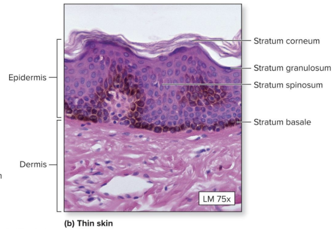
Thin skin
Covers most of the body
four layers of keratinocytes

Thick Skin
Covers the palms of the hands and soles of the feet
five layers of keratinocytes
“hairless'“ skin

The five strata of keratinocytes
From basal lamina (deep) to free surface (superficial)
1. Stratum basale
2. Stratum spinosum
3. Stratum granulosum
4. Stratum lucidum*
5. Stratum corneum
What is the most superficial strata?
Stratum corneum
What is the 2nd most superficial strata?
Stratum lucidum
What is the 3rd strata?
Stratum granulosum
What is the 2nd most deep strata?
Stratum spinosum
What is the most deep strata?
Stratum basale
Stratum Basale consist of
Epidermal ridges (form fingerprints)
Dermal papillae
Stratum Basale
not smooth/flat (wavy)
adds surface area to connections
Dermal papillae
Strengthen attachment between epidermis and dermis
The Stratum Basale has many basal cells
germinative cells (mitosis begins)
The Stratum Basale also contains
Melanocytes
Stratum Spinosum
“spiny layer”
Eight to ten layers of keratinocytes in the stratum spinosum are bound by
desmosomes
In the stratum spinosum cells shrink
cytoskeletons stick out (spiny)
When cells continue to divide in the stratum spinosum, the thickness of the epithelium _________
increases
The stratum spinosum contains what kind of cells?
Dendritic cells
Stratum Granulosum
“grainy layer”
The Stratum Granulosum is where cells
stop dividing
The Stratum Granulosum starts producing
Keratin
Keratohyalin
Keratohyalin forms:
Dense granules
Cross-link keratin fibers
The Stratum Granulosum is the layer where
Eventually cells die
Stratum Lucidum
“clear layer”
Stratum Lucidum is found only in what skin?
thick skin
Stratum Lucidum covers the
stratum granulosum
Stratum Lucidum contains
3-5 layers of dead keratinocytes
Stratum Corneum
“horn layer”
The Stratum Corneum is the
exposed surface of skin
The Stratum Corneum contains
15 to 30 layers of dead keratinized cells
The Stratum Corneum is resistant to what?
water
The Stratum Corneum sheds and replaces itself every:
2 weeks
What is keratinization (cornification)?
formation of a layer of dead, protective cells filled with keratin
Where does Keratinization occur?
on all exposed skin surfaces except eyes
Keratinization takes how many days from the stratum basale to the stratum corneum?
15–30 days
Skin Color is Influenced by Two Pigments
Carotene
Melanin
Carotene pigment color:
yellowish/brown (Orange-yellow pigment)

Melanin pigment color:
brownish/black (Yellow-brown or black pigment)
Blood circulation (red blood cells)
Hemoglobin
What does Hemoglobin contributes to?
red color
Where is Carotene found?
orange vegetables
Carotene is nonpolar
hydrophobic; interact with lipids/dissolve in lipids
Carotene gets converted into
vitamin A
vitamin A acts in the
Maintenance of epithelia
Photoreceptor pigments

What is melanin produced by?
melanocytes in stratum basale
Where is melanin stored?
in vesicles - melanosomes
Keratinocytes incorporate melanosomes by
endocytosis

Keratinocytes eat melanin/incorporate it into cells; this leads to melanin getting packed together
develops a dark ring/layer and absorbs UV radiation, which gets absorbed into dead cells
Function of Melanocytes
Melanin protects skin from sun damage
Ultraviolet (UV) radiation
Skin color depends on melanin production, not number of melanocytes
What does Melanin protects skin from?
sun damage
Ultraviolet (UV) radiation causes
DNA mutations and burns that lead to cancer and wrinkles
Skin color depends on
melanin production, not number of melanocytes
(all skin has the same number of melanocytes)
Blood contributes to skin color
Erythema
Cyanosis
Pallor
Hematoma
Hemoglobin is a
red pigment
Erythema
increased blood flow in the dermis
Blush/Flush, reddish
Cyanosis
low oxygenated blood; bluish tint due to light reflecting off it
Pallor leads to
Pale, low blood flow to skin
Pallor indicates
reduced blood flow
Hematoma
Bruising, black->blue->green->yellow
What is jaundice caused by?
bilirubin buildup when body breaks down RBC
Jaundice
Yellow, first in the eyes
Pituitary tumor
Excess MSH (melanocyte stimulating hormone)
Addison’s disease
bronzing (tanning of the skin)
Vitiligo
Autoimmune destruction of melanocytes
Melasma etc.
“Mask of pregnancy”
Albinism
Lack of melanin production
UV Damage can cause
actinic keratosis
What is actinic keratosis?
scaley sun damaged areas
Skin cancers
Basal cell carcinoma – #1
Squamous cell carcinoma - #2
Malignant melanoma