Spinal Pathology
 Call Kai
 Learn
Learn Practice Test
 Spaced Repetition
 Match
Match Flashcards
 Knowt Play
1/28

There's no tags or description
Looks like no tags are added yet.
Name | Mastery | Learn | Test | Matching | Spaced | Call with Kai | Chat |
|---|
No analytics yet
Send a link to your students to track their progress
29 Terms
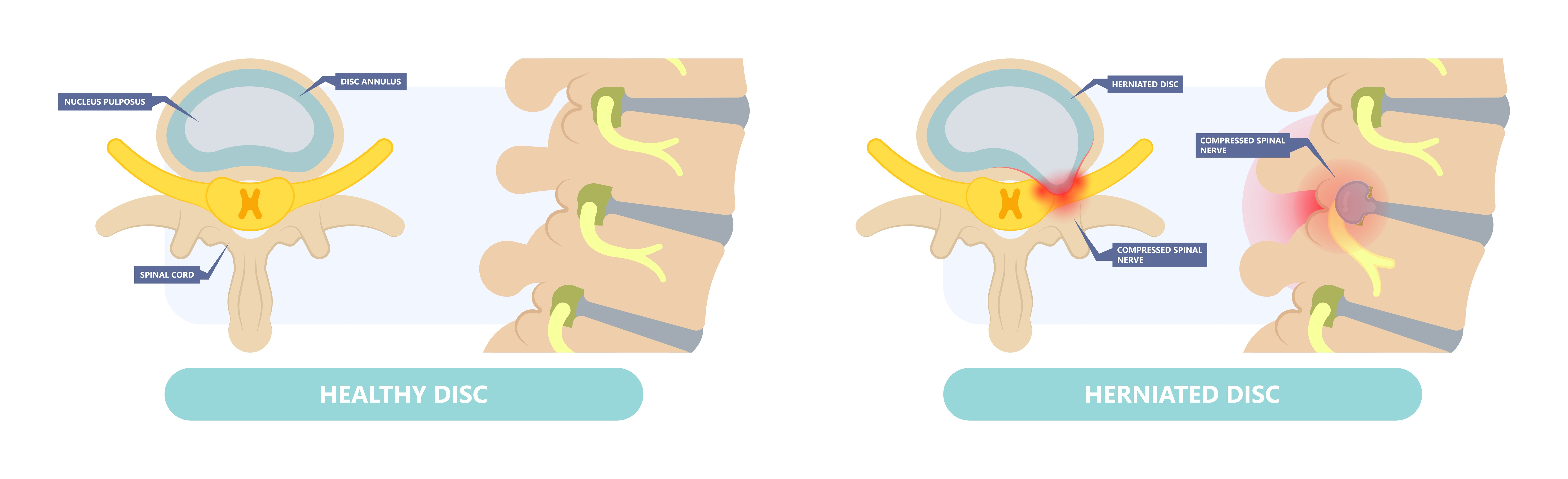
herniated disc
also called a ruptured disc. This occurs when the entire nucleus pulposus protrudes through a torn annulus fibrosis. This can put pressure on the spinal cord. A herniated disc can also be called a HNP (herniated nucleus pulposus).
Reasons for herniated disc include:
•
Trauma
•
Strain
•
Joint degeneration
Appearance on MRI
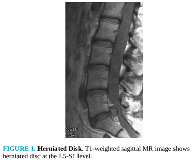
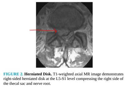
Imaging characteristics include disc degeneration, compression of the thecal sac, and compression of the nerve root.
Spinal stenosis
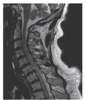
a narrowing of the spinal canal. Spinal stenosis is a degenerative disease that affects the bone, the disk space, and ligaments. Spinal stenosis mostly occurs in the cervical and lumbar regions of the spine.
Appearance on MRI
On MRI it may demonstrate narrowing of the spinal canal, bulging discs, increased size of facet joints and ligamentum flavum, and spondylolisthesis.
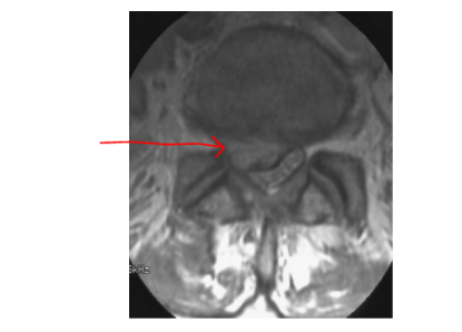
Spondylolisthesis
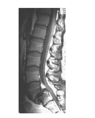
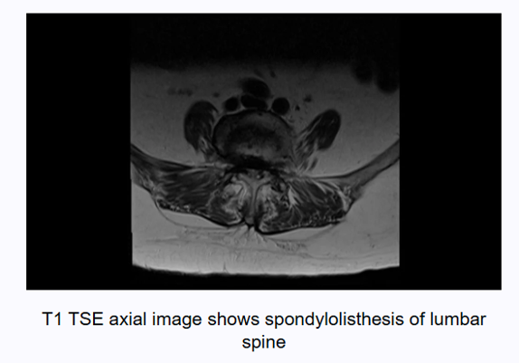
a slipping of a vertebra either anteriorly (anterolisthesis) or posteriorly (posterolisthesis). Spondylolisthesis is classified into types that grade the percentage of displacement.
Spondylolisthesis may occur due to trauma or may be due to degenerative changes.
Appearance on MRI
On sagittal MRI the vertebra will appear out of alignment. Degenerative disc disease and spinal stenosis may also be present.
Osteomyelitis
an inflammation of the bone caused by infection. In the spine the infection is usually affecting the disc space. Osteomyelitis can occur at any level of the spine. It is common in people who have diabetes, are immunocompromised or utilize intravenous drugs.
Appearance on MRI
•
On MRI, T1-weighted images show low-signal intensity.
•
T2-weighted images show high-signal intensity.
•
Postcontrast, fat-suppressed, T1-weighted images show enhancement in bone due to abscess formation and juxtacortical soft tissue enhancement.
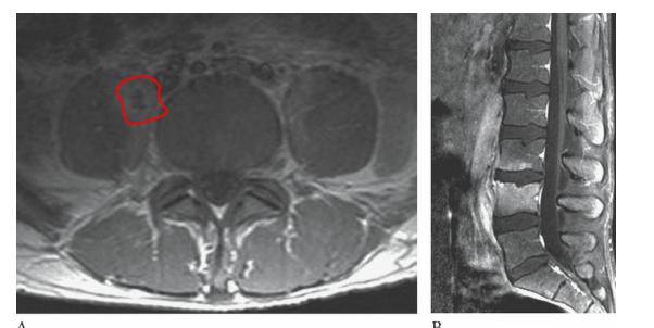
osteomyletis
Discitis
an infection in the intervertebral disc space, usually from a bacterial or viral infection. Discitis can lead to inflammation causing swelling of the intervertebral disc space.
Appearance on MRI
•
Reduced disc height due to disc destruction from infection.
•
T2-weighted images show the disc becomes hyperintense (bright) due to edema and inflammation.
•
T1-weighted images show the disc becomes hypointense (dark).
•
Post contrast T1 will show enhancement of the disc and surrounding endplates
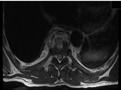
Discitis
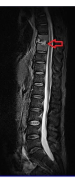
Discitis
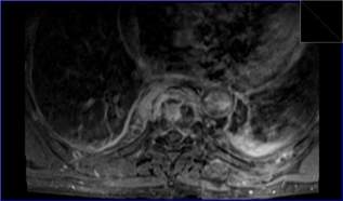
Discitis
Metastatic disease
when a primary cancer spreads. Metastasis can affect any part of the spine, and the thoracic spine is the most common. Common primaries that are known to spread to the spine are breast and lung carcinomas.
Appearance on MRI
MRI may demonstrate spinal cord compression and signal changes to the affected vertebra
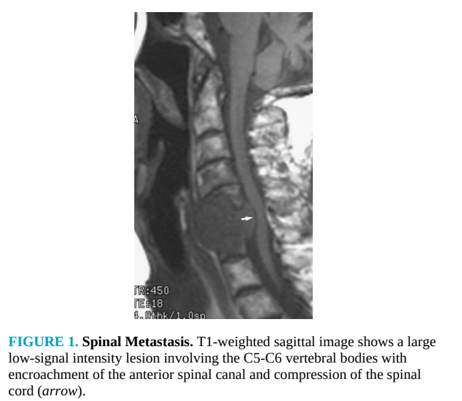
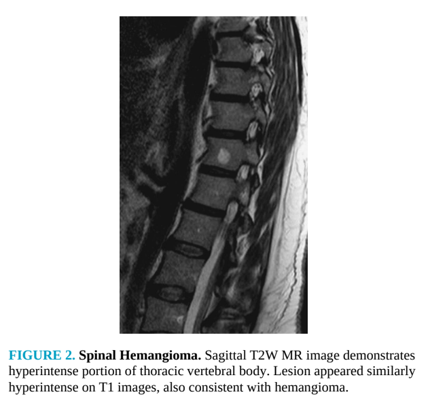
Spinal Hemangioma
the most common benign lesion of the spine. This is often an incidental finding. These are slow growing and rarely symptomatic, but they can create an expansion within the vertebral body.
Appearance on MRI
T1-weighted images will appear hyperintense due to the presence of fat or blood product.
T2-weighted images will appear hyperintense.
Burst Fracture
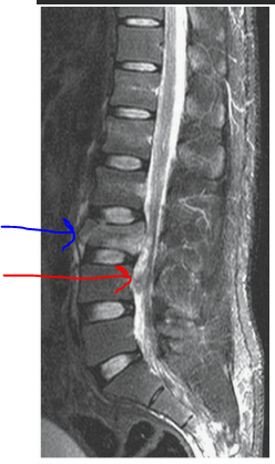
is described as: Imagine the vertebral body bursting like a water balloon.
burst fracture
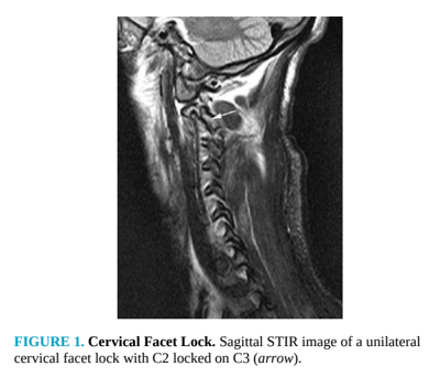
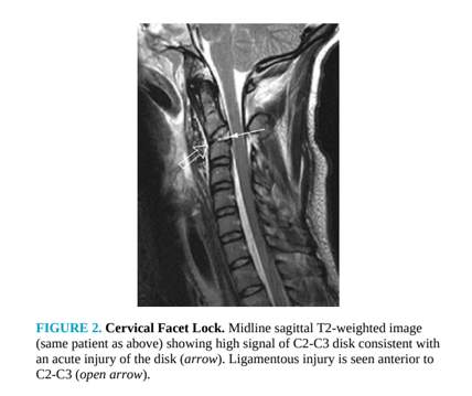
Cervical Facet Lock
occurs when a zygapophyseal or facet joint becomes dislocated. This occurs from a flexion or rotation type of injury.
compression fracture
usually the result of axial loading, and these fractures are common in geriatric patients with osteoporosis. T2 with fat suppression or STIR helps differentiate edema from normal bone marrow signal intensity
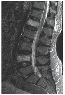
compression fracture at l1 and l4
Spinal subluxation
the dislocation of vertebrae as a result of trauma. MRI is helpful in evaluating ligament involvement or spinal cord injury due to subluxation
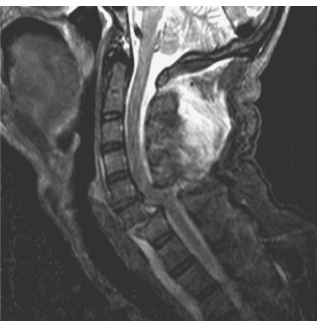
spinal subluxation of c6-c7 ligamentous injury and spinal card hemmorrhage