Brain and Behaviour - Chapter 3: Anatomy and Research Methods
 Call Kai
 Learn
Learn Practice Test
 Spaced Repetition
 Match
Match Flashcards
 Knowt Play
1/113

Earn XP
Description and Tags
Vocabulary practice flashcards covering nervous system terminology, spinal cord and brain structures, cerebral cortex lobes, and neuroimaging research methods.
Name | Mastery | Learn | Test | Matching | Spaced | Call with Kai | Chat |
|---|
No analytics yet
Send a link to your students to track their progress
114 Terms
The thing about the nervous system’s operations
Brain areas and neurons have specialized functions, but they perform those roles by connections with other areas
i.e Like how in society each person has a specialty (e.g. teacher, farmer, or nurse) but no one performs any function without the cooperation of many others
Central Nervous System (CNS)
The major division of the nervous system consisting of the brain and the spinal cord.
Peripheral Nervous System (PNS)
The division of the nervous system that connects the brain and spinal cord to the rest of the body.
Somatic Nervous System
A part of the peripheral nervous system that controls voluntary muscles and conveys sensory information to the central nervous system.
Autonomic Nervous System
A part of the peripheral nervous system that sends and receives messages to regulate the automatic behaviours of the body,
E.g. heart rate, blood pressure, respiration, and digestion.
Enteric nervous system
The set of neurons that control digestion and the digestive system
Dorsal
Anatomical direction meaning toward the back, away from the ventral (stomach) side
Dorsal (con’t)
The top of the human brain is considered dorsal.
Ventral
Anatomical direction meaning toward the stomach, away from the dorsal (back) side.
Anterior
Anatomical direction meaning toward the front end.
Posterior
Anatomical direction meaning toward the rear end.
Superior
Above another part
Inferior
Below another part
Medial
Toward the midline, away from the side
Proximal
Located close (approximate) to the point of origin or attachment
Distal
Located farther from the point of origin or attachment
Ipsilateral
On the same side of the body
e.g. Two parts on the left or two on the right
Contralateral
On the opposite side of the body
e.g. One on the left and one on the right
Coronal Plane (frontal plane)
An anatomical plane that shows brain structures as seen from the front.
Sagittal Plane
An anatomical plane that shows brain structures as seen from the side.

Horizontal Plane (transverse plane)
An anatomical plane that shows brain structures as seen from above.
Spinal cord
A part of the central nervous system that communicates with all the sense organs and muscles except those of the head
Spinal cord (con’t)
It is a segmented structure, and each segment has on both its left and right sides a sensory nerve and a motor nerve
Spinal cord (III)
Each segment of the spinal cord sends sensory information to the brain and receives motor commands from the brain
Column
A set of cells perpendicular to the surface of the cortex and laminae, sharing similar properties.
e.g. If one cell in a column responds to touch on the palm of the left hand, then the other cells in that column do

Tract (projection)
A set of axons within the central nervous system
Nerve
A set of axons in the periphery, extending either from the CNS to a muscle or gland or from a sensory organ to the CNS.
Nucleus
A cluster of neuron cell bodies located within the central nervous system.
Ganglion
A cluster of neuron cell bodies, usually located outside the central nervous system.
Gyrus
A protuberance or bump on the surface of the brain.
Sulcus
A fold or groove that separates one gyrus from another on the brain surface.
Dorsal Roots (axon bundles)
Structures entering the spinal cord that carry sensory information from the peripheral sense organs.
Dorsal Roots (axon bundles) [con’t]
The dorsal root on each side conveys sensory information to the spinal cord
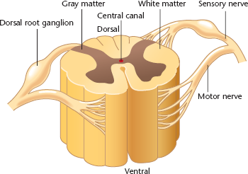
Ventral Roots
Structures exiting the spinal cord that carry motor commands to muscles and glands.
Dorsal Root Ganglia
Clusters of sensory neuron cell bodies located outside the spinal cord.
Gray Matter
Tissue located in the center of the spinal cord that’s densely packed with cell bodies and dendrites.
White Matter
Tissue composed mostly of myelinated axons

White Matter (con’t)
Carries information from the gray matter to the brain or other areas of the spinal cord.
Thoracic and lumbar areas
Chains of ganglia just to the left and right of the spinal cord’s central regions
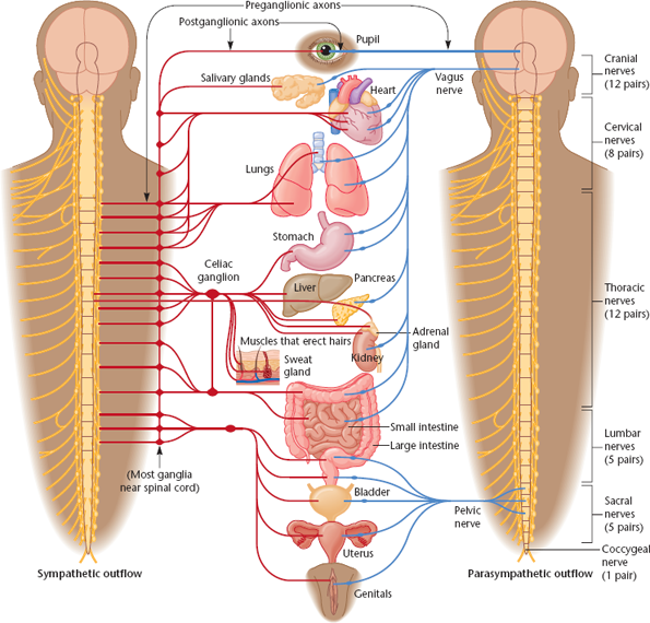
Sympathetic Nervous System
A network of unified, autonomic nerves that prepares organs for rigorous activity (the 'fight or flight' response)
e.g. By increasing breathing and heart rate and decreasing digestive activity.
Sympathetic Nervous System (con’t)
Uses norepinephrine as its primary postganglionic neurotransmitter
Systems exclusive to the sympathetic nervous system
Sweat glands
Adrenal glands
Muscles that constrict blood vessels
Muscles that erect the hairs of the skin
Parasympathetic Nervous System (Craniosacral system)
A network of autonomic, unconnected nerves that facilitates vegetative and non-emergency responses during relaxed states
e.g. Rest and digest, sexual arousal
Parasympathetic Nervous System (Craniosacral system) [con’t]
Primarily releases acetylcholine from postganglionic axons.
Parasympathetic Nervous System (rest and digest system) [con’t]
Is the opposite of the sympathetic nervous system and its activities
e.g. The sympathetic nervous system increases heart rate, and the parasympathetic nervous system decreases it.
Forebrain (prosencephalon)
The most anterior and prominent part of the mammalian brain, consisting of the diencephalon and telencephalon.
Amygdala
A temporal lobe structure important for evaluating emotional information
Midbrain (mesencephalon)
The middle division of the brain containing the tectum, tegmentum, superior colliculus, inferior colliculus, and substantia nigra.
Midbrain (mesencephalon) [con’t]
Is between the hindbrain and the forebrain
Tectum
The midbrain’s roof
Superior colliculus
The swelling on either side of the tectum that contributes to visual processing
Inferior colliculus
Swelling on the surface of the tectum that contributes to hearing
Substantia Nigra
A midbrain structure that gives rise to a dopamine-containing pathway facilitating readiness for movement.
Tegmentum
The intermediate level of the midbrain
Hindbrain (rhombencephalon;)
The posterior portion of the brain that’s composed of the:
Medulla
Pons
Cerebellum
Brainstem
The medulla, pons, midbrain, and central structure of the forebrain

Medulla (medulla oblongata)
A hindbrain structure and extension of the spinal cord that controls vital reflexes
E.g. Breathing, heart rate, vomiting, salivation, coughing, and sneezing.

Cranial Nerves
12 pairs of nerves that allow the medulla to control head sensation and muscle movement, as well as many parasympathetic outputs.
Pons
A hindbrain structure lying ventral and anterior to the medulla, where axons cross between the two brain hemispheres and opposite sides of the body.
Cerebellum
A highly folded hindbrain structure that regulates:
Timing
Motor movement
Balance
Coordinations
Shifting attention between auditory and visual stimuli.
Cerebellum (con’t)
It’s one of the first brain areas to start developing and one of the last to complete development

Limbic System
An interlinked set of forebrain structures surrounding the brainstem (including the olfactory bulb, hypothalamus, hippocampus, amygdala, and cingulate gyrus) involved in motivation and emotion.
Thalamus
A subcortical, central forebrain structure that acts as a sensory relay station and serves as the main source of sensory input to the cerebral cortex for all senses (except olfaction).
Hypothalamus
A small subcortical forebrain region ventral to the thalamus near the base that regulates motivated behaviours, and signals the pituitary gland to release hormones.
E.g. Eating, drinking, sexual activity

Basal Ganglia
Subcortical structures comprising the:
Caudate nucleus, putamen, and globus pallidus
Are essential for planning motor movement, learning habits/skills, and aspects of memory.
Nucleus Basalis
A ventral basal forebrain structure receiving input from the hypothalamus and basal ganglia that releases acetylcholine to the cerebral cortex
Nucleus Basalis (con’t)
Promotes arousal, wakefulness, and attention.
Hippocampus
A large forebrain structure situated between the thalamus and cerebral cortex that is critical for storing memories of individual events.
Ventricles
Four fluid-filled cavities within the brain that are all connected

Cerebrospinal Fluid (CSF)
A clear fluid that fills the central canal and ventricles of the brain, cushioning the CNS and providing nutrition and hormones.
Cerebrospinal Fluid (CSF) [con’t]
Can be accessed through the choroid plexus
Meninges
Membranes containing pain receptors that surround the brain and spinal cord
Meninges (con’t)
Inflammation causes meningitis and swollen meningeal blood vessels cause migraines.
Cerebral cortex
Layers of cells on the outer surface of the cerebral hemisphere of the forebrain
Cerebral cortex (con’t)
The cells on the outer surface of the cerebral cortex are gray matter, and their axons extending inward are white matter
Corpus callosum & Anterior commissure
Bundles of axons that connect the two hemispheres of the cerebral cortex
Laminae
Six distinct layers of cell bodies that are parallel to the surface of the cerebral cortex and separated from each other by layers of fibres

Occipital Lobe
The posterior region of the cerebral cortex containing the primary visual cortex (striate cortex) specialized for processing visual input.
Occipital Lobe (con’t)
The eyes provide the stimulus, and the visual cortex provides the experience
Parietal Lobe
The region of the cerebral cortex containing the postcentral gyrus (primary somatosensory cortex), responsible for touch sensations, body positioning, and spatial/numerical information.
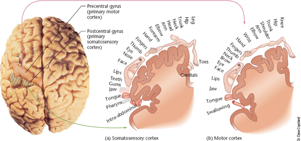
Parietal Lobe (con’t)
Lies between the occipital lobe and the central sulcus (one of the deepest grooves in the surface of the cerebral cortex)
The four bands of cells parallel to the central sulcus
2 of the bands receive mostly light-touch information
1 receives deep-pressure information
1 receives a combination of both
Temporal Lobe
The lateral portion of the cortical region near the temples that processes:
Auditory information
Spoken language
Complex visual movement and recognition
Aspects of emotion and motivation
Klüver-Bucy Syndrome
A condition characterized by visual agnosia and amnesia resulting from damage to the temporal lobe.
Temporoparietal junction.
The area where the parietal lobe and the temporal lobe meet
Temporoparietal junction (con’t)
Handles:
Attention
Body awareness
Social cognition
Temporoparietal junction (III)
It’s conveniently located to receive input from vision, hearing, and body senses
Frontal Lobe
A section of cerebral cortex that extends from the central sulcus to the anterior limit of the brain
Frontal Lobe (con’t)
Contains the precentral gyrus (primary motor cortex) in the back for fine motor control and the prefrontal cortex for cognitive integration.
e.g. Moving a finger
Prefrontal Cortex
The most anterior portion of the frontal lobe, responsible for higher functions
e.g. Abstract thinking, decision-making, planning, working memory, and evaluating potential outcomes.
Prefrontal Cortex (con’t)
Species with a larger cerebral cortex devote a larger percentage of it to the prefrontal cortex
e.g. It occupies almost twice as high a percentage of the human brain as it does of a monkey brain
The three major regions of the prefrontal cortex
The posterior portion
The middle zone
The anterior zone
Default network
The areas that dominate activity when you don’t need to concentrate on anything in particular
e.g. When daydreaming, or thinking as a whole

Prefrontal Lobotomy
A surgical procedure involving disconnection of the prefrontal cortex from the rest of the brain, historically leading to apathy, planning deficits, memory loss, and blunted emotions.
Binding Problem (large-scale integration problem)
The question of how distinct visual, auditory, and sensory processing areas in the brain combine activity to produce the perception of a unified object.
How does binding occur?
It occurs if you perceive two sensations as happening at the same time at approximately the same place.
e.g. When a skilled ventriloquist says something and makes the dummy’s mouth move at the same time, you perceive the sound as coming from the dummy
Ablation
The surgical removal of a specific brain area, usually with a knife
Lesion
Controlled damage to a brain area, commonly performed in laboratory research with specialized equipment

Stereotaxic Instrument
A laboratory device used to precisely target, create lesions, or insert electrodes into deep internal structures of the brain.
Gene-knockout approach
Directs a mutation to a gene that regulates one type of cell, transmitter, or receptor