Thoracic Anatomy: Compartments, Structures, and Clinical Relevance
 Call Kai
 Learn
Learn Practice Test
 Spaced Repetition
 Match
Match Flashcards
 Knowt Play
1/332

There's no tags or description
Looks like no tags are added yet.
Name | Mastery | Learn | Test | Matching | Spaced | Call with Kai | Chat |
|---|
No analytics yet
Send a link to your students to track their progress
333 Terms
What are the three major compartments of the thoracic cavity?
The left pleural cavity, right pleural cavity, and the mediastinum.
What structure closes the inferior thoracic aperture?
The diaphragm.

What is the shape of the thorax?
An irregularly shaped cylinder.
It acts as a conduit for structures passing through the thorax and connects organs in the thorax to other body regions.
Function of the mediastinum
What major structures are contained within the mediastinum?
The heart, esophagus, trachea, major nerves, and major systemic blood vessels.
How are the pleural cavities separated?
by the mediastinum.
The apex of each lung extends into _____.
the root of the neck.
What is the primary function of the thorax?
Breathing.
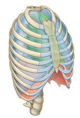
What musculoskeletal elements make up the thoracic wall?
Twelve thoracic vertebrae, ribs, 3 layers of muscles, and the sternum.
It alters the volume of the thoracic cavity to facilitate air movement into and out of the lungs.
Function of diaphragm in breathing
The joint where the manubrium of the sternum meets the body of the sternum, forming the sternal angle.
Manubriosternal joint
What are floating ribs?
Ribs XI and XII, which do not articulate with other ribs or the sternum.
The structure formed by the distal cartilaginous ends of ribs VII to X.
Costal margin
What is the anatomical position of the superior thoracic aperture?
It is open and allows continuity with the neck.
What structures pass through the mediastinum?
The esophagus, vagus nerves, and thoracic duct.
What is the anterior component of the thoracic wall?
The sternum, consisting of the manubrium, body, and xiphoid process.
All ribs articulate with ____, with most (____) having ____ articulations.
thoracic vertebrae posteriorly; ribs II to IX; three
What is the significance of the diaphragm's upward domed shape?
offers protection to some abdominal viscera.
What is the relationship between the thoracic wall and respiratory muscles?
The thoracic wall provides extensive attachment sites for muscles that assist in breathing.

Which structures form the margin of the inferior thoracic aperture?
Bone, cartilage, and ligaments
What is the anatomical significance of rib I?
It articulates with the manubrium and is involved in the structure of the superior thoracic aperture.
What is the role of costal cartilage?
It contributes to the mobility and elasticity of the thoracic wall.
What is the position of the first ribs relative to the diaphragm?
They slope inferiorly from their posterior articulation with vertebra TI to their anterior attachment to the manubrium.
What is the relationship between the thoracic cavity and the neck?
Structures that pass between the neck and thorax go through the superior thoracic aperture.
What is the significance of the intervertebral disc between vertebrae TII and TIII?
It is in the same horizontal plane as the superior margin of the manubrium.
What happens to the thoracic cavity during breathing?
The volume changes due to the movements of the diaphragm and ribs.

What anatomical feature is used by clinicians during physical examinations of the thorax?
The sternal angle formed by the manubrium and body of the sternum.
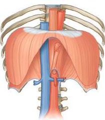
What is the diaphragm's shape?
The diaphragm is not flat; it 'balloons' superiorly, forming domes.
Which dome of the diaphragm is higher?
The right dome is higher than the left, reaching as far as rib V.
What structures penetrate the diaphragm?
The esophagus and inferior vena cava penetrate the diaphragm; the aorta passes posterior to it.
What is the role of the diaphragm during contraction?
As the diaphragm contracts, the height of the domes decreases and the volume of the thorax increases.
What separates the thorax from the abdomen?
The diaphragm separates the thorax from the abdomen.
A thick midline partition extending from the sternum to the thoracic vertebrae.
The mediastinum.
What are the two types of pleura?
The parietal pleura lines the thoracic cavity, while the visceral pleura covers the lungs.
What is the costodiaphragmatic recess?
The costodiaphragmatic recess is the largest recess located between the thoracic wall and diaphragm.
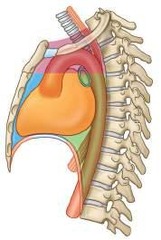
What divides the mediastinum into superior and inferior parts?
A horizontal plane passing through the sternal angle and the intervertebral disc between vertebrae TIV and TV.
What is contained in the middle mediastinum?
The pericardium and heart constitute the middle mediastinum.
What is the significance of the axillary inlet?
The axillary inlet serves as a gateway to the upper limb and is formed by rib I, the clavicle, and the scapula.
At which vertebral level does the inferior vena cava pierce the diaphragm?
The inferior vena cava pierces the diaphragm at vertebral level TVIII.
At which vertebral level does the esophagus penetrate the diaphragm?
The esophagus penetrates the diaphragm at vertebral level TX.
What is the role of the brachial plexus in relation to the thorax?
The proximal parts of the brachial plexus pass between the neck and upper limb through the axillary inlet.
What is the anatomical relationship of the trachea and esophagus in the thorax?
The trachea lies immediately anterior to the esophagus in the midline.
What structures pass through or posterior to the diaphragm?
The inferior vena cava, esophagus, and aorta pass through or posterior to the diaphragm.
What is the function of the pleural cavities?
The pleural cavities accommodate changes in lung volume during breathing.
What is the significance of the sternal angle?
The sternal angle marks the horizontal plane that separates the superior and inferior mediastinum.
What are the anterior and posterior mediastinum?
The anterior mediastinum lies between the sternum and pericardium; the posterior mediastinum lies between the pericardium and thoracic vertebrae.
What is the role of lymphatic vessels in the breast?
Lymphatic vessels from the medial part of the breast drain into the parasternal nodes.
What arteries supply the breast?
Branches from the internal thoracic arteries and veins supply the breast.
What is the anatomical significance of vertebral levels in thoracic anatomy?
Vertebral levels help determine the position of important anatomical structures within body regions.
What is the apex of each triangular axillary inlet formed by?
The apex is formed by the medial margin of the coracoid process.
What is the relationship between the thorax and the neck?
The superior thoracic aperture opens directly into the root of the neck.
What is the significance of the costal margins?
The costal margins are formed by the distal cartilaginous ends of ribs VII to X.
What is the aortic hiatus?
The aortic hiatus is where the aorta passes posterior to the diaphragm at vertebral level TXII.
What is the role of the internal thoracic artery?
The internal thoracic artery supplies blood to the anterior thoracic wall and the breast.
What major veins drain into the right side of the heart?
The superior and inferior venae cavae.
What separates the superior mediastinum from the inferior mediastinum?
The sternal angle.
Where does the left brachiocephalic vein deliver blood?
It delivers blood from the left side of the head and neck, the left upper limb, and part of the left thoracic wall into the superior vena cava.
What anatomical landmark marks the beginning and end of the aortic arch?
The sternal angle.
What veins drain the posterior and lateral parts of the left thoracic wall?
The hemiazygos and accessory hemiazygos veins.
What does the trachea bifurcate into?
Right and left main bronchi.
What is the role of the internal thoracic arteries?
They supply the anterior thoracic wall.
From where do the arteries supplying the thoracic wall arise?
From the thoracic aorta and the subclavian arteries.
What is the highest thoracic dermatome on the anterior chest wall?
T2.
What spinal nerves carry preganglionic sympathetic fibers?
Spinal nerves T1 to L2.
How does rib elevation affect the thoracic wall?
It moves the anterior thoracic wall forward and expands the thoracic wall laterally.
What nerves innervate the diaphragm?
The phrenic nerves originating from C3, C4, and C5.
What happens to diaphragm movement with spinal cord injuries below the level of the phrenic nerve origin?
Movement of the diaphragm is not affected.
What separates the breast from the deep fascia?
A layer of loose connective tissue known as the retromammary space.
What anatomical structures are included in the cylindrical thorax?
A wall, two pleural cavities, the lungs, and the mediastinum.
What causes pitting of the skin in carcinoma of the breast?
Tension on the ligaments of the breast.
What is the primary component of breasts in nonlactating women?
Fat.
What is more abundant in lactating women compared to nonlactating women?
Glandular tissue.
What is the anterior limit of the breast?
The sternum.
What is the lateral limit of the breast?
The midaxillary line.
What is the vertical extent of the breast?
From ribs II to VI.
What is the primary function of the diaphragm?
To facilitate breathing by changing the volume of the thoracic cavity.
What anatomical feature marks the superior limit of the pulmonary trunk?
The sternal angle.
What is the relationship between intercostal nerves and the thoracic wall?
Intercostal nerves innervate the thoracic wall and related parietal pleura.
What is the anatomical significance of the thoracic sympathetic trunk?
It carries preganglionic sympathetic fibers from the spinal cord.
What happens to the thoracic cavity during inspiration?
The diaphragm descends, increasing thoracic capacity.
What structures are housed in the thorax?
The heart and lungs.
What is the role of the thoracic wall?
It protects the heart and lungs and supports the upper limbs.
Which arteries supply the pectoral region laterally?
Vessels from the axillary artery: superior thoracic, thoraco-acromial, lateral thoracic, and subscapular arteries.
What arteries supply the pectoral region medially?
Branches from the internal thoracic artery.
What is the function of the second to fourth intercostal arteries?
They provide blood supply via branches that perforate the thoracic wall and overlying muscle.
How is the breast innervated?
Via anterior and lateral cutaneous branches of the second to sixth intercostal nerves; the nipple is innervated by the fourth intercostal nerve.
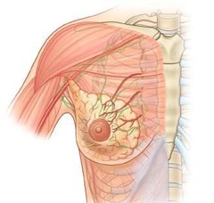
What percentage of lymphatic drainage from the breast goes to axillary nodes?
Approximately 75%.
What are the mammary glands classified as?
Modified sweat glands.
What is the function of the suspensory ligaments of the breast?
They support the breast and are continuous with the dermis of the skin.
What is the axillary tail of the breast?
A projection of the upper lateral region of the breast around the lateral margin of the pectoralis major muscle into the axilla.
What is a common malignancy in women?
Breast cancer.
What are the sites of origin for breast cancer?
Cells of the acini, lactiferous ducts, and lobules of the breast.
What is the first step in diagnosing breast cancer?
A biopsy and histological evaluation.
What does staging a tumor involve?
Defining the size, exact site, number and sites of lymph node spread, and organs to which the tumor may have spread.
What imaging technique is used to check for pulmonary metastases?
Computed tomography (CT) scanning.
What is a mastectomy?
Surgical removal of the breast tissue.
What can happen if the long thoracic nerve is damaged during a mastectomy?
It can result in paralysis of the serratus anterior muscle, producing a winged scapula.
What are the primary muscles of the pectoral region?
Pectoralis major, pectoralis minor, and subclavius.
Where do the pectoralis major and minor muscles originate?
From the anterior thoracic wall.
What is the retromammary space?
The loose connective tissue space separating the breast from the pectoralis major muscle.