antr 350 CNS, PNS, and nerves
 Call Kai
 Learn
Learn Practice Test
 Spaced Repetition
 Match
Match Flashcards
 Knowt Play
1/85

There's no tags or description
Looks like no tags are added yet.
Name | Mastery | Learn | Test | Matching | Spaced | Call with Kai | Chat |
|---|
No analytics yet
Send a link to your students to track their progress
86 Terms
dermatomes
an area of skin supplied by a single spinal nerve
myotomes
a group of muscles supplied by one spinal nerve
PAR
posterior - axillary, radial
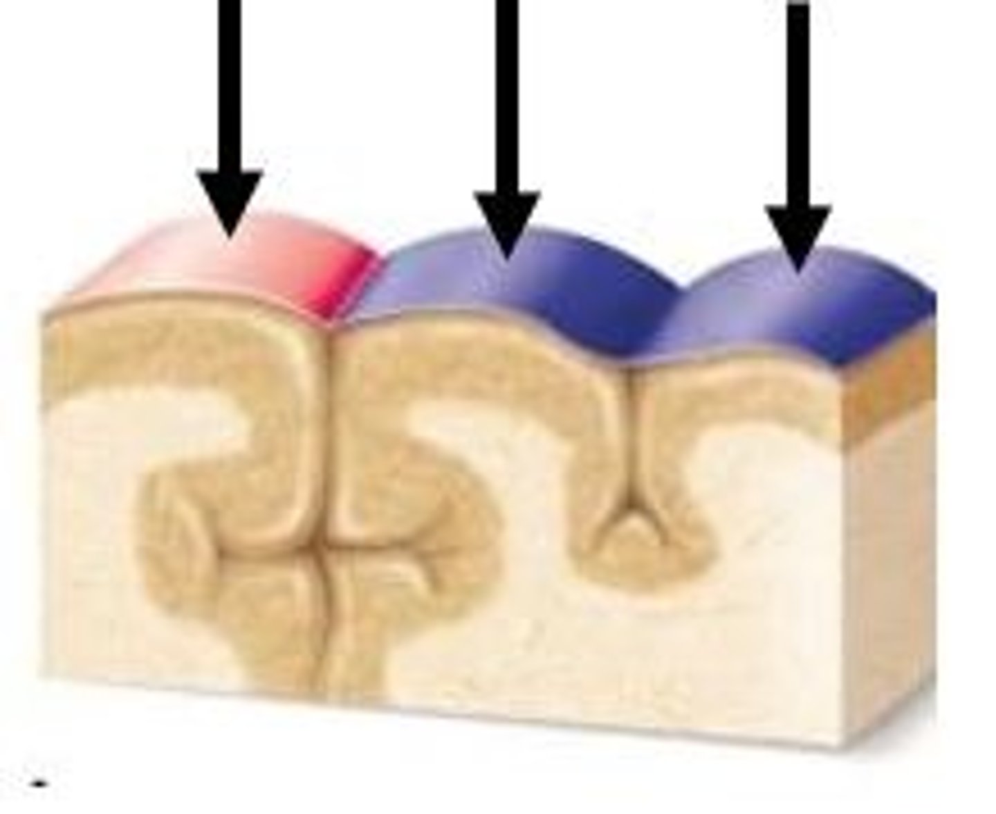
gyrus (gyri)
elevated ridges or hills
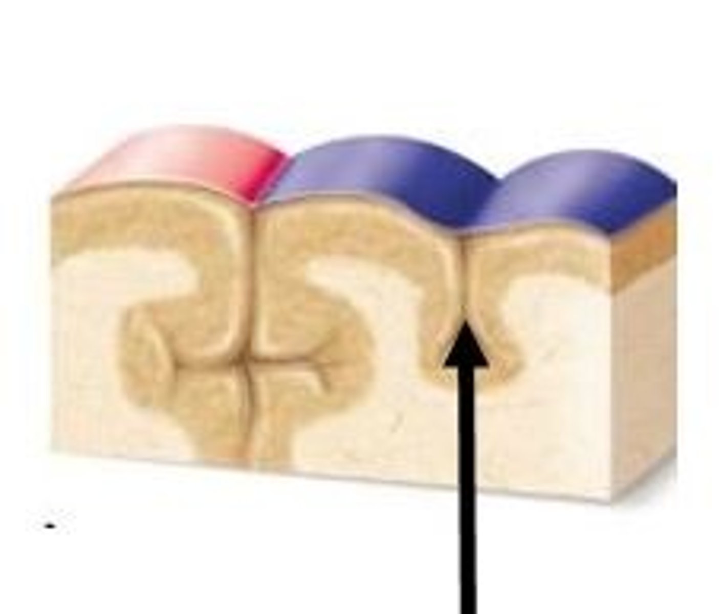
sulcus (sulci)
small grooves or valleys
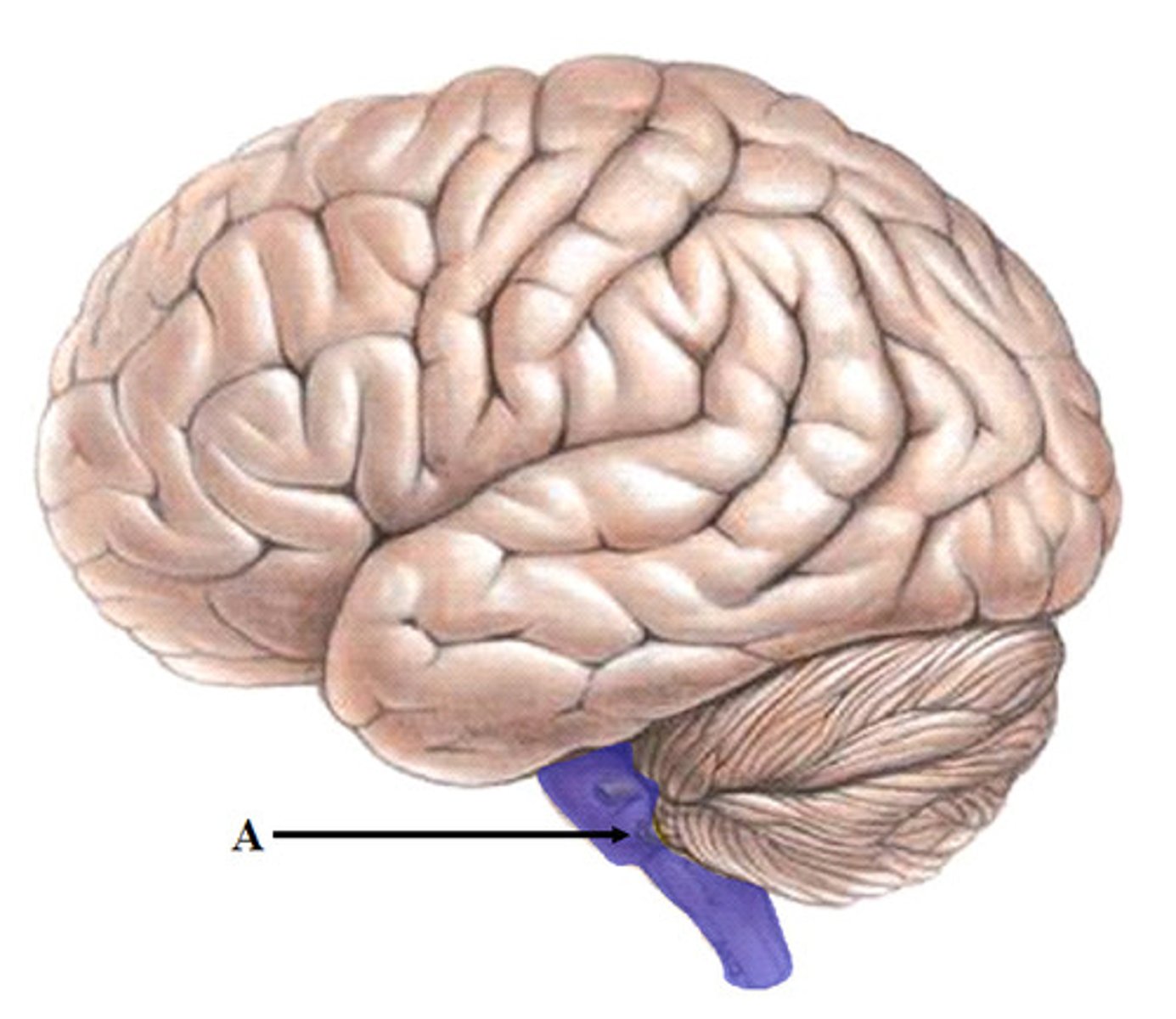
Brainstem
The part of the brain connecting the cerebrum with the spinal cord; controls autonomic functions.
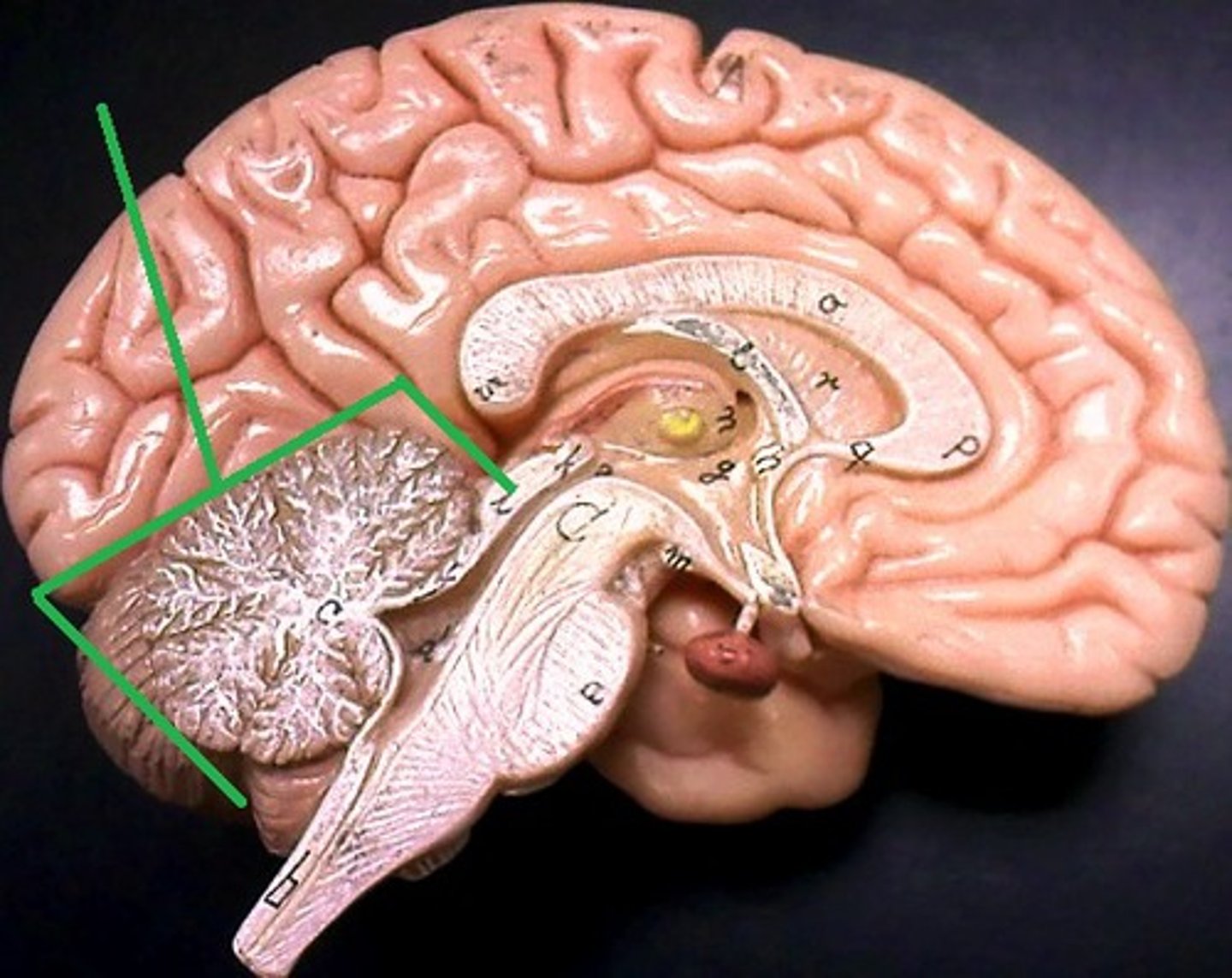
Cerebellum
controlling coordination, balance, and motor skills.
Cerebrum
The largest part of the brain, responsible for voluntary activities, sensory perception, and thought.
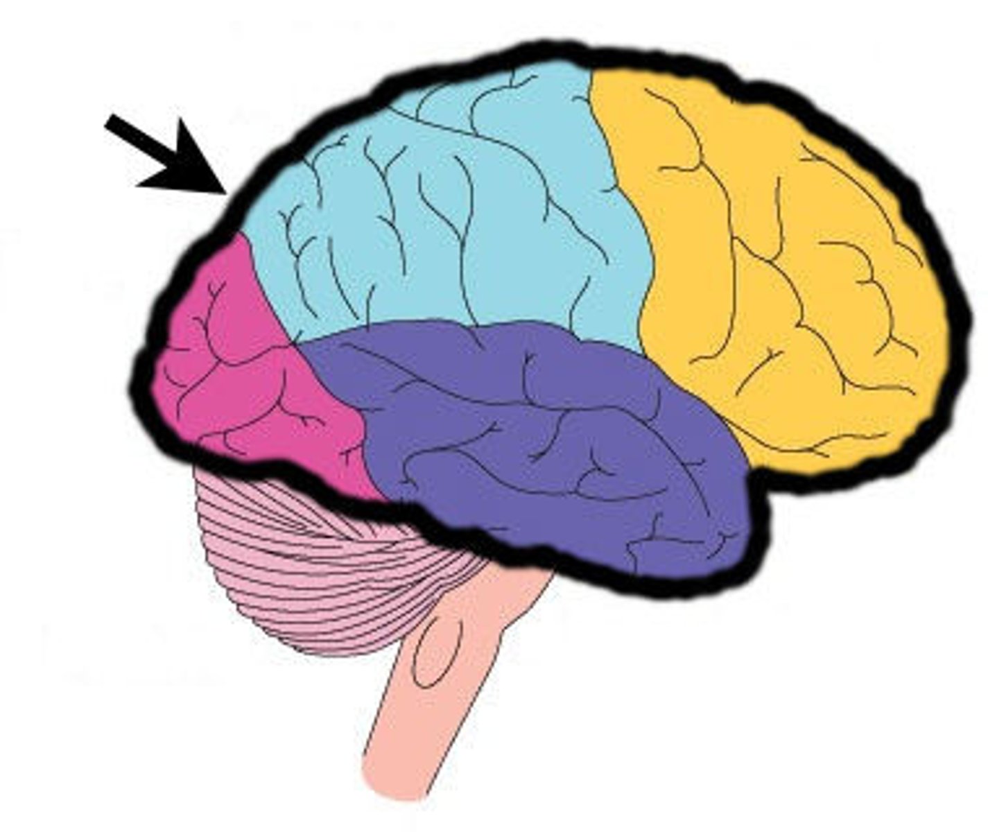
Central sulcus
A prominent groove separating the frontal lobe from the parietal lobe.
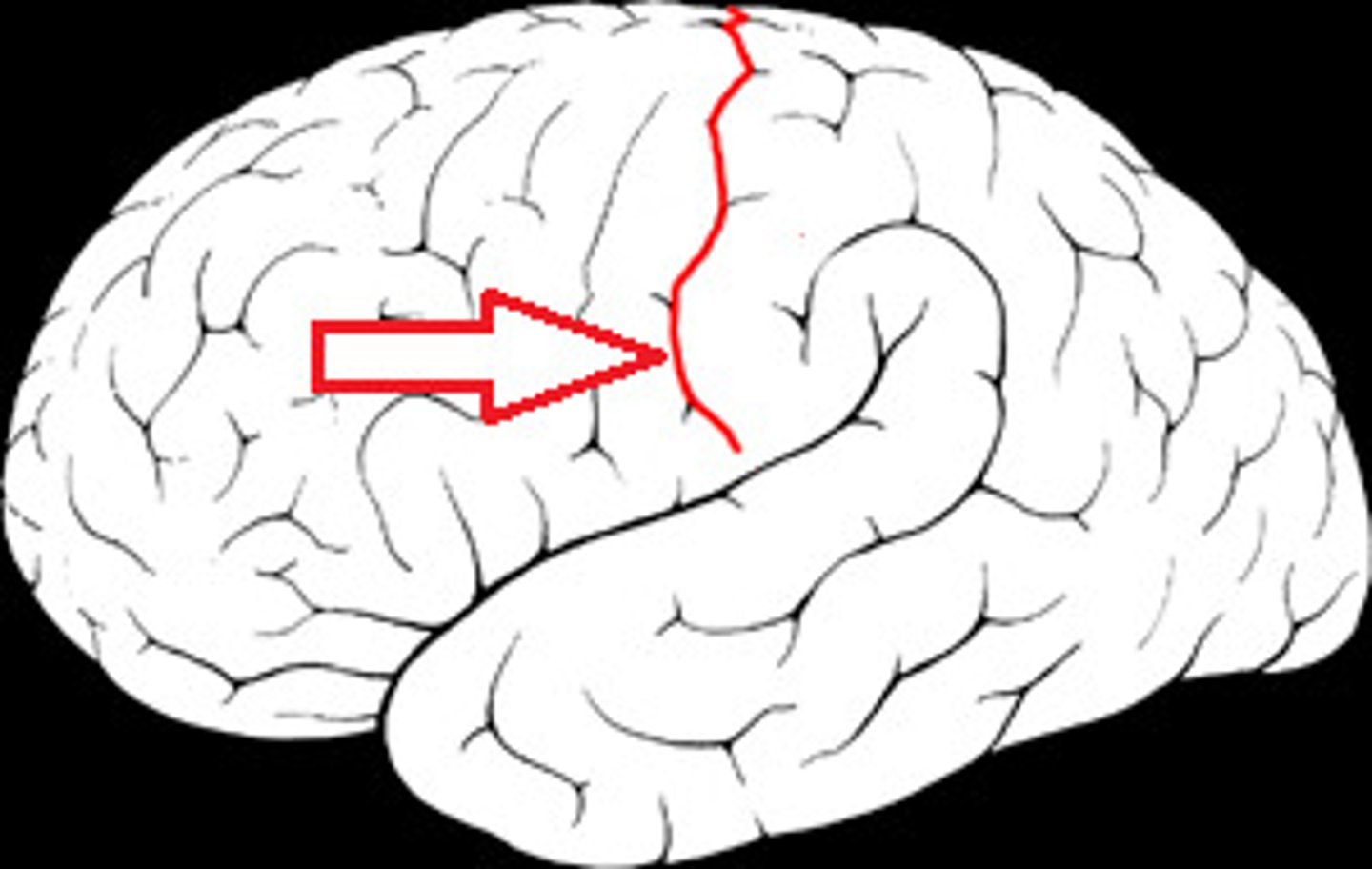
Lateral fissure
A deep groove separating the temporal lobe from the frontal and parietal lobes.
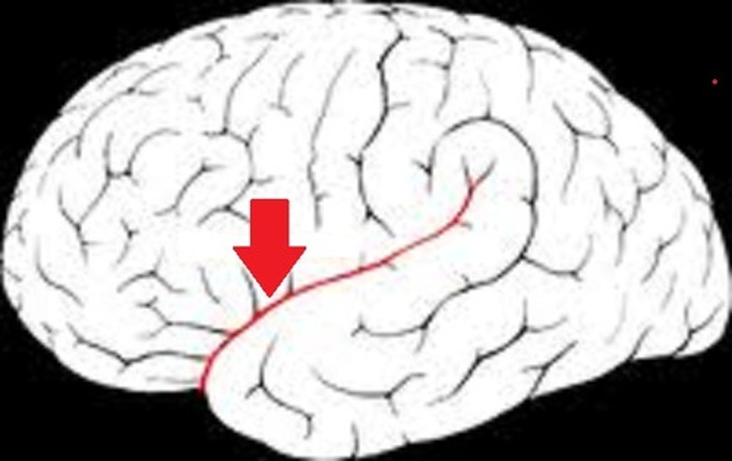
Longitudinal fissure
The deep groove that separates the left and right cerebral hemispheres.
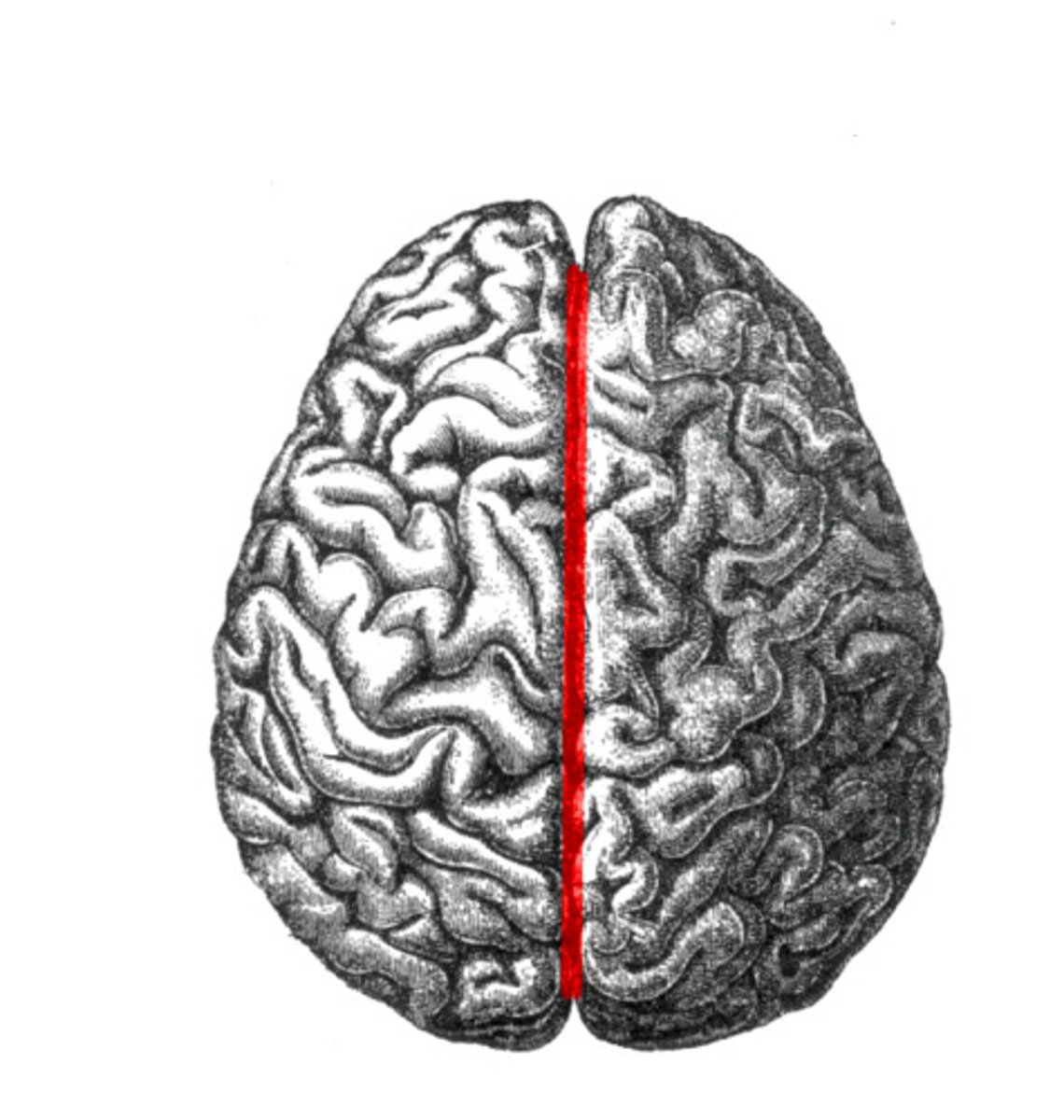
Corpus callosum
A broad band of nerve fibers joining the two hemispheres of the brain.
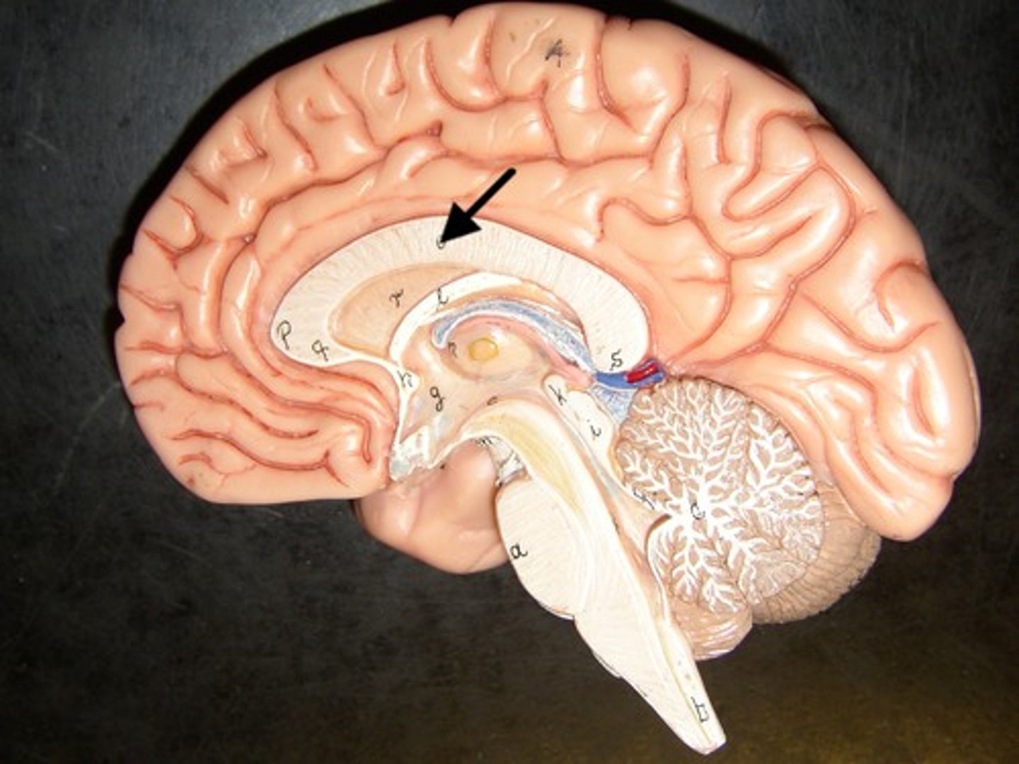
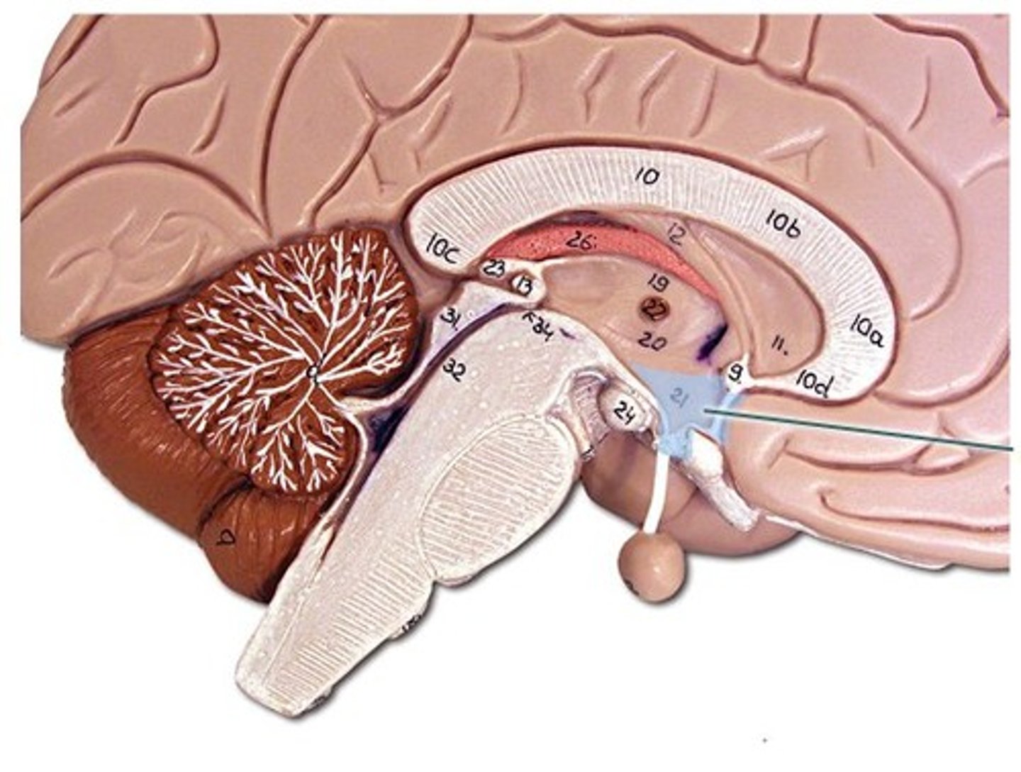
Hypothalamus
Brain region controlling body temperature, hunger, thirst, and the endocrine system.
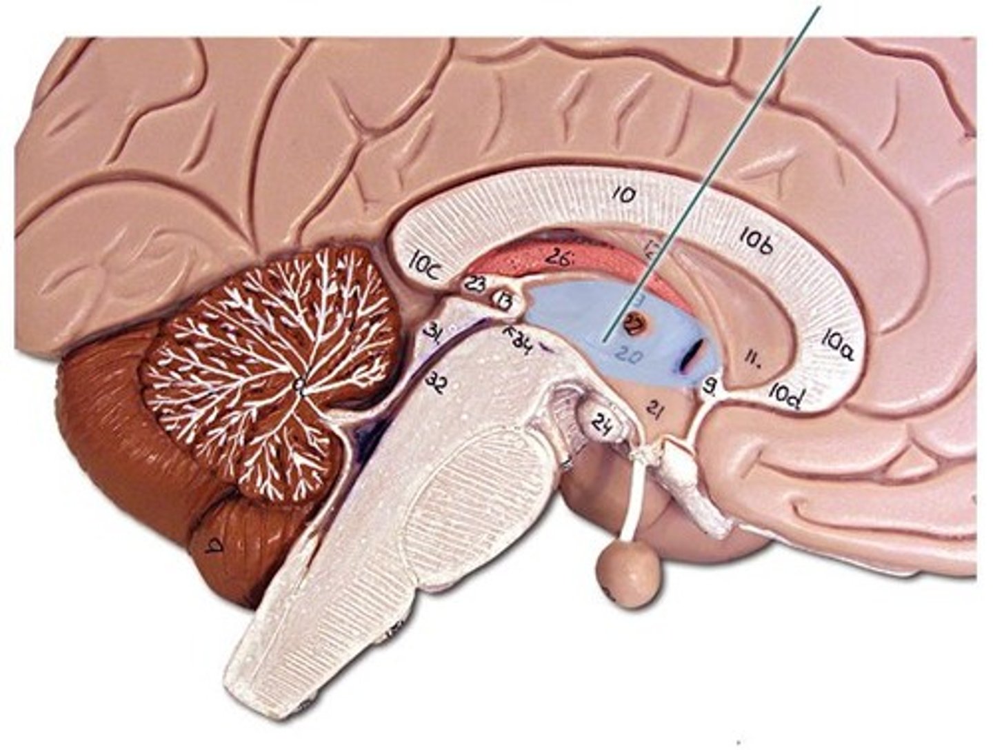
Thalamus
The brain's sensory control center, routing sensory messages to the cortex.
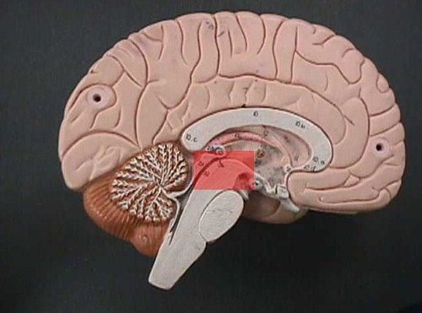
Midbrain
The uppermost part of the brainstem, involved in visual and auditory reflexes.
Medulla oblongata
The lower part of the brainstem, regulating heart rate, breathing, and blood pressure.
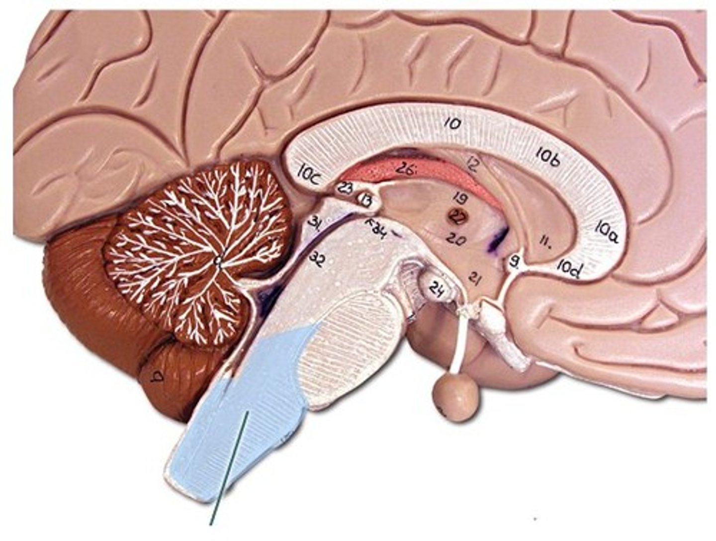
Pons
Part of the brainstem linking the medulla and thalamus, involved in sleep and arousal.
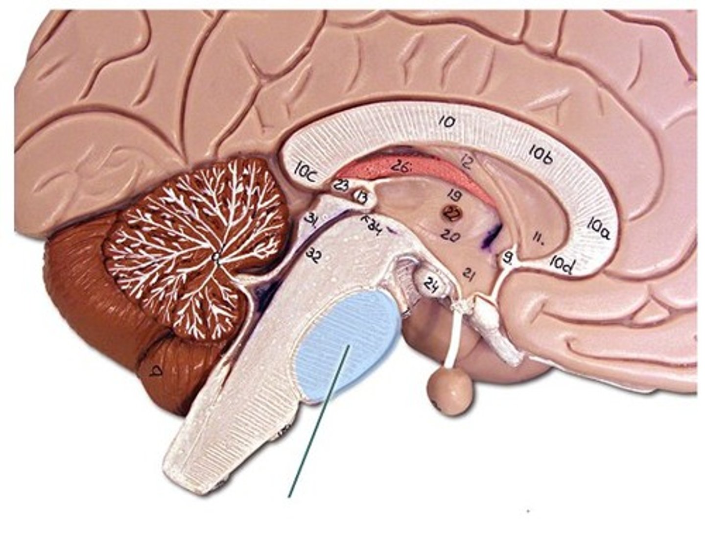
Spinal cord
The bundle of nerve fibers connecting the brain to the rest of the body.
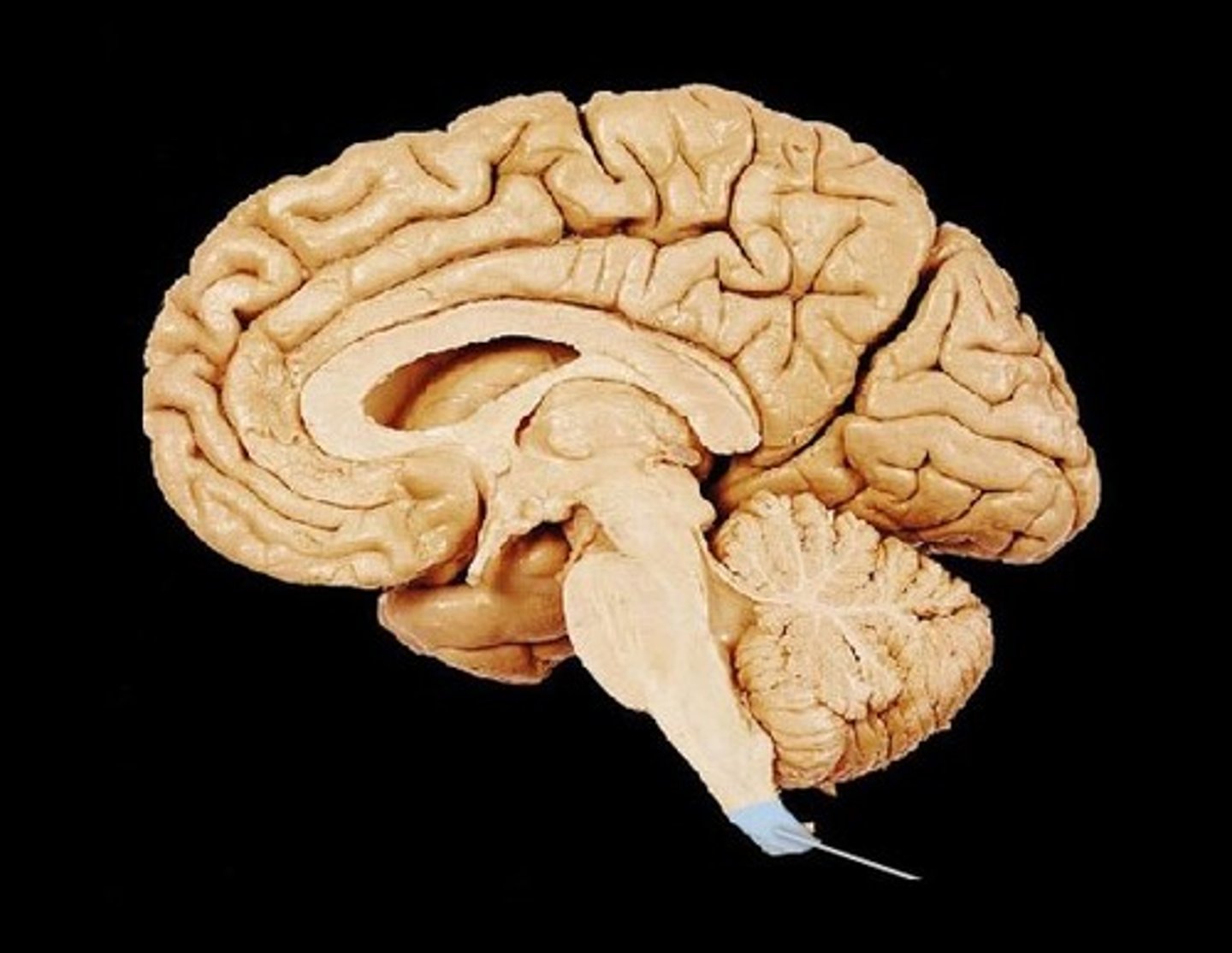
Amygdala
memory and emotion, particularly fear and aggression.
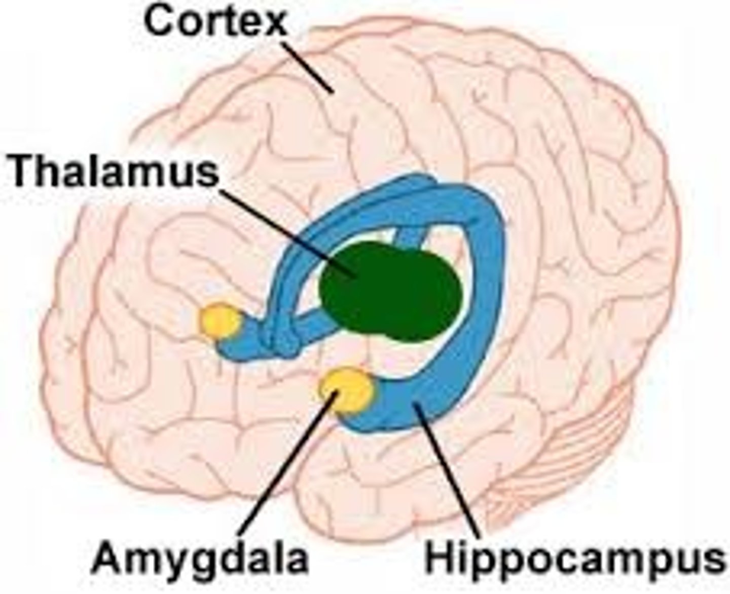
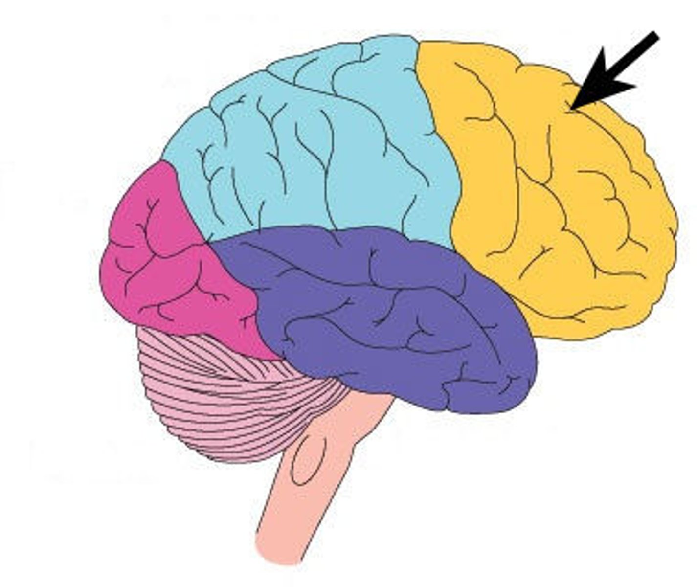
Frontal lobe
Cerebral lobe responsible for voluntary movement, decision-making, and higher-level executive functions.
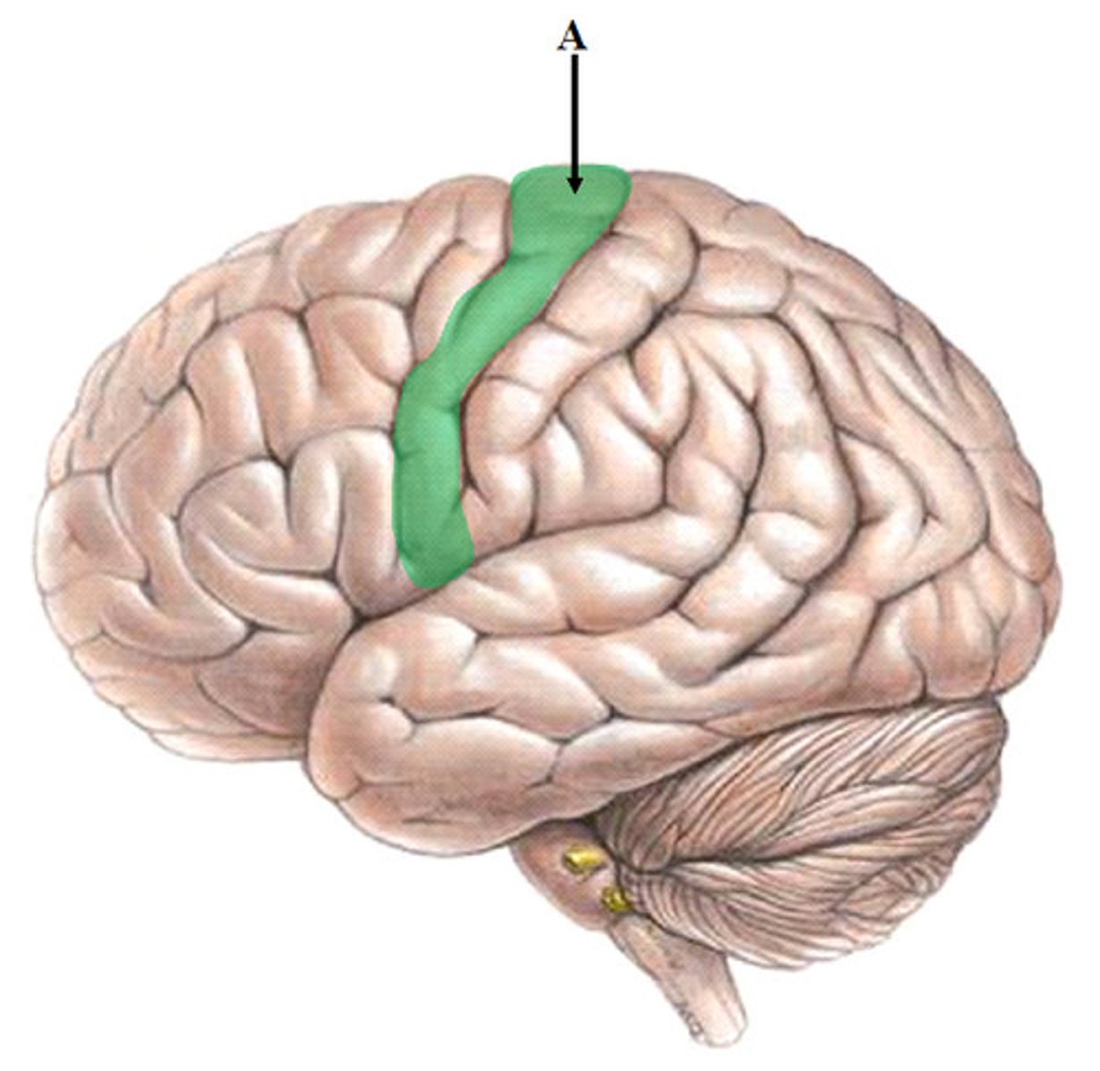
Precentral gyrus
The location of the primary motor cortex, responsible for controlling voluntary movements.
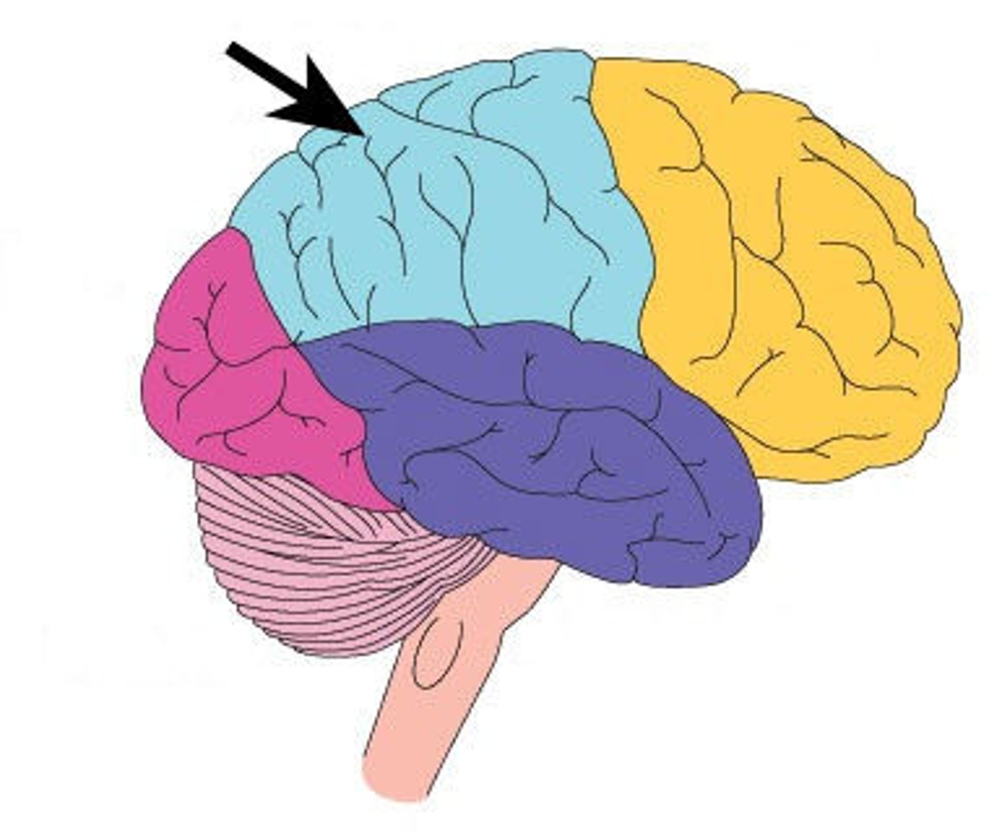
Parietal lobe
Cerebral lobe responsible for processing sensory information like touch, pressure, and temperature.
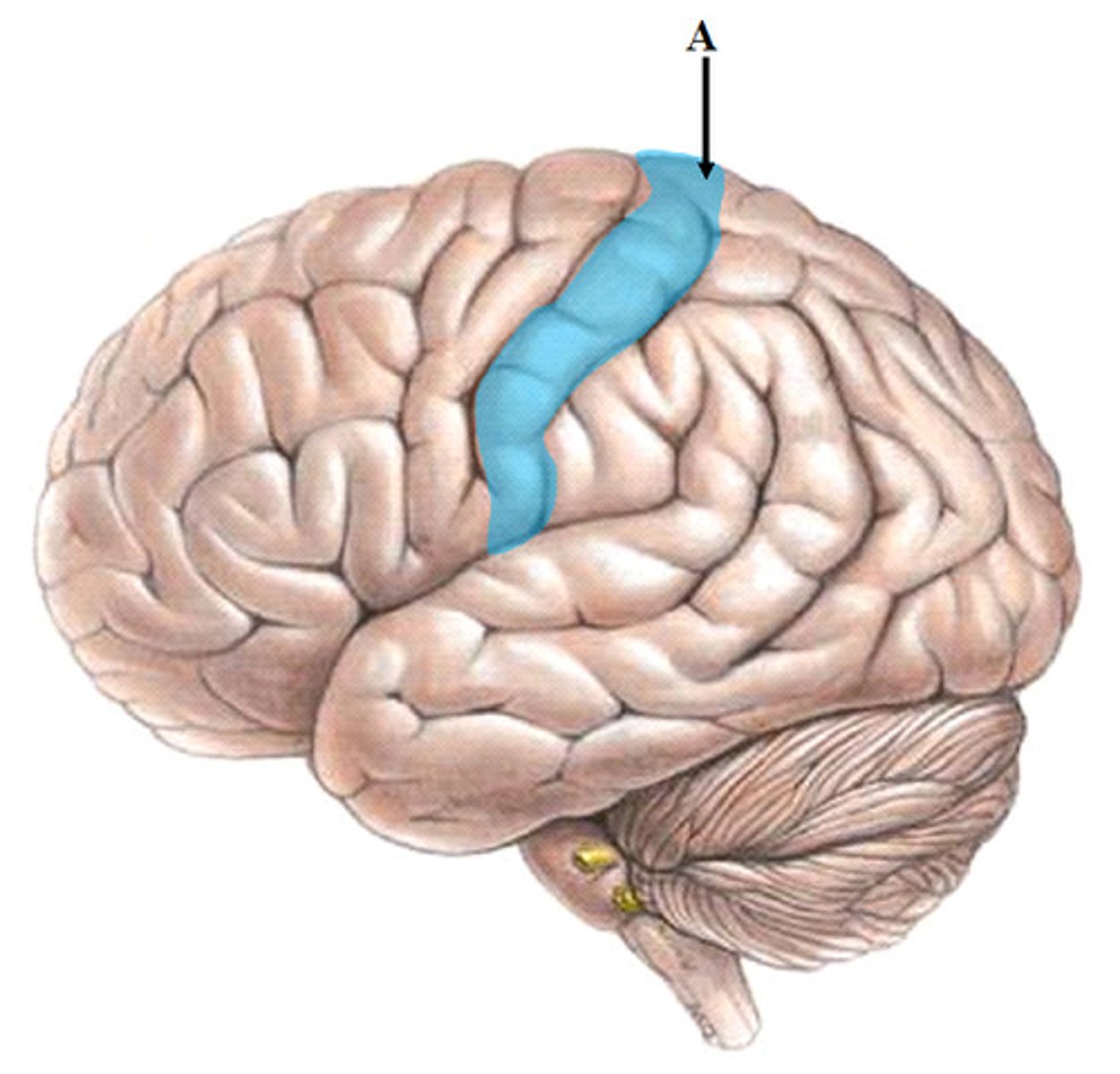
Postcentral gyrus
The location of the primary somatosensory cortex, processing sensory signals from the body.
Temporal lobe
Cerebral lobe responsible for processing auditory information, hearing, and memory encoding.
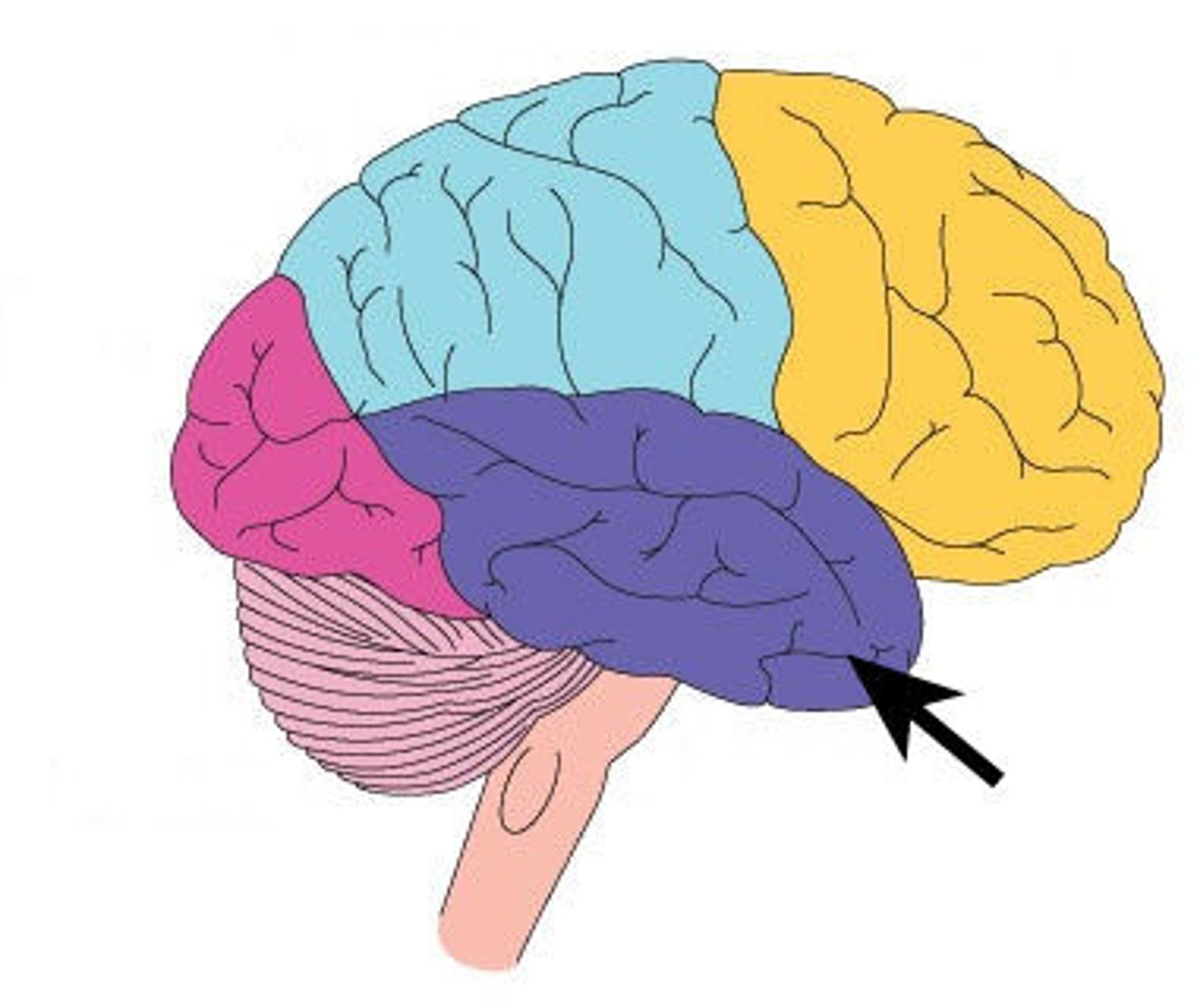
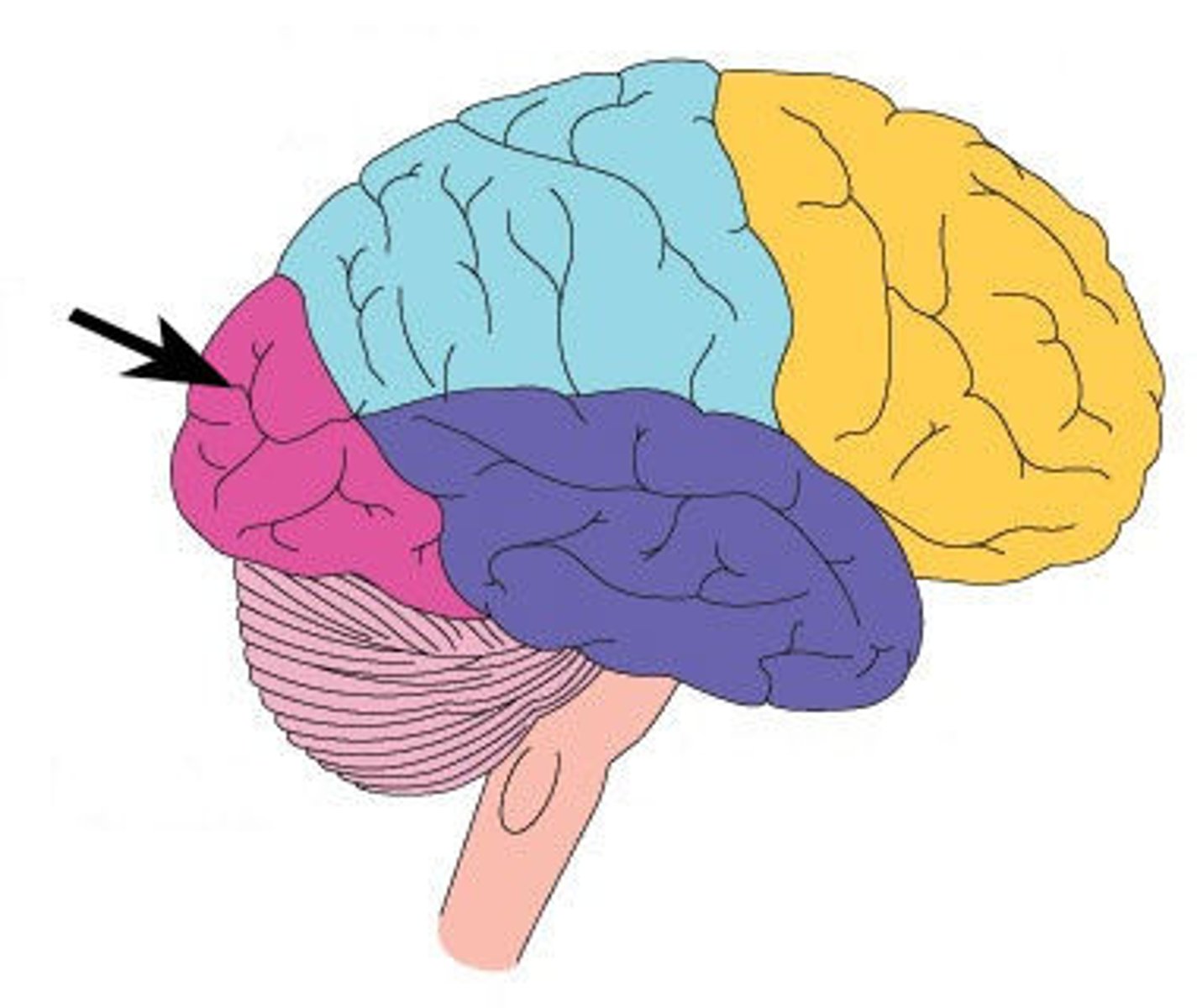
Occipital lobe
Cerebral lobe primarily responsible for visual processing and interpretation.
Broca's area
A region in the frontal lobe involved in speech production and language processing.
Wernicke's area
A region in the temporal lobe involved in the comprehension of written and spoken language.
Primary motor cortex
The region of the cerebral cortex involved in the planning, control, and execution of voluntary movements.
Primary somatosensory cortex
The region of the cerebral cortex that processes sensory input from the body, including touch and temperature.
Thalamus damage
Can cause sensory loss, altered consciousness, or chronic pain (thalamic pain syndrome).
Hypothalamus homeostasis
Controls body temperature, hunger, thirst, fatigue, sleep, and circadian rhythms.
Medulla oblongata injury
Can be instantly fatal due to its control over breathing and heart rate.
Pons role
Works with the medulla to regulate the rate and depth of breathing.
Midbrain reflexes
Controls pupillary light reflex, eye movements, and auditory startle responses.
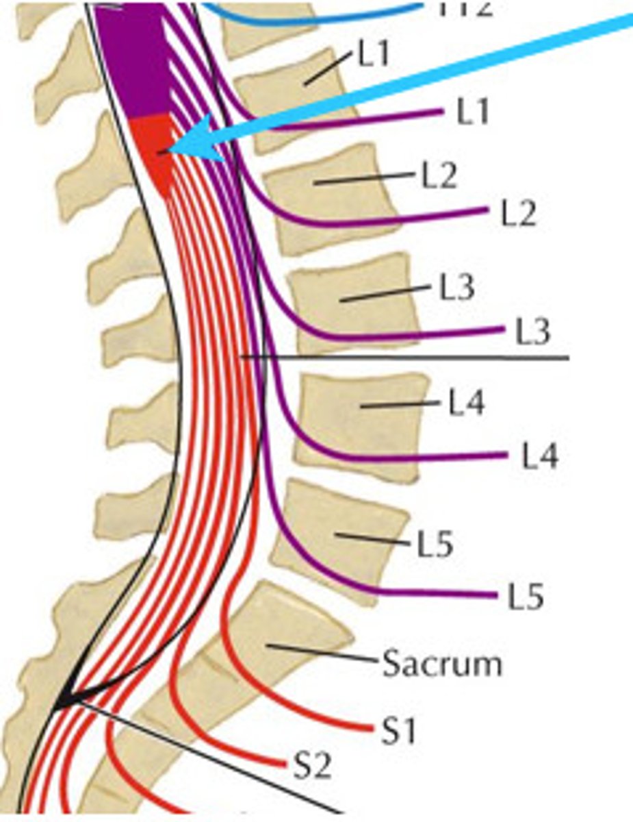
Conus medullaris
The tapered, conical lower end of the spinal cord.
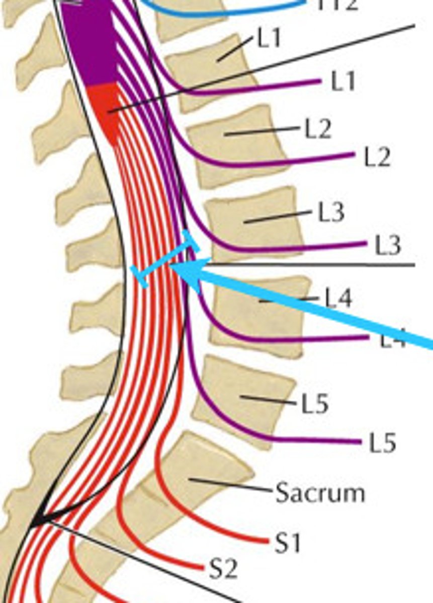
Cauda equina
horse tail bundle of spinal nerve roots extending downward from the conus medullaris.
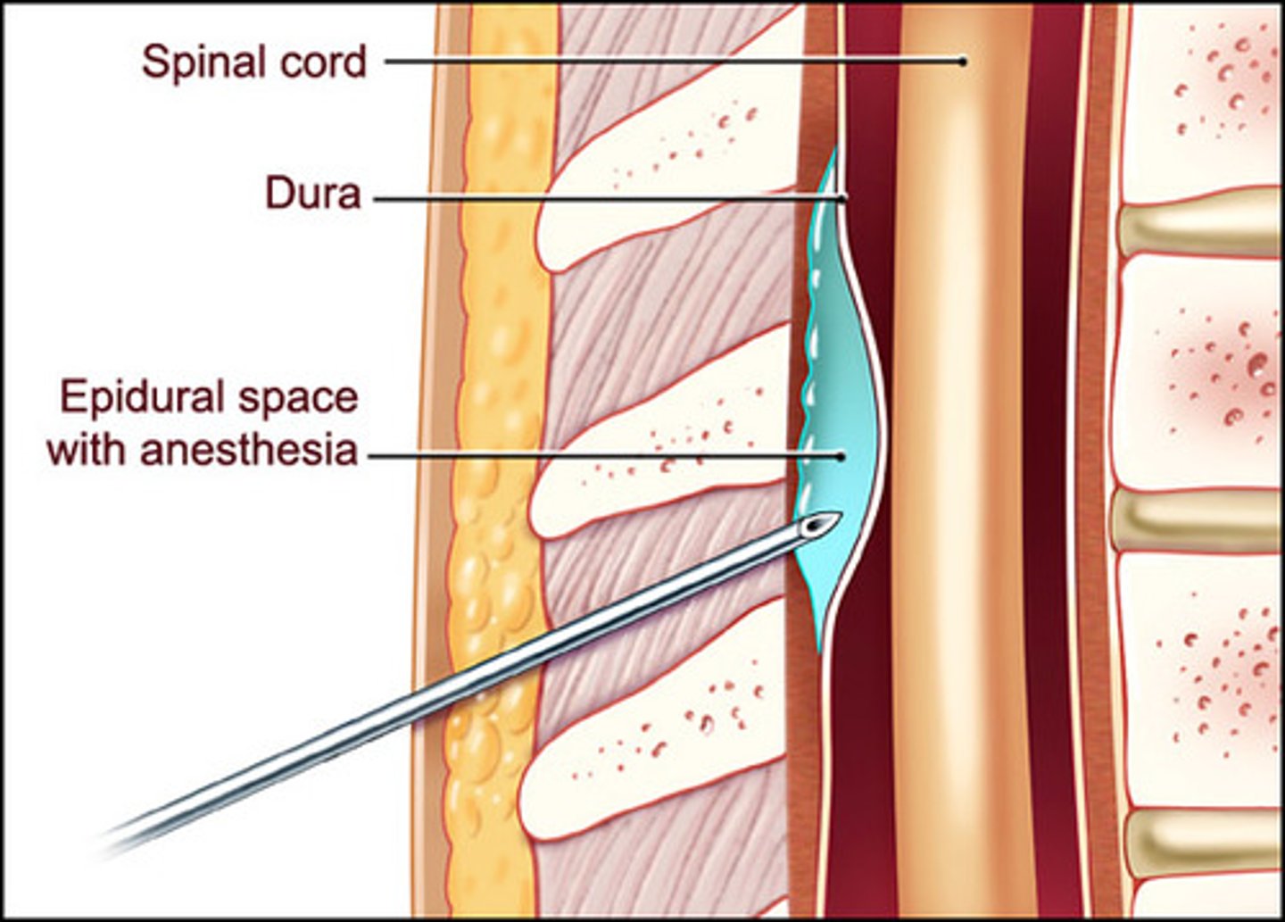
Epidural space
The space between the dural sheath (dura mater) and the vertebral canal.
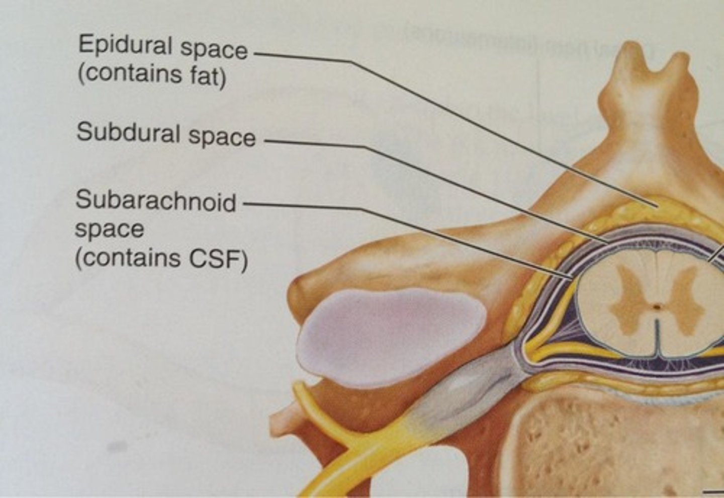
Subdural space
The potential space between the dura mater and the arachnoid mater.
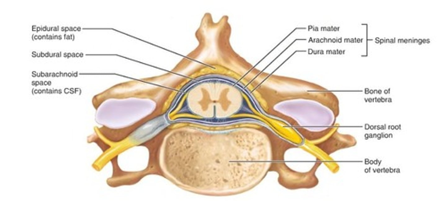
Subarachnoid space
The space between the arachnoid mater and pia mater, containing cerebrospinal fluid.
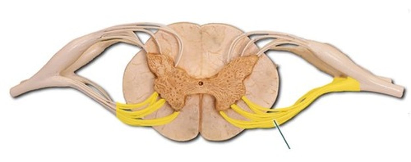
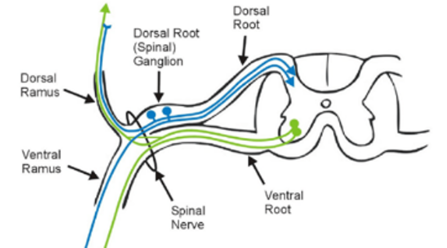
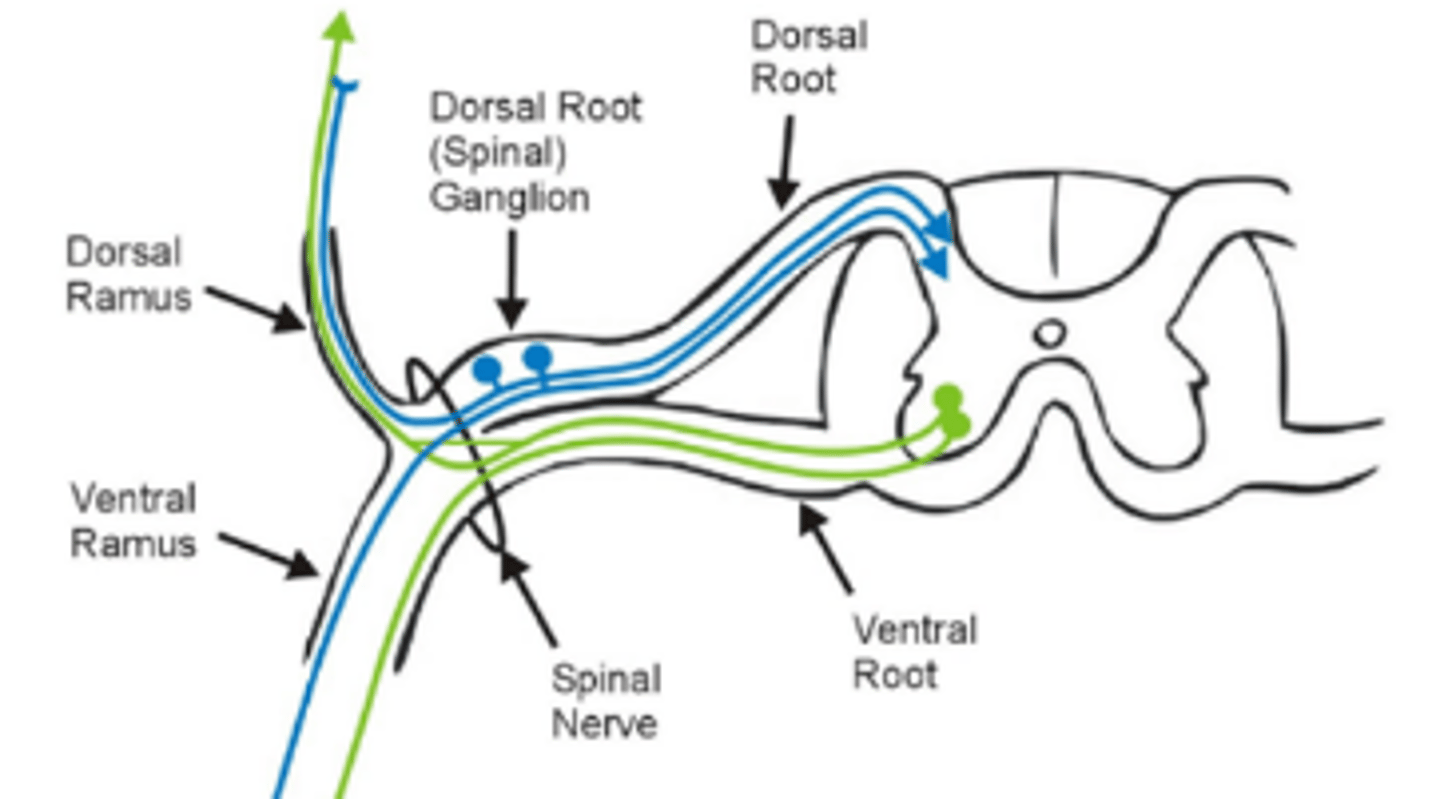
Ventral root
The motor root of a spinal nerve, carrying efferent fibers from the spinal cord.
Dorsal root
The sensory root of a spinal nerve, carrying afferent fibers to the spinal cord.
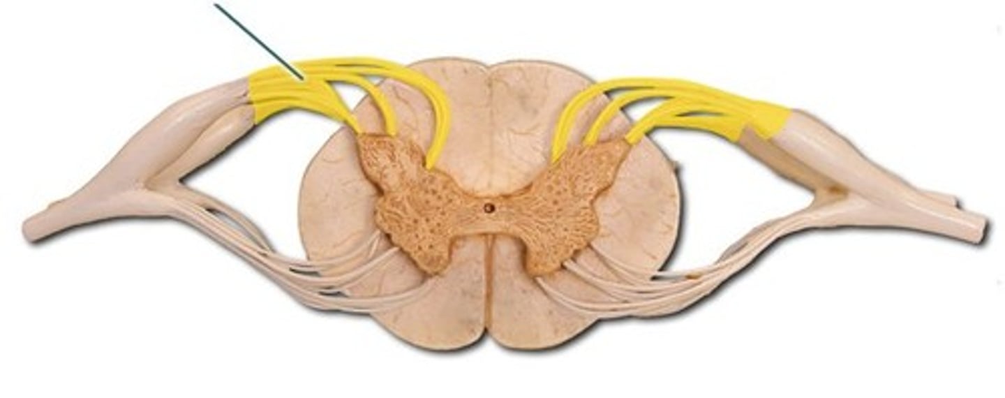
Dorsal root ganglion
A cluster of sensory neuron cell bodies located on the dorsal root.
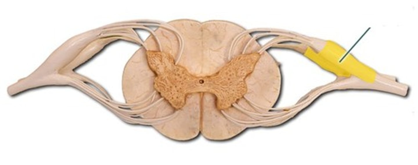
Spinal nerve
A mixed nerve formed by the junction of the dorsal and ventral roots.
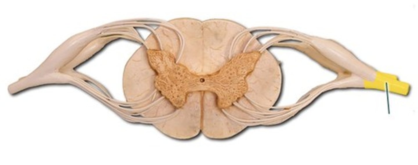
Dorsal primary ramus
what posterior branch of a spinal nerve splits into, innervating deep back muscles and skin.
Ventral primary ramus
what anterior branch of a spinal nerve splits into, innervating the limbs and anterior trunk.
T4 dermatome level
At the level of the nipples
T10 dermatome level
At the level of the umbilicus (belly button)
T12/L1 dermatome level
At the level of the inguinal / groin crease
brachial nerve plexus
ventral primary rami of C5 to T1
lumbar nerve plexus
ventral primary rami of L1 to L4
sacral nerve plexus
ventral primary rami of L4 to S4
phrenic nerve
ventral primary rami of C3, C4, and C5 and descends along each side of the neck and enters the thorax
Ulnar nerve
Innervates most intrinsic hand muscles and skin of the medial one and a half fingers.
Median nerve
Innervates most anterior forearm muscles, thenar muscles, and lateral palmar skin.
Musculocutaneous nerve
Innervates anterior arm muscles and skin of the lateral forearm.
Axillary nerve
Innervates deltoid and teres minor muscles, and skin over the shoulder.
Radial nerve
Innervates posterior arm and forearm muscles, and skin of the posterior arm/forearm.
Sciatic nerve
Innervates posterior thigh muscles and branches into the tibial and common fibular nerves.
Tibial nerve
Innervates posterior compartment leg muscles, plantar foot muscles, and skin of the sole.
Common fibular nerve
Branches into superficial and deep fibular nerves, innervating lateral and anterior leg.
Femoral nerve
Innervates anterior thigh muscles (quadriceps) and skin of anterior thigh and medial leg.
Obturator nerve
Innervates medial thigh adductor muscles and skin of the medial thigh.
Superficial fibular nerve
Innervates lateral leg compartment muscles and skin on the dorsum of the foot.
Deep fibular nerve
Innervates anterior leg compartment muscles and skin of the first web space.
patellar reflex (knee jerk)
tests femoral nerve and L2-L4 spinal segments
foramen ovale
foramen rotundum
CN V2 (maxillary nerve)
optic canal
optic nerve
foramen magnum
spinal cord
hypoglossal canal
Hypoglossal nerve (CN XII)
jugular foramen
CN IX, X, XI
CN I
Olfactory nerve; sensory nerve responsible for the sense of smell.
CN II
Optic nerve; sensory nerve responsible for vision.
CN III
Oculomotor nerve; motor nerve controlling most eye movements and pupil constriction.
CN IV
Trochlear nerve; motor nerve controlling the superior oblique muscle of the eye.
CN V
Trigeminal nerve; mixed nerve for facial sensation and muscles of mastication (chewing).
CN VI
Abducens nerve; motor nerve controlling the lateral rectus muscle of the eye.
CN VII
Facial nerve; mixed nerve for facial expression, taste on anterior tongue, and lacrimation/salivation.
CN VIII
Vestibulocochlear nerve; sensory nerve responsible for hearing and equilibrium (balance).
CN IX
Glossopharyngeal nerve; mixed nerve for taste, swallowing, and parotid gland secretion.
CN X
Vagus nerve; mixed nerve regulating visceral organs, heart rate, digestion, and speech.
CN XI
Accessory nerve; motor nerve controlling the sternocleidomastoid and trapezius muscles.
CN XII
Hypoglossal nerve; motor nerve controlling muscles of the tongue.
CN V1
Ophthalmic nerve; sensory to forehead, eyes, and nose.
CN V2
Maxillary nerve; sensory to mid-face, upper teeth, and maxillary sinus.
CN V3
Mandibular nerve; sensory to lower face/teeth, motor to muscles of mastication.