Comparative Anatomy- Final Exam
1/125
There's no tags or description
Looks like no tags are added yet.
Name | Mastery | Learn | Test | Matching | Spaced | Call with Kai |
|---|
No study sessions yet.
126 Terms
Ovaries are paired, except in…?
hagfish, birds, and bats- only have 1
birds and bats- decreased mass for flight
What are oocytes?
produce eggs
develop inside a follicle
egg = ovum
= yolk → made of egg cell and lipids
eggs released into coelom
process = ovulation
When do ovaries regress?
after breeding (except in placental mammals)
Describe oviducts.
muscular and glandular
ostium = opening (where egg goes in)
infundibulum = funnel
end at cloaca in most
Describe the shark oviducts.
produce large ovum
single ostium
1. shell gland
produce albumin = “egg white” → protein plus water
secretes leathery shell for egg layers
laying eggs = oviparity
2. uterus
for offspring to develop
get nutrients from yolk OR uterine lining
few viviparous species
viviparous = give birth to live young
Describe bony fish oviducts.
ovarian cavity continuous with oviduct (so eggs not released into coelom)
carried directly to genital pore
no cloaca
Describe amphibian oviducts.
some have glandular oviducts
secrete jelly around eggs
prevents desiccation
ovisacs
= caudal expansion of oviduct to store eggs
function: hold eggs until environmental conditions are right (water and temp)
Describe the bird oviduct.
one functional ovary and oviduct
regionalized for producing shelled egg
magnum = secretes albumin
uterus/shelled gland = secretes egg shell and pigment added
What is spermathecae?
folds in lining of oviduct or cloacal lining
sperm storage
seen in salamanders, turtles, squamates, birds
Describe monotremata reproduction.
platypus and echidna
only oviparous mammals
lack nipples
have cloaca
one ovary and oviduct → 2 uteri
eggs retained in uterus
eggs incubated briefly outside
Describe theria reproduction.
marsupials and placentals
true nipples
viviparity
all have placenta to nourish embryo
marsupials
embryo has short internal development and long external development in pouch
placentals
embryo has much longer internal development
reproductive tracts
oviduct- short and lined with cilia
uterus
highly vascularized lining = endometrium
egg implants here
muscular wall = myometrium
gives contractions for child birth
vagina
unpaired, terminal opening
opens into
a. urogenital sinus (which also receives urethra)- most common
b. vestibule of vulva
primates
urethra separate from vagina
cervix = muscular opening between vagina and uterus
What are the types of uteri in therians?
1. duplex
2 separate reproductive tracts
marsupials
most have two uterine horns that fuse distally
2. bipartite
slight partition
rabbits and hamsters
3. bicornuate
horns connected but with no partitions
ungulates, carnivores
4. simplex
complete fusion of uterine horns into single uterus
primates and armadillos
What is the functional unit of the nervous system?
neuron
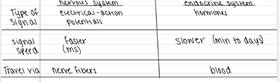
What is the type of signal, signal speed, and travel route for the nervous system and endocrine system?
Which body system coordinates body functions with endocrine system?
nervous system
What are the parts of the central nervous system?
brain and spinal cord
What are the parts of the peripheral nervous system?
cranial nerves, spinal nerves, autonomic nerves
Describe the parts of the neuron.
1. cell body
nucleus and organelles
2. dendrites- many receivers
conduct signals toward cell body
3. axon
conduct signals away from cell body
axon hillock → terminal branches
4. schwann cells
insulating cells around axon
make myelin: layers of lipids that impede nerve impulses
nodes of ranvier: areas of exposed axon
saltatory conduction: nerve impulses move faster because they jump from node to node
“nerve fiber” = usually a myelinated axon
5. synapse
gap between neurons where nerve impulse is transferred
neurotransmitters: chemicals that carry nerve impulse across a synapse
What is a nerve?
1 or more nerve fibers wrapped in fibrous sheath and supplied by blood vessels
What 3 things are needed for the nerve circuit?
sensory input
integration
motor output
What are the steps of the nerve circuit?
1. sensory input
collect info from sensory receptors
conduct signals to integration centers
sensory/afferent neurons
sensory info types:
a. external (ex: tactile info, temp, vision, auditory)
b. internal (ex: osmotic pressure of blood, body temp)
2. integration
signal info processed
appropriate response needs to be determined
done by interneurons
3. motor output
conduct response signal to effector cells for response (ex: muscle contracts)
ex of simple reflective response in spinal cord
most integration happens in brain
What nerves are part of the peripheral nervous system?
somatic
visceral
Describe somatic nerves
voluntary actions
skeletal muscle
a. sensory nerves (afferent)
carry signals TO CNS
b. motor nerves (efferent)
carry signals from cns to effector cells
c. mixed nerves
contain both sensory and motor fibers
common in verts
Describe visceral nerves and the autonomic nervous system
involuntary actions
signals to:
organs
glands
smooth muscle
etc.
mostly involves motor neurons
= autonomic nervous system
a. sympathetic division
energy expenditure and arousal
b. parasympathetic division
energy and self maintenance
para and sym connect to one organ = antagonistic effects
examples of autonomic system
1. heart rate
sympathetic- increased rate
para- decreased rate
2. stomach and intestines
sym- decreased digestive activity
para- increased digestive activity
What is the difference between the sympathetic and parasympathetic nervous systems?
sympathetic: get ready for action- lots of oxygen and shut off digestion in short term
parasympathetic: maintain body function and energy- slow heart rate and stimulate digestion
What is the function of the central nervous system?
integration and processing
What does the spinal cord use?
receives sensory signal → motor response signal
simple, reflective responses
pathway for signals to travel to and from the brain
What does the brain do?
complex integration and processing
ex: homeostasis, learning, memory
sensory info from spinal cord or directly from cranial nerves
What is the meninges?
connective tissue that covers brain and spinal cord
Describe the fish meninges.
1 layer = primitive meninx
vascularized membrane
Describe the amphibian and reptile meninges.
2 layers
a. leptomeninx
inner vascularized membrane
b. dura mater
tough outer membrane
Describe the meninges of the birds and mammals.
3 layers
a. pia mater
inner vascularized
b. arachnoid
middle- connective tissue
subarachnoid space: between layers and fills up with cerebrospinal fluid (CSF)
c. dura mater
tough outer layer
Describe the basic spinal cord structure.
occupied vertebral canal
neurocoel: internal cavity filled with CSF
cerebrospinal fluid
white matter = contains myelinated axons
gray matter = unmyelinated axons and cell bodies
ganglion = group of cell bodies (outside of CNS)
interneurons: conduct signals to and from brain
bundles of interneuron nerve fibers = tracts
ascending tract = carries to brain
descending tract = carry impulses down from brain
most vertebrates
2 spinal cord enlargements
cervical
lumbar
What is the function of CSF?
protective cushioning (or shock absorption)
remove waste products and pH buffer
buoyancy of brain
What is the amniote spinal cord pattern?
dorsal root = contains sensory nerve fibers and ganglia
ventral root = contains motor neuron fibers
2 roots fuse distally to become spinal nerve
Why do most vertebrates have spinal cord enlargements?
lots of nerves to innervate pectoral and pelvic appendages
Describe the cross section of the agnathan spinal cord.
no blood vessels in brain
only have gray matter (no myelin until jawed fish)
Describe the spinal cord cross section in other verts
round or square
X or H shaped gray matter
tetrapods- folded surface
What are some variations in the dorsal and ventral roots of the spinal cord
don’t fuse in lampreys = separate spinal nerves
non-amniotes may also have motor neurons in dorsal root
Describe the jawed fish spinal cord.
urophysis = caudal swelling of spinal cord with endocrine function
contain neurosecretory neurons = neurons in the CNS that produce neurohormones and secrete them into the blood
urophysis produces urotensins: raise blood pressure
Describe spinal nerves.
1 pair per segment or vertebra
emerge through intervertebral foramina
cauda equina
long caudal spinal nerves
go through vertebral canal before exiting foramen
occur because spinal cord grows slower than vertebral column
occurs in mammals, frogs, and a few fish
rami: division of spinal nerve close to roots
a. dorsal ramus: connects to muscles and skin of back
b. ventral ramus: goes to lateral-ventral muscles and skin
plexus
2 or more nerves unite into common trunk then redistributed
2 major- at paired appendages
Describe the rami of mammals.
rami communicantes
connect to sympathetic trunk = part of ANS
signals with visceral function
Describe early development of the brain.
3 parts
1. prosencephalon = forebrain
sensory processing, endocrine function and higher integration
2. mesencephalon = midbrain
sensory processing (visual and auditory)
3. rhombencephalon = hindbrain
coordination of movement and autonomic functions
Describe later development of the brain.
3 parts → 5 parts
prosencephalon = forebrain
1. telencephalon and 2. diencephalon
3. mesencephalon
rhombencephalon
4. metencephalon and 5. myelencephalon
Describe the internal development of the brain
forms from neural tube
sheet of ectoderm that folds up
4 cavities remain (=ventricles)
ventricles 1 and 2 = lateral ventricles
in right and left half of telencephalon
ventricle 3 in diencephalon
ventricle 4 in metencephalon and myelencephalon
aqueduct of sylvius = passageway between 3rd and 4th ventricles
Describe the internal structure of the brain
choroid plexuses: projections of pia mater and cells lining ventricles
mostly in 3rd and 4th ventricles
produce CSF
minimal to no lymphatic vessels in brain
collects excess water from brain
diffuses into venous system
mammals
collects in subdural venous sinuses
nucleus (nuclei): groups of cell bodies in CNS
neuroglia (or glial) cells: interstitial cells that have a supportive function
What are the parts of the myelencephalon?
medulla oblongata
Describe the medulla oblongata.
coordinates involuntary actions (ex: heart rate, respiration, blood pressure)
little size variation- why?
all groups need same functions
one exception: vagal lobe
large nucleus off medulla, found in bottom feeding fish (ex: catfish, carp, sucker)
receive signals from numerous chemoreceptors
Describe the cerebellum.
coordinates movement of skeletal muscles and equilibrium
equilibrium: sensory input from semicircular canals
variation- size correlated with complexity of movements
larges in mammals and birds
complex movement on land and flying
Describe the pons
pons = fiber tracts between cerebellum and cerebrum
carries motor signals
connect to pyramids of medulla = fiber tracts from pons through medulla to spinal cord and carries motor signals
What are the parts of the metencephalon?
cerebellum
ventral side (birds and mammals only)
pons
What are the parts of the mesencephalon?
optic lobes
auditory lobs
Describe the mesencephalon.
1. optic lobes
visual input
largest in lampreys and birds
rely heavily on vision for foraging
2. auditory lobes
input from ear
well developed in most amniotes
both 1 and 2 process basic reflexes and relay signals to cerebral hemispheres
amniotes
brainstem = brain minus cerebral hemispheres and cerebellum
2 sets of lobes (optic and auditory) = corpora quadrigemina
ventral side
tegmentum = basal plate
deep layer (not visible)
cerebral peduncles = bulges
= additional fiber tracts that communicate between telencephalon and hindbrain- spinal cord and carry motor signals
in most amniotes
What are the parts of the diencephalon?
epithalamus
thalamus
hypothalamus
Describe the epithalamus.
pineal organ
= photoreceptor in agnatha
= endocrine role in jawed verts
function: regulates biological rhythms
habenulae = nuclei receiving olfactory fibers and tracts from forebrain
reflex responses to olfactory stimuli
large in olfactory foragers
ex: well developed in sharks
absent in aquatic mammals (can’t smell in water)
Describe the thalamus.
important relay center of brain
all ascending fibers synapse here and sent to telencephalon
intermediate mass = nuclei bulge into 3rd ventricle
mammals only
ventral side
hypothalamus
Describe the hypothalamus.
optic chiasma = where optic nerves cross
infundibulum = ventral projection that holds the pituitary gland
pituitary gland = endocrine gland
hypothalamic nuclei: center for homeostasis
osmotic monitoring
regulates appetite and temperature
some autonomic control
contains neurosecretory neurons
produce neurohormones that control pituitary gland and gonads
What are the parts of the telencephalon?
olfactory bulb and tract
2 cerebral hemispheres = cerebrum
Describe the olfactory bulb.
receives nerves from the cells lining olfactory epithelium
variation:
poorly developed in birds and some mammals (especially true of bats and some primates)
well developed in lower vertebrates
development is related to importance in foraging and communication
What are ventricles in the brain?
open area with CSF
Describe the fish cerebrum.
1. primitive pallium
process sensory input
olfactory cues directly from olfactory tract
other sensory info relayed up from thalamus
2. globus pallidis (aka subpallium)
process motor signals
Describe the amphibian cerebrum.
additional nuclei overall
globus pallidis expands- why?
because now have appendages and more muscles for walking on land- requires more processing capability
non-amniotes → large olfactory bulbs and large part of cerebrum devoted to olfaction (“olfactory brains”)
Describe the cerebrum of reptiles and birds.
major expansion
new error = dorsoventricular ridge forms
receives and processes sensory info from thalamus
some direct processing and motor signals sent out to body but other info sent to globus pallidus
Describe the cerebrum of birds and mammals.
major expansion
flexure: forebrain becomes angles to rest of brain
cerebrum covers diencephalon and mesencephalon
otherwise, bird brains are similar to reptiles
Describe the cerebrum of mammals.
reorganization of pallium into neocortex on outside
other changes?
surface folded
ridge = gyrus
grooves = sulcus
why? increased surface area of neurons
lacking in monotremes, marsupials and some rodents
internal structural changes
internal capsule: fiber tract to brainstem
corpus callosum: fiber tract between hemispheres
where epilepsy comes from
basal ganglia
= globus pallidus + 3 new nuclei
caudate nucleus, putamen, and amygdoloid nucleus
processing of motor signals
relay motor signals
neocortex < - > thalamus
parkinson’s disease
neocortex
divided into 4 lobes:
major processing functions
sensory
temporal → auditory and olfaction
occipital → vision
parietal → touch
voluntary motor activity → frontal lobe
problem solving → frontal and parietal
memory → frontal and temporal
frontal → control over social behavior, personality
hippocampus
inner temporal lobe
spatial and short term memory
navigation
alzheimer’s disease
other verts also have hippocampus but not in neocortex
bird hippocampus- chickity bird does seed cacheing
in fall, hippocampus grows by 1/3
What is a lobotomy? Who was the first to do it on humans?
procedure that destroys part of the neocortex to alter behavior
treat severe human psychiatric problems
Gottlieb Burkhardt (1st to use it on humans)- europe
What is a leucotomy?
antonio egas moniz- europe
destroyed neocortex and parts of thalamus
won nobel prize
Who found the ice pick lobotomy? What is it?
uses ice pick
frontal lobotomy: destroys frontal lobe or prefrontal cortex
cognition, personality, decisions
What are the sensory cranial nerves (PNS)?
#0- terminal
#1- olfactory
VN- vomeronasal
#2- optic
epiphyseal (e)
profundus (p)
ALL- anterior lateral line
PLL- posterior lateral line
#8- vestibulocochlear
Describe the terminal cranial nerve.
#0
sensory
in gnathostomes (jawed vertebrates)
goes between olfactory mucosa → forebrain
pher0mone receptors
mating and reproductive behavior
Describe the olfactory nerve.
#1
sensory
goes from olfactory epithelium → bulb
Describe the vomeronasal nerve.
sensory
cranial nerve (PNS)
goes from vomeronasal organ → olfactory bulb
VN organ = accessory chemoreceptor (for pheromone detection)
isolated in nasal cavity
not in humans
some mammals, amphibians, squamates
flenmen behavior (mammals)
forked tongue in squamates
Describe the optic nerve.
sensory
retina → chiasma
fibers cross to opposite sides of brain = 2 separate images
produces monocular vision (eyes don’t overlap)
some mammals- only some fibers cross and continue to opposite sides = 1 image and binocular vision
eyes must overlap
birds- also have binocular vision → visual signs relayed throughout brain
Describe the epiphyseal nerve.
sensory
pineal organ or parapineal connected → habenulae
lamprey and some bony fish
lampreys - photoreceptor
some frogs, toads, and lizards: parapineal organ (also a photoreceptor)
Describe the profundus nerve.
extends to skin of snout- head
separate nerve
some jawed fishes
fuses with trigeminal in other vertebrates (is the ophthalmic of trigeminal)
Describe the anterior lateral line and posterior lateral line.
sensory nerves
connect lateral line to hindbrain
lateral line detects motion in water
merge with cranial nerve #7,9,10
aquatic non-amniotes
Describe the vestibulocochlear nerve.
sensory
connects medulla oblongata → inner ear
2 major branches
a. vestibular- goes to semicircular canals
b. cochlear- goes to organ of the inner ear that converts vibrations to nerve impulses
fish = lagena
tetrapods = cochlea
What are the motor cranial nerves?
#3- oculomotor
#4- trochlear
#6- abducens
#11- spinal accessory
#12- hypoglossal
Describe the oculomotor, trochlear, and abducens nerves.
motor nerves
connect eye muscles → mid and hindbrain
oculomotor- autonomic connections for vision
controls iris and size of pupil
Describe the spinal accessory nerve.
motor
only in tetrapods
connects hindbrain to pharynx and soft palate, as well as superficial neck and shoulder muscles in mammals only
Describe the hypoglossal nerve.
motor
only in tetrapods
= spinal nerve locked in cranium
during development, spinal nerves have a dorsal and ventral root (lacking in cranial nerves)
goes to muscle of tongue
What are mixed nerves? Which nerves fall under this category?
contain sensory and motor fibers and may contain visceral fibers of autonomic nervous system
all mixed nerves emerge from hindbrain
#5 (trigeminal), #7 (facial), #9 (glossopharyngeal), #10 (vagus) = branchiomeric
only in fish
go to branchial arches and jaws
ventilating gills and feeding
Describe the trigeminal nerve.
mixed nerve
sensory-tactile reception
skin of head and teeth
covers linings of oral and nasal cavities
anterior tongue
motor- muscles of jaws
Describe the facial nerve.
mixed nerve
sensory
taste receptors
fish- pharynx
tetrapods- anterior tongue
motor
muscles of hyoid arch
in all vertebrates
tetrapods- face muscles
Describe the glossopharyngeal nerve.
mixed nerve
fish- sensory and motor
3rd pharyngeal arch
tetrapods
many fibers lost along with gills
motor- neck muscle
sensory- posterior tongue
taste, touch, temp
also have visceral fibers to one salivary gland
Describe the vagus nerve.
mixed nerve
fish- sensory and motor
pharyngeal arches
tetrapods- motor
neck muscles- swallowing
all vertebrates
sensory- oral cavity
taste and touch
also have visceral factors to heart and many of our visceral organs
What are the nerves that are involved with the tongue?
4 major nerves
motor → hypoglossal
sensory
touch → trigeminal (anterior), glossopharyngeal (posterior)
taste → facial (anterior), glossopharyngeal (posterior)
visceral salivary glands → facial and glossopharyngeal
What nerves comprise the autonomic nervous system?
entirely visceral motor neurons
sensory fibers may share a common pathway
What are the two components of the autonomic nervous system?
a. sympathetic
branches spinal cord and spinal nerves
diagram: black nerves = sympathetic, red nerves = parasympathetic
b. parasympathetic
branches from brain and spinal cord (sacral)
What neurons are needed to reach effector cells?
a. preganglionic neuron
CNS → autonomic ganglion
b. postganglionic neuron
autonomic ganglion → effector cell
some go through rami communicantes: connects spinal nerves to sympathetic trunk
What are the three types of ganglia present in the ANS?
a. sympathetic
sympathetic trunk near spinal cord
b. collateral
near head or abdominal aorta
c. terminal
in visceral organ
Are effector cells innervated by sympathetic or parasympathetic nerves?
most effector cells are innervated by both
exception: skin only has sympathetic
What is the endocrine system?
glands that produce chemicals that influence specific target cells
gland: tissue producing a secretion
hormones: chemical messengers of endocrine system
travel through blood
hormones only bind with cells with specific receptors
except general metabolic hormones
influence all cells
ex: insulin
tropic hormones: stimulate other endocrine glands
What are the major endocrine glands?
brain:
hypothalamus
pituitary
pineal
around body:
adrenal
gonads
thyroid
parathyroid
pancreas
What do glands do?
diffuse endocrine tissue
What hormone does the stomach produce?
gastrin: stimulates cells to produce acid
Describe the hypothalamus gland.
= intermediary for endocrine and nervous systems
works closely with pituitary gland
receives info from brain
initiates hormone production
hormones either:
1. stored in pituitary
2. activate pituitary
to produce other hormones
pituitary
production and movement of neurohormones
What parts is the pituitary divided into?
a. neurohypophysis
b. adenohypophysis
What are neurohormones?
hormones produced in CNS
Describe production and movement of neurohormones in the hypothalamus.
neurosecretory hormone
neurohormones made at cell body
cell body → axon
axon terminal → blood sinusoid
sinusoid → circulation
axon terminal and sinusoid come together = neurohemal organs