immuno 2
 Call Kai
 Learn
Learn Practice Test
 Spaced Repetition
 Match
Match Flashcards
 Knowt Play
1/120

There's no tags or description
Looks like no tags are added yet.
Name | Mastery | Learn | Test | Matching | Spaced | Call with Kai | Chat |
|---|
No analytics yet
Send a link to your students to track their progress
121 Terms
serologic testing, an assay involving
serum constituents
Immunologic testing, testing related to
antigens and antibodies
blood specimen preparation, preanalytical variables specimen types
most immunology assays are done on serum; tube should be allowed to clot before centrifugation occurs; some assays are done on body fluids (cerebrospinal fluid or urine)
blood specimen preparation, preanalytical variables delay in testing
in most cases, immunology testing should be done within 72 hours; if not, serum should be frozen
blood specimen preparation, preanalytical variables excessive heat
coagulates proteins
blood specimen preparation, preanalytical variables bacterial contamination
alters protein molecules
blood specimen preparation, preanalytical variables strong acid or alkali solutions
must be avoided because these substances have a denaturing effect on serum proteins and make the specimens useless for serologic testing
blood specimen preparation, preanalytical variables lipemia, hemolysis, or icterus
can make a specimen unacceptable
complement in body fluids can be inactivated by heating at
56C for 30 min
manual pipettes - graduated pipette
delivers the amount of liquid contained between two calibration marks on the pipette, required draining between two calibration marks (less accurate)
manual pipettes - serologic pipette
similar to the graduated pipette, but it is calibrated to the tip, contains a frosted ring at the non-calibrated end
Manual pipettes - to contain (TC)
glassware designed to contain like graduated cylinders and volumetric flasks are usually marked with a TC, when liquid is poured from a piece of glassware a small amount remains behind clinging to the slides of the vessel, a 100 ml volumetric flask is designed to hold exactly 100 mL but if the liquid is poured out it will actually deliver a little less than 100 mL
manual pipettes - to deliver (TD)
glassware designed to deliver like pipettes and burets are marked with a TD, these pieces of glassware account for the small amount of liquid that remains behind, a 100 mL pipette contains a little more than 100 mL of liquid but when the liquid is drained from a pipette exactly 100 mL is delivered
steps in using piston type automatic micropipette
A: attached proper tip size for range of pipette volume and twisting tip as it is pushed onto pipette to give airtight continuous seal B: holding pipette before use C: follow instructions for filling and emptying pipette tip
If testing cannot be done without _____ hours of collection, a serum specimen should be frozen at -20C
72
complement can be inactivated in human serum by heating to
56C
solution (in dilutions)
the mixture of one substance (solute) into another (solvent)
dilution (in dilutions)
reduces the concentration of the original solute in the solution
dilutions
a dilution is the number of parts of the substance in the total number of parts of the final diluted solution
if we make a ½ dilution of blood using saline
1 mL of blood + 1 mL of saline =1/2
a patient blood sample is ran on the instrument and reads > 500 mg/dL for glucose. A 1/3 dilution with saline is required in order to get the reading in the dynamic range. How many mL of patient’s blood would be used to prepare a 1/3 dilution in which the total volume is 6 mL
2 mL
a patient blood sample is ran on the instrument and reads >500 mg/dL for glucose. A 1/3 dilution with saline is required in order to get the reading in the dynamic range. If the analyzer gives a result of 300 mg/dL how should the patient’s glucose level be reported
900 mg/dL
serial dilutions
the stepwise decreasing strength of a substance in solution
example of serial dilution
each sample gets 1 ml of saline, 1 patient serum is 1:2 final dilution, take 1 mL of first into second (1 of 1:2) to get final dilution of 1:4, (1:8, 1:16, 1:32 etc)
testing for antibody levels - acute phase specimen
serum from one with a current infection for which bacterial or viral specific immunoglobulins are measured
testing for antibody levels - convalescent phase specimen
serum from one who has recovered from an infectious disease and considered to be especially rich in antibodies against the infectious agent
antibody titer refers to
the concentration of an antibody, the antibody titer is defined as the reciprocal of the highest dilution of the patients serum which the antibody is still detectible (if serum is being tested for antibody levels of specific infection one should be drawn during acute phase and the other during convalescent)
determination of titer
for most pathogenic infections, an increase in the patient’s titer of two double dilutions, or from a positive result of 1:8 to a positive result of 1:32 over several weeks, is an infection of a current infection (this is known as the four-fold rise in the antibody titer)
Stretpozyme testing is preformed on an initial patient serum dilution of 1:100 the dilution procedure is
10 ul of patient serum is added to 990 ul of saline diluent
serum for detection of antibodies should be drawn during
acute and convalescent phases of illness
precipitation and agglutination are
examples of unlabeled immunoassays, visible expressions of the aggregation of antigen particles or molecules alternating with antibody molecules
precipitation
aggregation of soluble test antigens; combination of soluble antigen with soluble antibody to produce a visible insoluble complex
agglutination
specific antigens (eg RBCs) aggregate to form larger visible clumps when the corresponding specific antibody is present in the serum
IgM is the best
at agglutination and precipitation and is almost always exclusively used as reagents
precipitation assays - double immunodiffusion technique
over time, antibody from the antibody well and antigen from the antigen from the antigen well will migrate toward each other to form a precipitation line at the zone of equivalence, used in fungal serology to detect antibodies in response to aspergillus, Blastomyces, Candida, coccidioides, histoplasma
precipitation assay - this precipitation band is called
line of identity (if the unknown serum contains antibody to the known antigen, a precipitation band forms at a point of optimal concentration - zone of equivalence - of each component)
precipitation assays - double immunodiffusion
patient samples must always be adjacent to positive control, rate of diffusion is affected by size - igG diffuses well in gel, igA is the predominate immunoglobulin in chronic cases and diffuses moderately well, igM does not diffuce well due to size and will negative in gel
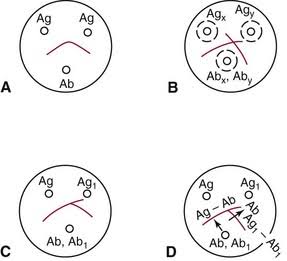
what is A’s reaction
reaction of identity (positive)
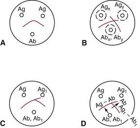
what is B’s reaction
reaction of nonidentity (negative)
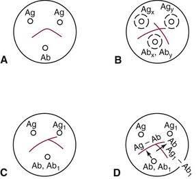
what is C’s reaction
reaction of partial identity (spur, multiple infections)
precipitation is the term applied to
aggregation of soluble test antigens
agglutination
observable clumping of insoluble antigen particles with soluble antibody, forms a framework in which antigen particles or molecules alternate with antibody molecules
Agglutination is the clumping of particles that have antigens on their surface, such as erythrocytes, by antibody molecules that form bridges between the antigenic determinants, this is the end point for
most tests involving erythrocyte antigens
particle agglutination reactions
artificial carrier particles such as latex or charcoal may be needed to indicate visibly that an antigen-antibody agglutination reaction has taken place, when carrier particles are used the assay is known to be passive, quality of results depends on: time of incubation with the antibody source, amount and avidity of an antigen conjugated to the carrier, conditions of the test environment
immunologic assays preformed by latex particle agglutination
C-reactive protein, igG rheumatoid factor, igM rheumatoid factor, rubella antibody
latex particles in C reactive protein assay results
if CRP is present in the serum, an antigen-antibody reaction takes place. This reaction causes a change in the uniform appearance of the latex suspension and a clear agglutination results
Stabilized sheep erythrocytes in Rheumatoid factor (RF) results
if gamma globulin is attached to a particular carrier the reaction of RF with gamma globulin becomes a visible agglutination
agglutination - liposome-enhanced testing
is a variation of latex agglutination to enhance visibility or agglutination
agglutination - direct bacterial agglutination
detects patient antibody; antigen is naturally on the particle itself (bacteria, fungus); the binding of patients antibody to antigen in a bacterial suspension causes the bacteria to clump together in visible aggregates, also called bacterial agglutination
agglutination - inhibitation agglutination
detects patient antigen (hCG - pregnancy), the blocking of the formation of clumps (agglutination) is the principle of the test
agglutination - passive agglutination
(passive hemaagglutination) detects patient antibody; a carrier such as red blood cells is used to absorb soluble antigen onto their surface; the red blood cells than aggluation in the presence of antiserum specific for the absorbed antigen
reverse passive agglutination
detects antigen (CRP fibrinogen); antibody is attached to the carrier particle
agglutination - flocculation tests for antibody detection
based on interaction of soluble antigen with antibody, which results in the formation of a precipitate of fine particles of visible masses (aggregates) indicating an antigen antibody reaction, these particles are macroscopically or microscopically visible only because the precipitated product is forced to remain in a confined space, examples include veneral disesase research labs (VDRL) test and rapid plasma reagin (RPR) for syphillus testing
hemagglutination
method of testing that detects antibodies to erythrocyte antigens
Hemagglutination direct
direct antiglobulin test (DAT or Coombs): detects antibodies directly bound to the RBC surface antigens; helps diagnose hemolytic disease of the newborn (HDN)
Hemagglutination indirect
Indirect antiglobulin test (IAT): detects circulating antibodies not yet bound but have potential to do harm
Hemagglutinin is influences by a number of factors and is believed to occur in two stages:
sensitization: physical attachment of antibody molecules to antigens on erythrocytes, which is influenced by: particles charge, electrocytes concentration and viscosity, antibody type, antigen-to-antibody ratio, antigenic determinants, physical conditions such as pH, temperature, and incubation
lattice formation: occurs when antigen particles and antibodies crosslink to form bridges, which is influenced by: factors such as zeta potential: electrostatic charge that repels adjacent particles
enzyme pretreatment of red blood cells - techniques to reduce zeta potential
removes negatively charged sialic acid residues from cell surface membrane
addition of colloids (e.g. albumin) - techniques to reduce zeta potential
increases electrical conductivity of environment
centrifugation - techniques to reduce zeta potential
mechanical process to force red blood cells closer together
other techniques to enhance hemagglutination include the use of
anti-human globulin (AHG), forms cross-links between antibodies that are bound to antigens on a carrier surface, AHG is incorporated into the procedure to link the antigens and antibodies close enough to cross-link
4+, 3+, 2+ 1+ mixed field aggluination reactions
all one solid, several large solids, medium solids clear supernatant, lots of small solids with turbid background, red background
prozone
if excessive antibody concentration is present, the prozone phenomenon occurs and can result in a false-negative reaction
postzone
if an excess of antigens occurs, the postzone phenomenon occurs, in which small aggregates (clumps) are surrounded by excess antigen and no lattice formation is established
nephelometry
antigens and antibody added to a tube. Visualize light scattered at 10-90 (usually 70) determine concentration using a standard curve
turbidometry
antigen and antibody added to a tube visualize that is directly across from the light source. Determine concentration using a standard curve
Immunogenic assays performed by nephelometry
acid a-glycoprotein, albumin, c1-8, transferrin
agglutination can be used to enhance reactions by all the following means
using enzyme treatment to alter the zeta potential, centrifugation, using colloids and anti-human globulin
anti-human globulin (AHG) is used to
form cross-links between antibodies that are bound to antigens on a carrier surface
the appropriate description of the prozone phenomenon is
an excessive antibody concentration produces a false-negative reaction
Major Histocompatibility complex - class I region
genes control HLA-A, B, C antigens found on most nucleated cells, MHC class I proteins functions to present endogenous antigen to CD8 T cytotoxic lymphocytes
Major histocompatibility complex - Class II region
genes code for DP, DQ, and DR antigen expressed primarily on B cells, macrophages, other antigen presenting cells, and activated T cells, MHC class II proteins function to present exogenous antigen to CD 4 T helper lymphocytes
Major histocompatibility complex - class III region
genes code for some of the complement components and cytokines
role of HLAs (human leukocytes antigens)
HLAs are the molecular basis for T cell discrimination of self from non-self, key applications for HLA typing include organ transplantation and disease association
HLA nomenclature - N
null allele (produces a non-functional protein)
HLA nomenclature - L
lower than normal cell surface expression
HLA nomenclature - S
soluble protein not found on cell surface
HLA nomenclature - Q
questionable (allele may affect normal expression)
HLA nomenclature - C
cytoplasm protein not present on the cell surface
HLA nomenclature - A
adherent expression (uncertain if protein is expressed)
HLA haplotypes inheritance
expressed codominantly, example of an HLA type: HLA-A10, A28, B7, Bw52, Cw2 (better donor - homogyzous, person receiving - hetero)
the major histocompatibility complex (MHC) HLA-II region codes for
class I molecules
the _____ antigen is a MHC class II antigen
HLA-DR
application of HLA typing disease association examples
HLA B27 is useful in diagnosing ankylosing spondylitis
reasons for a transplant
organ transplantation is widely viewed as the preferred treatment for end stage organ failure
reasons for kidney recipients
diabetes, glomerulonephritis, hypertensive nephrosclerosis, or polycystic kidneys
reasons for liver recipients
cirrhosis, cholestatic liver disease, biliary atresia, or hepatitis C infection
reasons for heart recipients
cardiomyopathy, congenital heart disease, valvular heart disease, or coronary heart disease
HLA crossmatching methods
the classic method to detect antibodies was complement dependent cytotoxicity, it has been gradually replaced by more sensitive solid phase immunoassays, such as enzyme linked immunosorbent assay (ELISA) and the bead-based technology, flow cytometry
autograph
graft transferred from one position to another in the same individual
syngraft
graft transplanted between different but identical recipient and donor
allograft (homograft)
graft between genetically different recipient and donor the same species; grafted donor tissue or organ contains antigens not present in recipient
xenograft (heterograft)
graft between individuals of different species
allogenic
stem cells come from a donor
syngeneic
stem cells from an identical twin
autologous
stem cells from the patients own stem cells
examples of diseases treatable by stem cell transplantations
acute leukemias, stem cell disorders, other malignancies
application of HLA typing includes all of the following
platelet transfusion, disease association, organ transplantation
graft between genetically different recipient and donor of the same species; the grafted donor tissue or organ contains antigens not present in the recipient
allograft
transplant rejection, role of T cells
graft rejection is primarily regulated by the interaction of the host’s T cells with the antigens in the graft