LAB 10 Proteolysis SDS Page and RP HPLC
 Call Kai
 Learn
Learn Practice Test
 Spaced Repetition
 Match
Match Flashcards
 Knowt Play
1/30

There's no tags or description
Looks like no tags are added yet.
Name | Mastery | Learn | Test | Matching | Spaced | Call with Kai | Chat |
|---|
No analytics yet
Send a link to your students to track their progress
31 Terms
proteolysis
process breaks down proteins or peptides into amino acids by action of enzymes
protease enzyme hydrolyze peptide bond to break it into smaller fragments
Non-enzymatic proteolysis occurs with use of mineral acids & heat
proteolysis
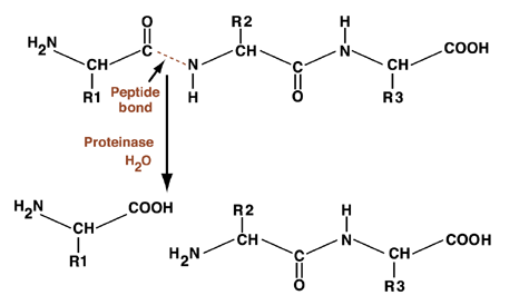
schematic representation
figure shows basic chemical structure of protein’s 2 amino acids, connected by peptide bond (represented by dotted line in figure). It’s at peptide bond that proteinase, with addition of water (H2O), cuts protein into 2 fragments.
trypsin
serine protease produced in pancreas, where it hydrolyses proteins
excreted as inactive protease trypsinogen
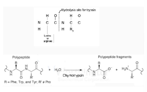
cleaves peptide chains at carboxyl side of amino acids lysine or arginine, except when followed by proline
trypsin 2
uses special serine amino acid in protein-cutting rxn, serine protease
serine proteases are diverse family of enzymes, all use similar enzymatic machinery
Trypsin & chymotrypsin that proteolyze proteins during digestion
Each has particular taste for protein chains: trypsin cuts next to lysine & arginine, chymotrypsin cuts next to phenylalanine & other large amino acids
protein separation
SDS PAGE Electrophoresis is 1 of 3 major techniques used to analyze & purify proteins (other 2 are chromatography & centrifugation)
matrix (stationary phase) is made of acrylamide polymer
By controlling % (from 3% - 30%) of polyacrylamide, precise pore sizes can be obtained for separating protein from 5 - 2,000 kDa
protein separation
details
Electric current provides force for migration
gel is run in running buffer containing electrolytes
Sodium dodecyl sulphate (SDS) is added to running buffer to mask charges
SDS-polypeptide complexes have same neg charge & shape & migrate through gel according to polypeptide size
Once individual proteins are separated, gel is stained with Coomassie blue (GelCode Blue) to visualize protein bands
protein separation
laemmli buffer
designed to dissociate proteins into individual polypeptide subunits
Sodium dodecyl sulfate (SDS) is ionic detergent which denatures protein complexes by wrapping around polypeptide backbone
Dithiothreitol (DTT) is thiol reagent that cleaves disulfide bonds
When protein sample is heated in presence of DTT & excess of SDS, proteins are fully dissociated into their subunits
protein separation
laemmli buffer
5X Lammeli sample buffer with DTT (200 mM)
Contents: 20% SDS 6 ml
Bromophenol blue 6 mg
Glycerol 4.7 ml
1M Tris pH 8 0.6 ml
DTT (per 5 ml of Laemmli buffer) 0.465 g
visualization of proteins
Protein bands are stained with dye to observe
Proteins are stained using a Gel Code Blue
GelCode Blue Safe Protein Stain is proprietary Coomassie blue based stain
Gel Code Blue stains gel which can be easily washed off by rinsing repeatedly with water
Note: coommassie blue is also used in coomassie blue protein assay (bradford assay)
videos
protein molecular marker
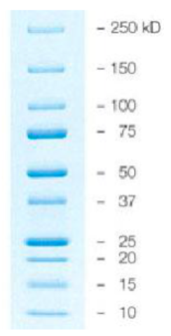
contains mixture of proteins of known molecular weight & loaded on gel to estimate size of proteins in sample
In today’s lab, “Precision Plus Protein Standard” containing range of protein sizes is used to size GFP
stationary phase & mobile phase
stationary phase
aka ‘matrix’
thin layer on solid support (glass/plastic) or packed in column
compounds are separated based on relative affinity to stationary & mobile phase
If compound has greater affinity for mobile phase as compared to stationary phase, compound will move with mobile phase
As a result, each compound in sample mixture moves at diff speeds through stationary phase
stationary phase & mobile phase
stationary phase
normal phase chromatography
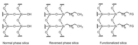
has more polar stationary phase & made of silica gel
Polar compounds bind strongly to matrix while hydrophobic compounds move quickly
Silica gel is acidic & offers poor separation of basic samples & causes deterioration of acid-labile molecules
stationary phase & mobile phase
stationary phase
reversed phase chromatography
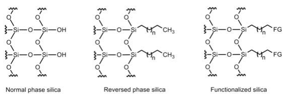
matrix particles (silica gel) coated with compounds with hydrophobic side chains (C18 chains)
matrix has higher affinity for hydrophobic compared to polar compounds
highly polar compounds from sample easily eluted using polar mobile phase (eluting solvent)
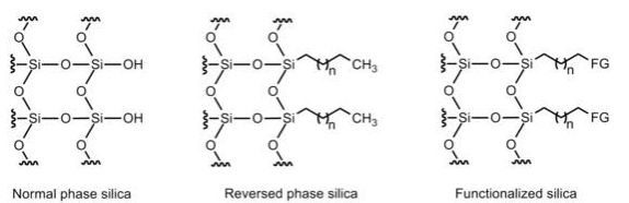
For separation of charged proteins & compounds, silica molecules are attached with charged functional groups
stationary phase & mobile phase
stationary phase
ion-exchange chromatography
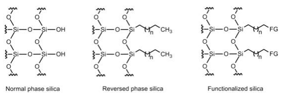
has charged matrix, which binds to molecules with opposite charge
Negatively charged proteins will bind to positively charged columns and vice versa
mobile phase
liquid or gas & moves through stationary phase via capillary action or pressure
2 or more solvents (polar/nonpolar) can be mixed together in varying proportions to obtain best polarity (or pH) for separation
Common mobile phases used in liquid chromatography are water (polar solvent), ethanol & methanol (less polar solvents)
For more sophisticated chromatographic separations, other organic solvents (aniline, octane) are used
how to read a chromatogram
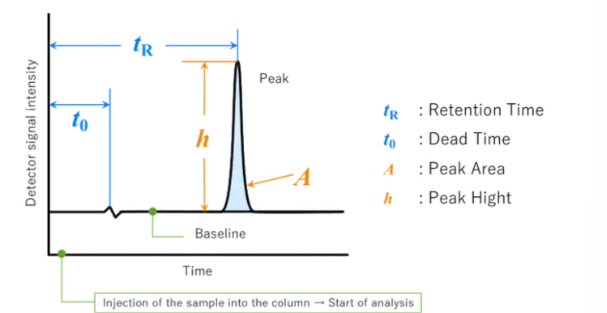
word "chromatogram" means a plot obtained via chromatography. See figure below
video
how to read a chromatogram
chromatogram is 2d plot with vertical axis showing conc in terms of detector signal intensity & horizontal axis representing analysis time
When no compounds are eluted from column, a line parallel to horizontal axis is plotted → called baseline
detector responds based on conc of target compound in elution band
obtained plot is more like shape of bell rather than a triangle = shape is “peak”.
how to read a chromatogram
retention (tR)
time interval between sample injection point & apex of peak
required time for non-retained compounds (compounds with no interaction for stationary phase) to go from injector to detector is dead time (t0).
how to read a chromatogram
peak height h
vertical distance between peak's apex & baseline, & peak area (A) colored in light blue is area enclosed by peak & baseline
results will be used for qualitative & quantitative analysis of sample's components
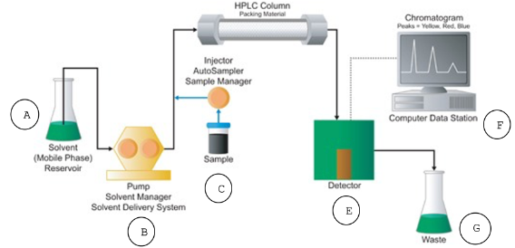
high performance (or pressure) liquid chromatography (HPLC)
Reverse phase HPLC with a C18 column used to separate samples using methanol
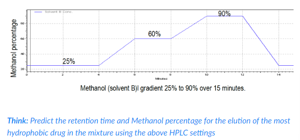
mobile phase is water: methanol mixture. This equipment will be demonstrated to you by your instructor.
Dual pump system & computer software (method) is used to provide varying concs (gradients) of water & methanol during each run (see below)
spectrophotometer is set to read at 254 nm wavelength.
high performance (or pressure) liquid chromatography (HPLC)
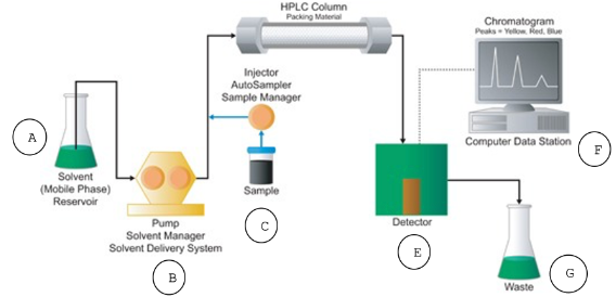
instrumentation
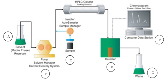
solvent reservoir
Stores the solvent (mobile phase).
high performance (or pressure) liquid chromatography (HPLC)
instrumentation
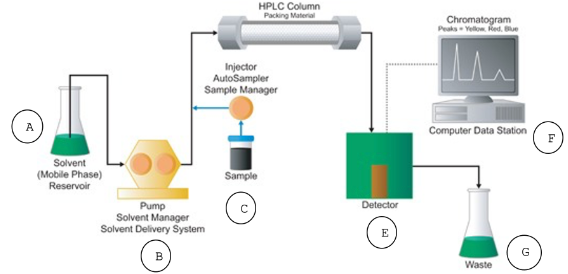
pump
High-pressure pump that generates a specific flow rate of mobile phase.
high performance (or pressure) liquid chromatography (HPLC)
instrumentation
sample injector
valve in which you inject your sample
Introduces sample into stream of mobile phase that will carry sample to HPLC column.
high performance (or pressure) liquid chromatography (HPLC)
instrumentation
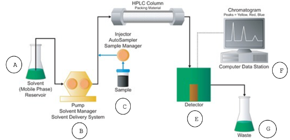
HPLC column
Stationary phase of HPLC
Contains packing material needed to separate injected sample
For today’s lab, column consists of silica coated in C18 chains.
high performance (or pressure) liquid chromatography (HPLC)
instrumentation
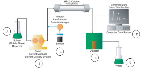
detector
Internal UV-Vis spectrophotometer used to visualize separated compounds as eluted from HPLC column
Sends info to computer data station.
high performance (or pressure) liquid chromatography (HPLC)
instrumentation
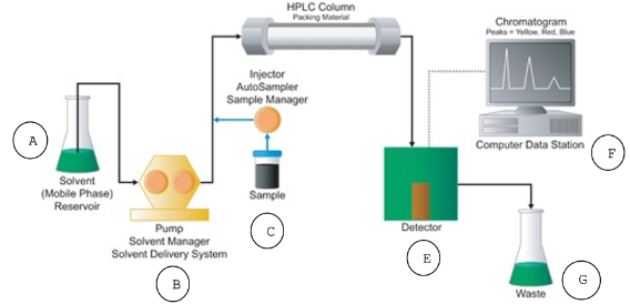
computer data station
Records electrical signal from detector & generates chromatogram
computer data station is also used to identify & quantify conc of sample
high performance (or pressure) liquid chromatography (HPLC)
instrumentation
waste
Collects mobile phase that’s eluted from column.