Fluids and Electrolytes
 Call Kai
 Learn
Learn Practice Test
 Spaced Repetition
 Match
Match Flashcards
 Knowt Play
1/110

There's no tags or description
Looks like no tags are added yet.
Name | Mastery | Learn | Test | Matching | Spaced | Call with Kai | Chat |
|---|
No analytics yet
Send a link to your students to track their progress
111 Terms
Total body water
60% of adult body weight (varies by age, gender, and adipose tissue)
Intracellular fluid
2/3 of body water (approx. 40% of body weight)
Primary cation: Potassium
Primary anion: Phosphate
Extracellular fluid
1/3 of body water (approx. 20% of body weight)
Primary cation: Sodium
Primary anion: Chloride
Intravascular
Type of ECF
Plasma (5% of body weight)
Interstitial
Type of ECF
Fluid surrounding cells/lymph (15% of body weight)
Transcellular
Type of ECF
Cerebrospinal, pericardial, synovial, pleural fluids (approx. 1%)
Osmosis
Movement of water across a semipermeable membrane from low solute concentration to high solute concentration
Diffusion
Movement of solutes from high concentration to low concentration
Filtration
Movement of water and solutes from high hydrostatic pressure to low pressure (e.g. arterial end of capillary bed)
Active transport
Movement against a concentration gradient requiring ATP (e.g. Sodium-potassium ATPase pump)
Isotonic solution
275-295 mOsm/L
Same osmolality as plasma
No net fluid shift
e.g. 0.9% NaCl, Lactated Ringer’s, D5W in the bag
Hypotonic solution
<275 mOsm/L
Lower osmolality than plasma
Drives fluid into cells leading to cellular swelling
e.g. 0.45% NaCl
Hypertonic solution
>295 mOsm/L
Higher osmolality than plasma
Pulls fluid out of cells leading to cellular shrinkage
e.g. 3% NaCl, D5NS, D10W
Isotonic solution
Osmolality vs Plasma: Equal
Cell effect: No volume change
Clinical indication: Fluid resuscitation, intravascular expansion
Hypotonic solution
Osmolality vs Plasma: Lower
Cell effect: Swells (fluid shift ICF)
Clinical indication: Cellular dehydration (e.g. DKA)
Hypertonic
Osmolality vs Plasma: Higher
Cell effect: Shrinks (fluid shifts ECF)
Clinical indication: Severe hyponatremia, cerebral edema
isotonic; hypotonic
D5W is _________, but it becomes __________ in the body once glucose is metabolized by the cells.
D5W
Cell swelling: Do not use in patients with increased intracranial pressure or head trauma, as fluid shifting into cells can worsen brain swelling
Not for resuscitation: It does not stay in the blood vessels long enough to treat acute blood loss or severe low blood pressure
Free water supply: Used to treat cellular dehydration and high sodium levels
Kidneys (primary regulator)
Filter approximately 180 L of plasma a day
Produce 1-2 L of urine/day
Regulate ECF volume, electrolyte levels, and pH
Heart and blood vessels
Pumping action maintains renal perfusion needed for filtration
Lungs
Exhale approx. 300-400 ml/day (insensible loss)
Maintain acid-base balance via CO2 excretion
Pituitary and hypothalamus
Synthesize and release antidiuretic hormone (ADH) to retain water
Adrenal cortex
Releases aldosterone in response to low serum sodium or elevated potassium, causing sodium and water retention and potassium excretion
Sensible loss
Loss that is measurable
e.g. Urine = 30 ml/hr minimum, bowel
Insensible loss
Loss that is non-measurable
e.g. Skin evaporation, lungs = approx. 500-1000 ml/day
Gerontologic
These people have reduced total body water (45-50%), diminished thirst sensation, reduced renal concentration ability, and decreased renin/aldosterone levels leading to high risk for rapid dehydration.
GI loss (vomiting, diarrhea, NGT suctioning)
Excessive sweating
Hemorrhage
Renal losses (diuretics, Diabetes Insipidus)
Third-space fluid shifts (burns, peritonitis, intestinal obstruction)
Etiology of fluid volume deficit/hypovolemia
Loss of ECF volume exceeds intake. Equal loss of solutes and water leads to isotonic fluid deficit, reducing intravascular volume and organ perfusion.
Pathophysiology of fluid volume deficit/hypovolemia
Acute weight loss (≥ 2% mild; ≥ 5% moderation; ≥ 8% severe)
Decreased skin turgor (tenting), dry mucous membranes
Oliguria (<30 ml/hr) or concentrated urine
Postural/orthostatic hypotension, weak and rapid thready pulse, flattened neck veins
Increased body temperature (unless in shock)
FVD signs and symptoms:
Elevated BUN out of proportion to creatinine (BUN:Cr ratio > 20:1)
Increased hematocrit (hemoconcentration)
Increased urine specific gravity (> 1.030) and urine osmolality
Diagnostic findings in FVD
Oral dehydration (isotonic fluids/water)
Medical management for mild FVD
Isotonic fluids (0.9% NS or LR) to restore vascular volume, followed by hypotonic solutions (0.45% NaCl) once normotensive
Medical management for severe FVD
Monitor I&O: Measure hourly if unstable; alert provider if urine output is <0.5 ml/kg/hr
Daily weights: Same scale, same time, same clothing (1 kg weight loss = 1L fluid loss)
Vital signs: Check for orthostatic changes (drop in SBP > 20 mmHg upon standing)
Safety: Fall precautions due to orthostatic hypotension
Nursing interventions for FVD
Heart failure
Renal failure
Cirrhosis of the liver
Excessive IV fluid administration (0.9% NaCl)
Hyperaldosteronism
Etiology of fluid volume excess/hypervolemia
Isotonic expansion of ECF due to abnormal retention of water and sodium in roughly equal proportions. Serum sodium remains near normal, but total body sodium and water expand.
Pathophysiology of fluid volume excess/hypervolemia
Peripheral edema (pitting) and sacral edema in bedridden patients
Distended neck veins (Jugular Venous Distention/JVD)
Crackles (rales) in lung bases, dyspnea, shortness of breath, orthopnea, cough
Bounding rapid pulse, elevated BP, elevated Central Venous Pressure (CVP)
Rapid weight gain over short duration
Signs and symptoms of FVE
Decreased hematocrit and hemoglobin (hemodilution)
Decreased BUN (hemodilution)
Low serum osmolality
Chest x-ray: pulmonary vascular congestion, pleural effusion
Diagnostic findings in FVE
Loop diuretics (Furosemide/Lasix)
Thiazide diuretics (Hydrochlorothazide)
Pharmacological medical management of FVE
Sodium restriction (1-2 g/day)
Fluid resuscitation if severe
Dietary medical management of FVE
Renal replacement therapy (Hemodialysis/peritoneal dialysis)
Medical management for severe renal impairment in FVE
Assess breath sounds q2-4h for worsening pulmonary edema
Position in semi-Fowler’s or high-Fowler’s to promote lung expansion
Monitor I&O, daily weight, and strict adherence to fluid resuscitations
Turn and reposition q2h to protect edematous tissue from pressure injury
Nursing interventions for FVE
Fluid volume deficit
BP: Hypotension/orthostatic
PR/Quality: Tachycardia/weak, thready
Jugular veins: Flat
Respirations: Normal or rapid
Hematocrit/BUN: Elevated (hemoconcentration)
Primary risk: Hypovolemic shock
Fluid volume excess
BP: Hypertension
PR/Quality: Tachycardia/bounding
Jugular veins: Distended (JVD)
Respirations: Dyspnea, crackles, tachypnea
Hematocrit/BUN: Decreased (hemodilution)
Primary risk: Pulmonary edema and heart failure
Daily weight (1 kg = 2.2. lbs = 1,000 ml)
What is the single most accurate indicator of fluid status?
Fluid challenge test
Used to distinguish pre-renal azotemia (dehydration) from intrinsic renal failure
Infuse 100-200 ml normal saline over 15 minutes
Increased urine output confirms hypovolemia
Third-spacing alert
Fluid moves into non-functional spaces (ascites, burn blisters)
The patient shows signs of severe hypovolemia despite high scale weight
135-145 mEq/L
Normal range of sodium
3.5-5.0 mEq/L
Normal range of potassium
8.5-10.5 mg/dl (total)
4.5 - 5.1 mg/dl (ionized)
Normal range of calcium
Total calcium
Measures all forms of calcium in the blood
Ionized calcium
Measures only the free, biologically active form of calcium
1.3-2.3 mEq/l
Normal range of magnesium
2.5-4.5 mg/dl
Normal range of phosphorus
98-106 mEq/l
Normal range of chloride
Hyperparathyroidism
Bone malignancies/metastasis
Prolonged immobilization
Thiazide diuretics
Etiology of hypercalcemia:
Elevated calcium reduces neuromuscular excitability, slowing cardiac, smooth, and skeletal muscle response
Pathophysiology of hypercalcemia
Bones: bones pain, pathologic fractures
Stones: renal calculi (kidney stones)
Groans: constipation, anorexia, nausea, vomiting, and paralytic ileus
Psychiatric overtones: confusion, lethargy, coma, depression
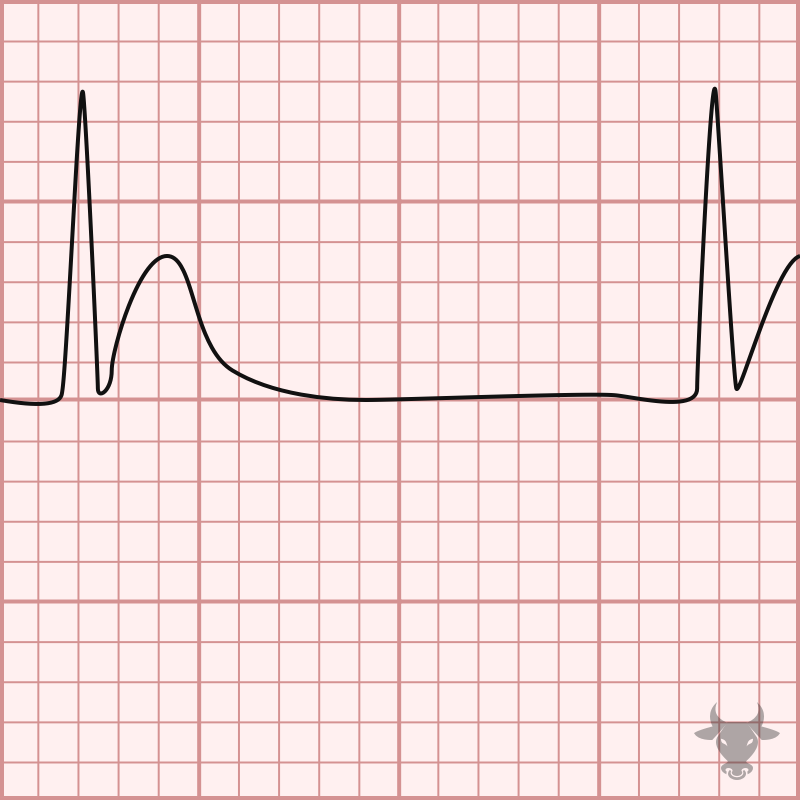
Cardiac: shortened QT interval, dysrhythmias
Signs and symptoms of hypercalcemia
SADH
Heart failure
Excessive administration of D5W IV
Vomiting
Diarrhea
Diaphoresis
Thiazide diuretics
Etiology of hyponatremia:
Low ECF sodium content lowers serum osmolality. Water shifts from ECF into ICF, causing cells (especially brain cells) to swell.
Pathophysiology of hyponatremia:
Neurological: Headache, confusion, lethargy, altered mental status, papilledema, seizures, coma
Musculoskeletal: Muscle cramps, weakness
GI: Anorexia, nausea, vomiting, abdominal cramps
Signs and symptoms of hyponatremia:
Serum sodium < 135 mEq/L
Serum osmolality < 280 mOsm/kg
Diagnostics for hyponatremia
Water restriction (primary therapy)
Medical management for hyponatremia when euvolemic or hypervolemic
0.9% normal saline IV
Medical management for hyponatremia when hypovolemic
Small doses of 3% hypertonic saline via central line
Medical management for hyponatremia when severe or symptomatic (< 120 mEq/L with seizures)
Seizure precautions (pad side rails, suction ready)
Monitor neurological status q1-2h
Nursing interventions for hyponatremia
Osmotic demyelination syndrome (Central pontine myelinolysis)
This occurs due to rapid correction of sodium (> 8-12 mEq/L in 24 hours) which causes irreversible neurological damage/paralysis. Correct serum sodium slowly!
Diabetes insipidus
Hypertonic tube feedings without adequate free water
Heat stroke
Hyperventilation
Water deprivation
Etiology of hypernatremia
High ECF sodium increases ECF osmolality. Water shifts from ICF into ECF, causing cellular dehydration and neuronal shrinkage.
Pathophysiology of hypernatremia
Neurological: Restlessness, agitation, irritability, hallucinations, hyperreflexia, seizures
Physical: Intense thirst, dry, sticky mucous membranes, flushed, dry skin, swollen, red tongue
Signs and symptoms of hypernatremia
Loop and thiazide diuretics (Furosemide)
GI losses (vomiting, diarrhea, NGT suctioning)
Metabolic alkalosis
Hyperaldosteronism
Persistent insulin administration
Etiology of hypokalemia
Low extracellular potassium impairs membrane resting potential, causing hyperexcitability followed by refractory unresponsiveness in neuromuscular and cardiac tissues
Pathophysiology of hypokalemia
Cardiac: Dysrhythmias, increased sensitivity to Digitalis toxicity
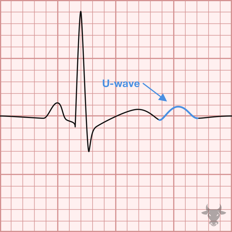
ECG changes: Flattened/inverted T waves, ST depression, prominent U waves
Neuromuscular: Muscle weakness, leg cramps, fatigue, paresthesias
GI: Decreased motility, constipation, paralytic ileus
Signs and symptoms of hypokalemia
Never IV push, IV bolus, or IM
Must be diluted
Infusion pump only (max rate: 10-20 mEq/hr)
Assess renal output before giving: Urine output must be at least 30 ml/hr (“No pee = No K+)
How is potassium administered?
Oral potassium replacement (take with food to avoid GI upset)
IV potassium chloride replacement for severe deficiency
Medical management for hypokalemia
Acute/chronic renal failure
Addison’s disease
Potassium-sparing diuretics (spironolactone)
Tissue trauma/burns (cell lysis)
Metabolic acidosis
Etiology of hyperkalemia
High ECF potassium partially depolarizes cell membranes, altering cardiac conduction speed and muscle contractility
Pathophysiology of hyperkalemia
Cardiac: Bradycardia, heart block, ventricular fibrillation, cardiac arrest
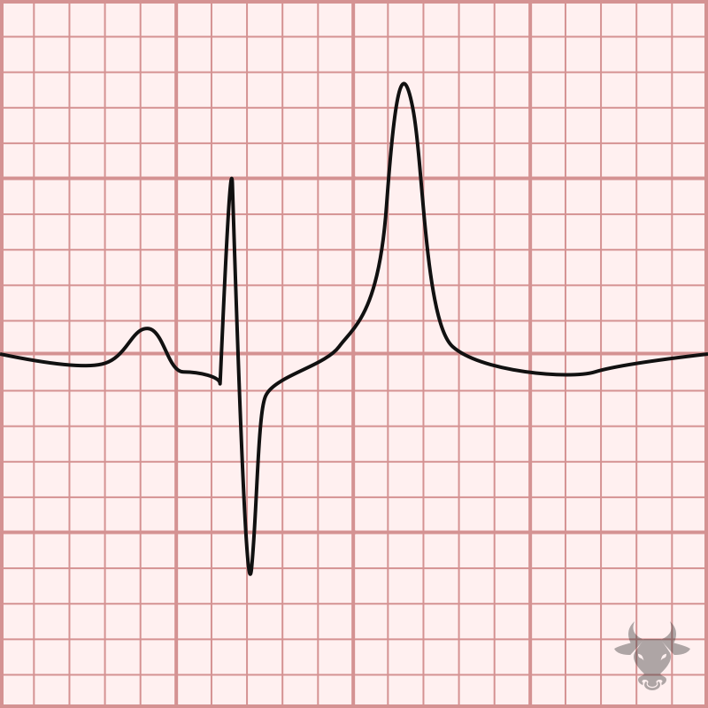
ECG changes: Tall, peaked T waves, wide QRS complex, prolonged PR interval, flat P wave
Neuromuscular: Paresthesias, muscle twitching → flaccid muscle paralysis
GI: Hyperactive bowel sounds, abdominal cramping, diarrhea
Signs and symptoms of hyperkalemia
IV calcium gluconate
Hyperkalemia emergency medical management
Antagonizes cardiac membrane excitability (protects heart)
Speed of onset is 1-3 minutes (immediate)
IV regular insulin + D50
Hyperkalemia emergency medical management
Shifts potassium into cells
Speed of onset is 15-30 minutes
Inhaled albuterol
Hyperkalemia emergency medical management
Also shifts potassium into cells
Speed of onset is also 15-30 minutes
Sodium bicarbonate
Hyperkalemia emergency medical management
Alkalinizes plasma, shifting potassium into cells
Speed of onset is 15-30 minutes
Sodium polystyrene sulfonate (Kayexalate)
Hyperkalemia emergency medical management
Binds potassium in bowel for fecal excretion
Speed of onset is hours (slow)
Loop diuretics/dialysis
Hyperkalemia emergency medical management
Removes potassium from the body
Speed of onset is variable/definitive
Hypoparathyroidism (accidental removal during thyroidectomy)
Vitamin D deficiency
Acute pancreatitis
Massive blood transfusions (citrate toxicity)
Chronic kidney disease
Etiology of hypocalcemia
Low serum calcium increases nerve cell membrane permeability to sodium, causing neuromuscular hyperexcitability and involuntary muscle contractions.
Pathophysiology of hypocalcemia
Chvostek’s sign
Trousseau’s sign
Tetany, circumoral paresthesia (tingling lips/fingers), laryngospasm
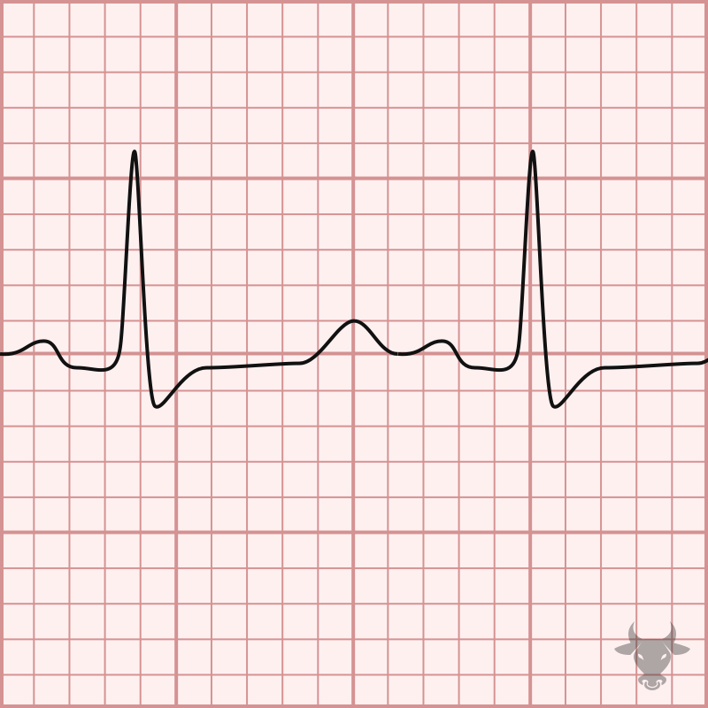
ECG: Prolonged QT interval → risk for Torsades de Pointes
Signs and symptoms of hypocalcemia
Chvostek’s sign
Facial twitching when tapping the facial nerve (CN VII) (anterior to ear)
Trousseau’s sign
Occluding brachial artery with BP cuff → ischemia-induced carpopedal spasm (flexion of wrist/thumb)
Tracheostomy tray and IV calcium gluconate
In post-thyroidectomy nursing care, what should you keep at the bedside due to immediate risk of severe hypocalcemia and life-threatening laryngospasm?
Chronic alcoholism
Malbasorption syndromes (Celiac, Crohn’s)
Aminoglycoside antibiotics
Propton pump inhibitors
Diarrhea
Etiology of hypomagnesemia
Magnesium controls the sodium-potassium pump and cellular energy (ATP). Deficiency leads to increased neuromuscular hyperexcitability (similar to hypocalcemia)
Pathophysiology of hypomagnesemia
Hyperactive deep tendon reflexes (DTRs, +3/+4)
Positive Chvostek’s and Trosseau’s signs
Neuromuscular: Tremors, tetany, seizures, altered mood
Cardiac: ECG shows prolonged PR/QT intervals, Torsades de Pointes (polymorphic VTach)
Signs and symptoms of hypomagnesemia
Hyperkalemia
Identify the electrolyte imbalance based on the ECG:
Peaked T wave
Wide QRS complex
Flat P wave
Hypokalemia
Identify the electrolyte imbalance based on the ECG:
Flat/inverted T wave
ST depression
U wave
Hypercalcemia
Identify the electrolyte imbalance based on the ECG:
Shortened ST segment
Shortened QT interval
Wide T wave
Hypocalcemia
Identify the electrolyte imbalance based on the ECG:
Prolonged ST segment
Prolonged QT interval
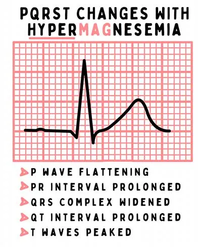
Hypermagnesemia
Identify the electrolyte imbalance based on the ECG:
Prolonged PR interval
Widened QRS complex
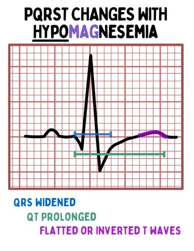
Hypomagnesemia
Identify the electrolyte imbalance based on the ECG:
Tall T wave
ST depressed
Prolonged QT interval
Renal failure
Excessive administration of magnesium during eclampsia treatment
Overuse of Mg-containing antacids/laxatives (milk of magnesia)
Etiology of hypermagnesemia
High magnesium depresses the central nervous system and neuromuscular junction, blocking acetylcholine release
Pathophysiology of hypermagnesemia