Lab 15 & 16 assignment 2 - SET DEFINITIONS FIRST
1/46
There's no tags or description
Looks like no tags are added yet.
Name | Mastery | Learn | Test | Matching | Spaced | Call with Kai |
|---|
No analytics yet
Send a link to your students to track their progress
47 Terms
d. Perilymph
The scala vestibuli contains
a. Blood
b. Lymph
c. Air
d. Perilymph
False
High-pitched sounds with short wavelengths displace the basilar membrane far from the oval window.
True/False
d. Displacement of the basilar membrane
Hair cells detect
a. Total volume of perilymph
b. Displacement of the oval window
c. Only low-pitched, long wavelength sounds
d. Displacement of the basilar membrane
a. Dampen vibrations in the perilymph
The function of the round window is to
a. Dampen vibrations in the perilymph
b. Receive vibrations of the middle ear bones
c. Support the hair cells
d. Produce perilymph
c. Axons of retinal ganglion cells
The optic nerves are composed of
a. Central processes of all cells in the neural retina
b. Central processes of cone cells
c. Axons of retinal ganglion cells
d. Central processes of rod cells
c. Refracts light through the pupil
The cornea
a. Is bi-concave
b. Is ineffective in redirecting light rays
c. Refracts light through the pupil
d. Is opaque
d. Retinal ganglion cells
In the neural retina, action potentials are generated by
a. Cone cells
b. Rod cells
c. Rod and cone cells
d. Retinal ganglion cells
a. Cone cells
High spatial resolution in vision is a function of
a. Cone cells
b. Rod cells
c. Retinal ganglion cells
d. Retinal pigment epithelium
d. Retinal pigment epithelium
Light scattering in the eye is prevented by the...
a. Lens
b. Shape of the eye
c. Cornea
d. Retinal pigment epithelium
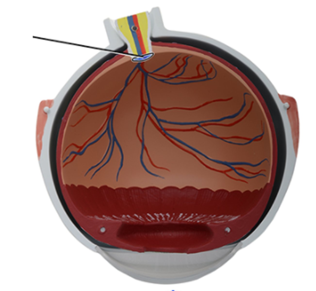
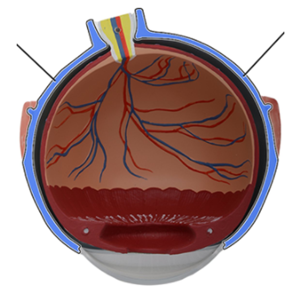
a. Optic nerve (CN II)
Which structure is highlighted and indicated by the leader line?
a. Optic nerve (CN II)
b. Retina
c. Central artery of retina
d. Choroid
e. Optic disc
f. Central vein of retina
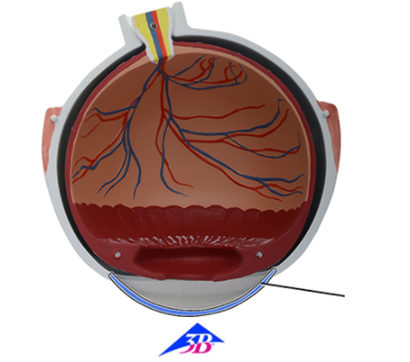
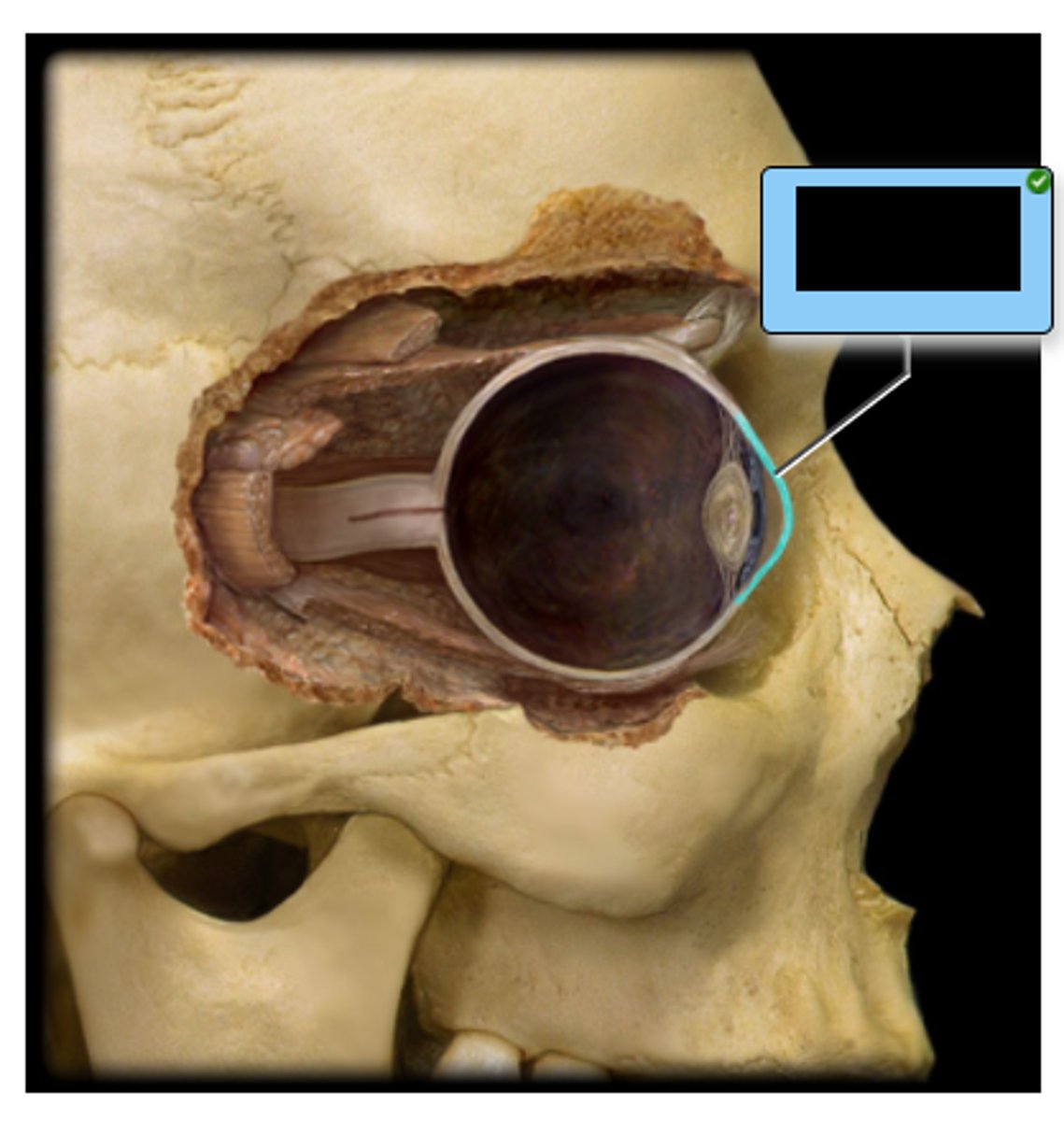
a. Cornea
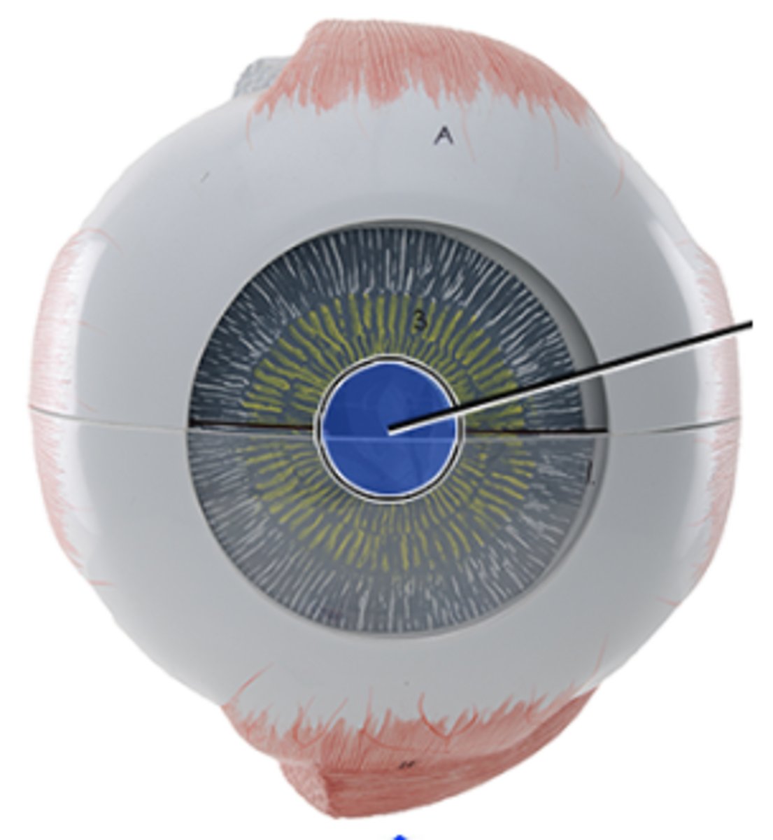
Which structure is highlighted and indicated by the leader line?
a. Cornea
b. Ciliary body
c. Sclera
d. Iris
e. Suspensory ligaments
f. Lens
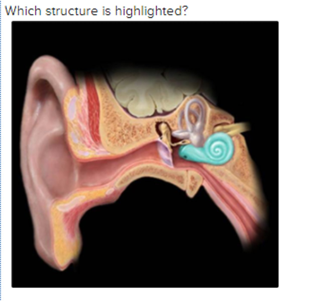
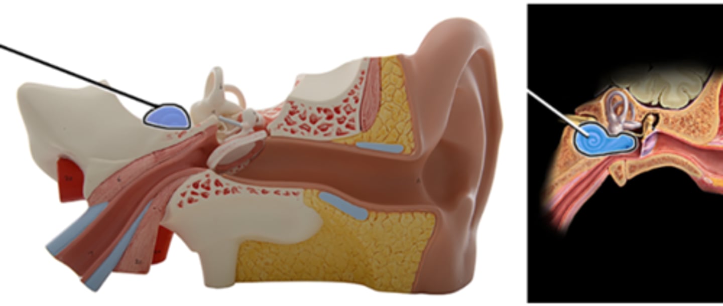
a. cochlea
Which structure is highlighted?
a. cochlea
b. vestibule
c. semicircular ducts
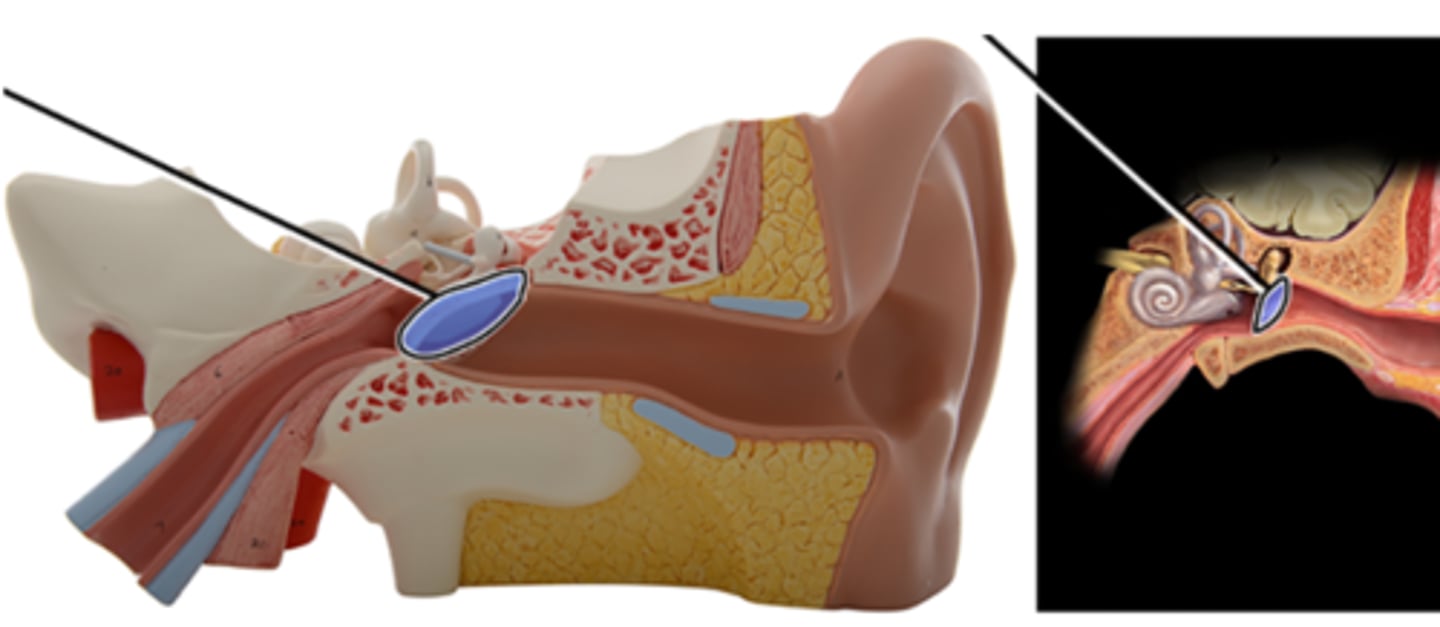
d. tympanic membrane
e. auditory ossicles
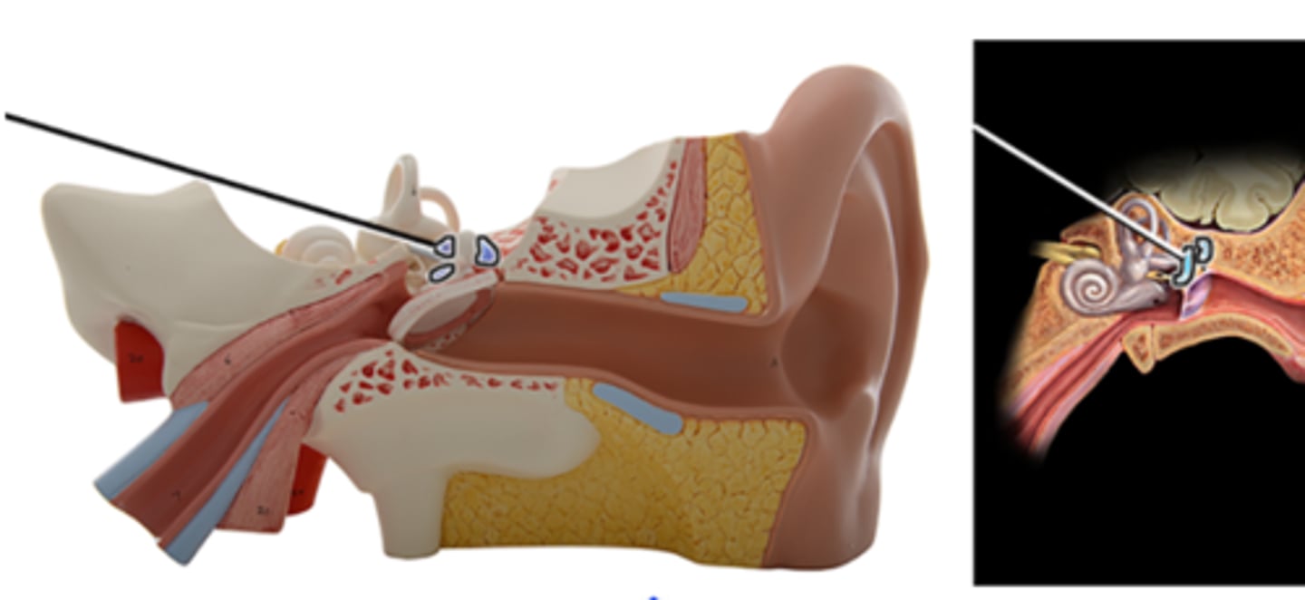
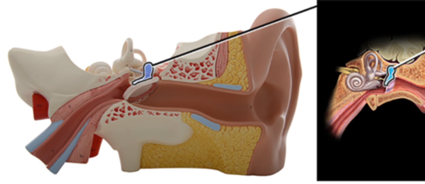
c. Incus
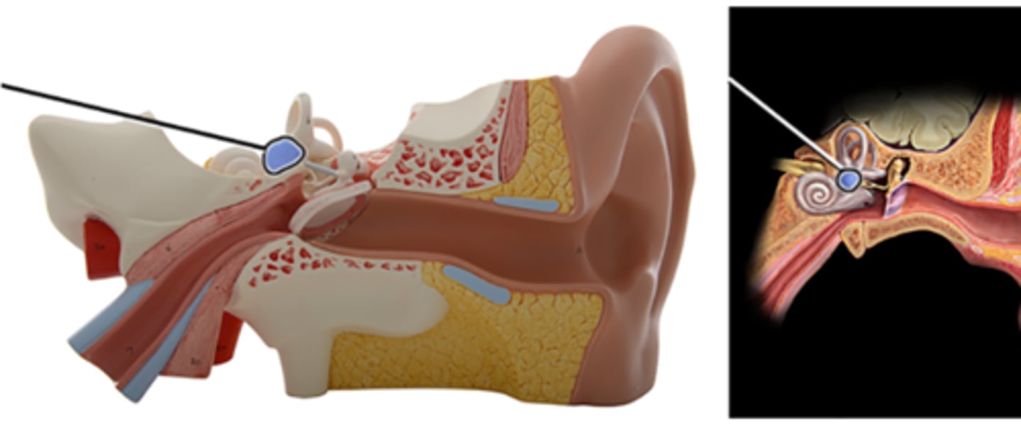
Which structure is highlighted and indicated by the leader line?
a. Stapes
b. Vestibule
c. Incus
d. Semicircular ducts
e. Cochlea
f. Malleus
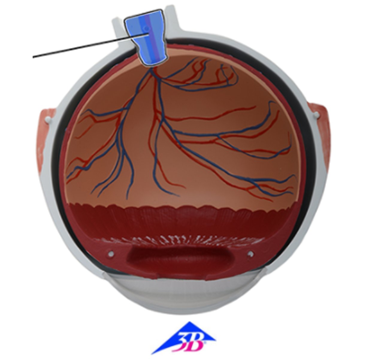
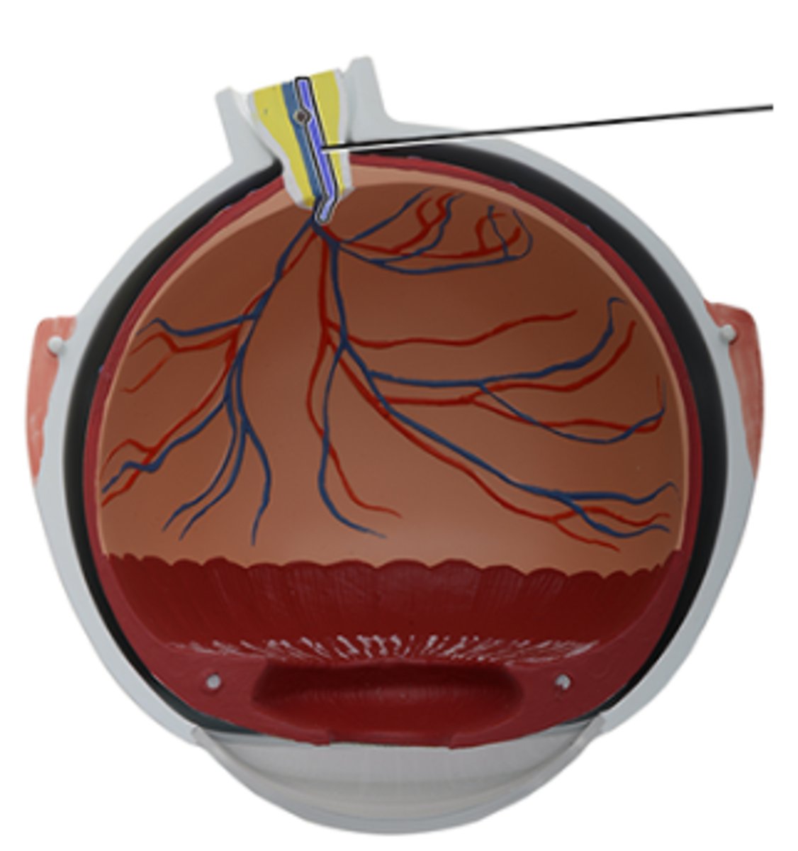
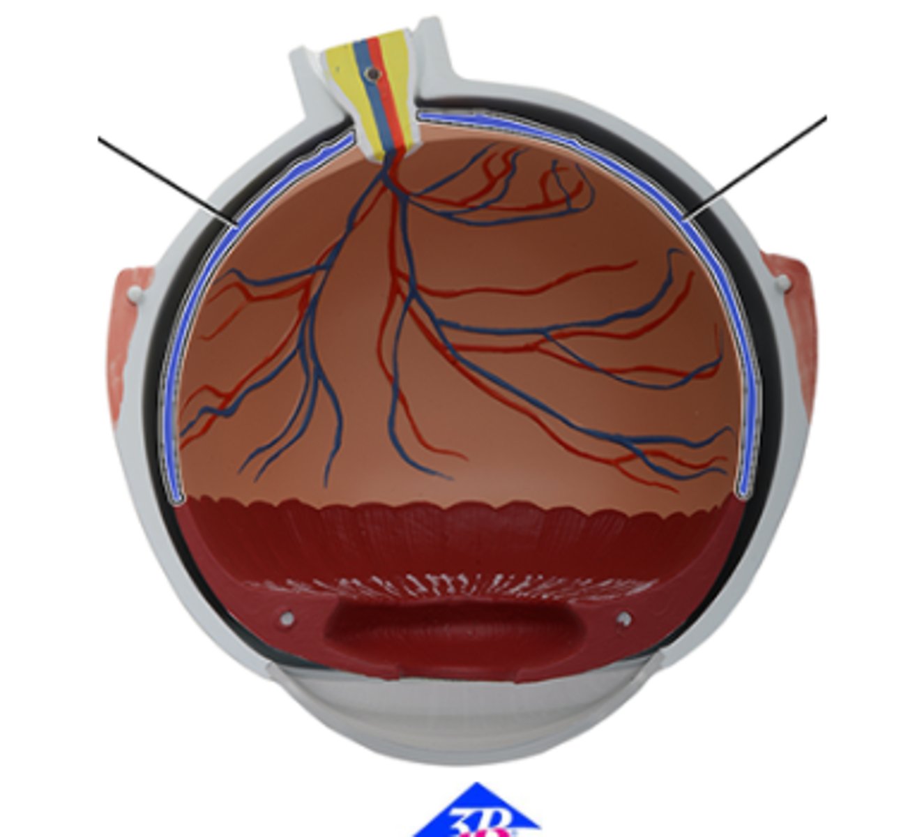
b. Central artery of retina
Which structure is highlighted and indicated by the leader line?
a. Optic nerve (CN II)
b. Central artery of retina
c. Central vein of retina
d. Choroid
e. Optic disc
f. Retina
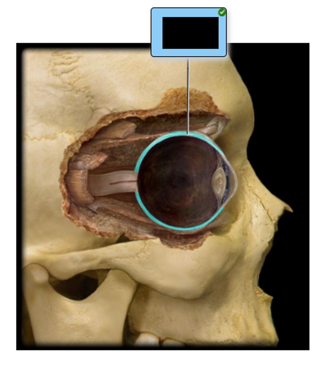
a. Sclera
Which structure is highlighted and indicated by the leader line?
a. Sclera
b. Choroid
c. Retina
d. Lens
e. Cornea
c. retina
Which structure is highlighted?
a. cornea
b. sclera
c. retina
d. choroid
e. lens
a. Optic disc
Which structure is highlighted and indicated by the leader line?
a. Optic disc
b. Choroid
c. Central artery of retina
d. Central vein of retina
e. Retina
f. Optic nerve (CN II)
f. Cornea
Which structure is comprised of transparent connective tissue?
a. Ciliary body
b. Choroid
c. Anterior chamber
d. Lens
e. Sclera
f. Cornea
e. Sclera
Identify the structure, which comprises the outer layer of the posterior five-sixths of the eye.
a. Retina
b. Choroid
c. Ora serrata
d. Cornea
e. Sclera
b. vestibule of ear
Which structure is highlighted?
a. cochlea
b. vestibule of ear
c. auricle
d. tympanic membrane
e. semicircular ducts
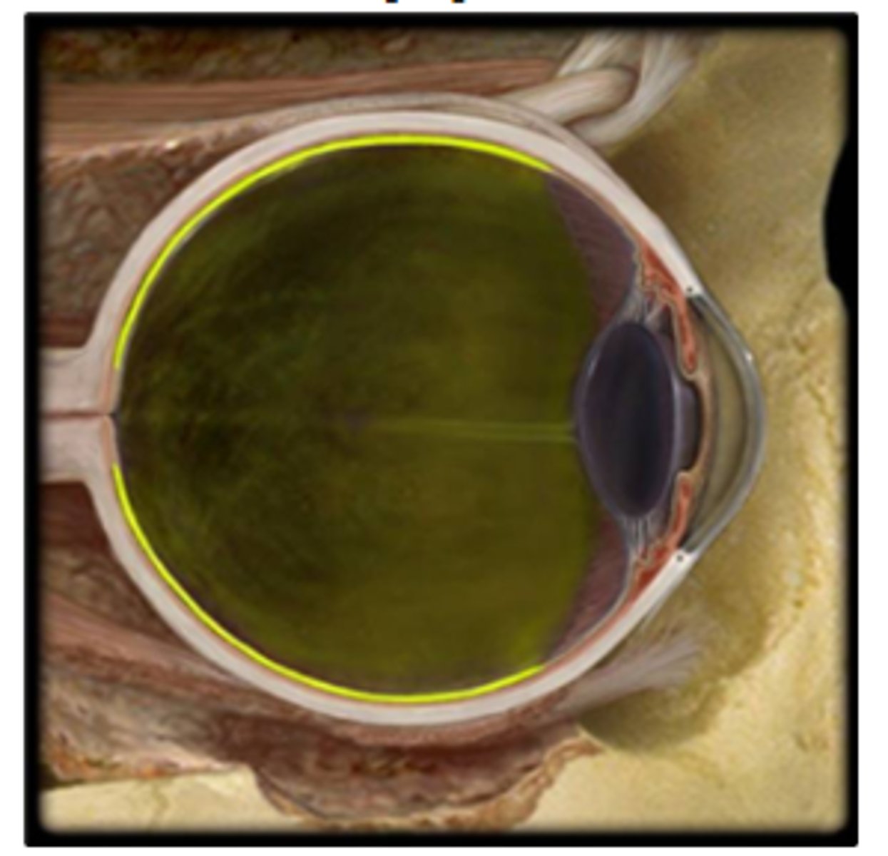
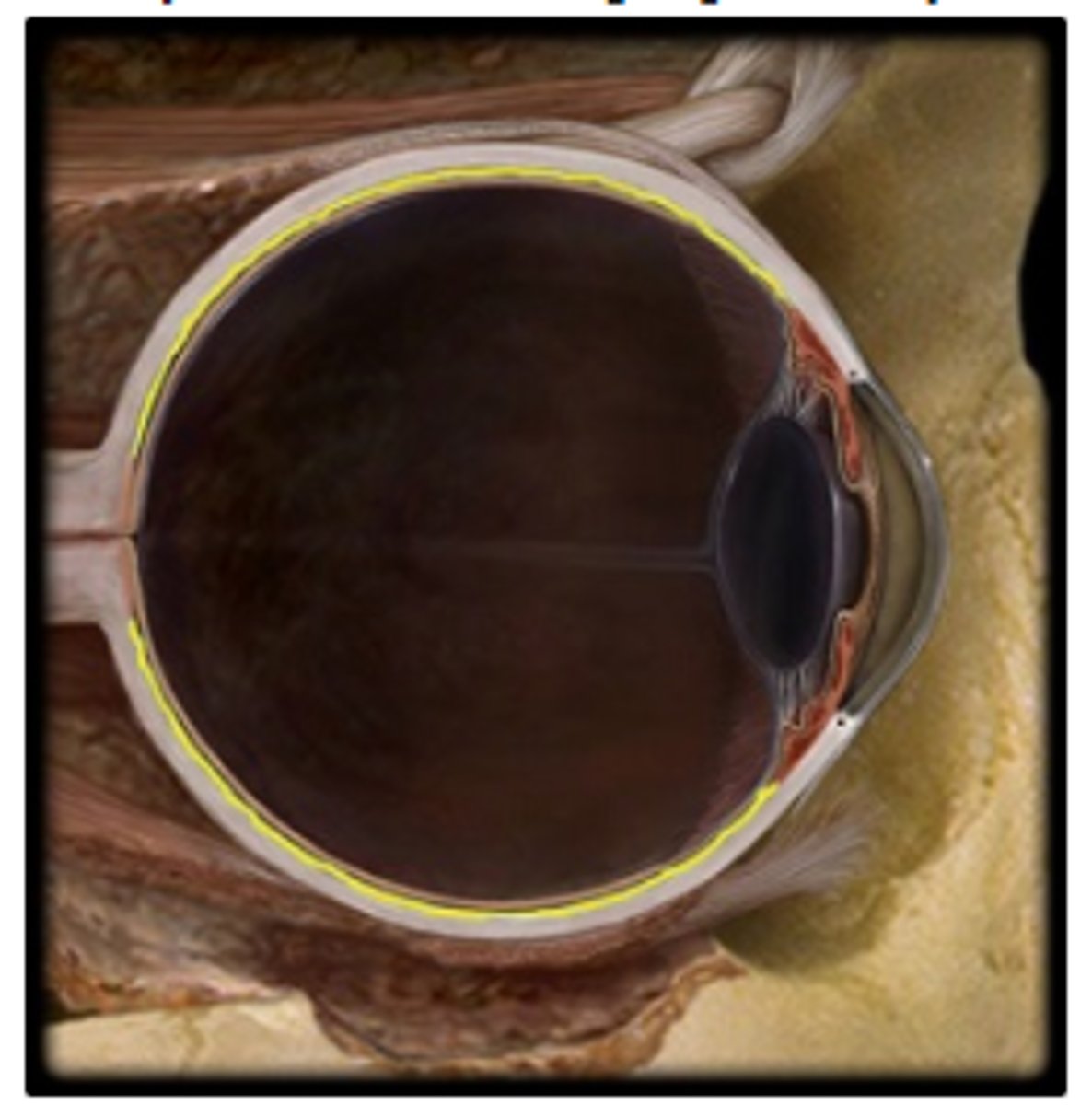
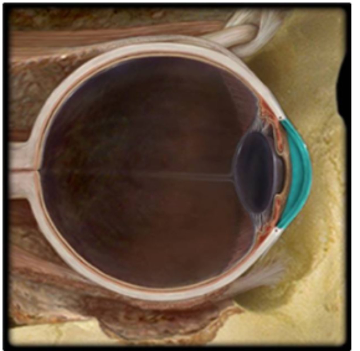
c. choroid
Identify the structure highlighted in yellow.
a. lens
b. sclera
c. choroid
d. cornea
e. iris
f. Choroid
Which structure is highlighted and indicated by the leader line?
a. Optic nerve (CN II)
b. Central vein of retina
c. Optic disc
d. Central artery of retina
e. Retina
f. Choroid
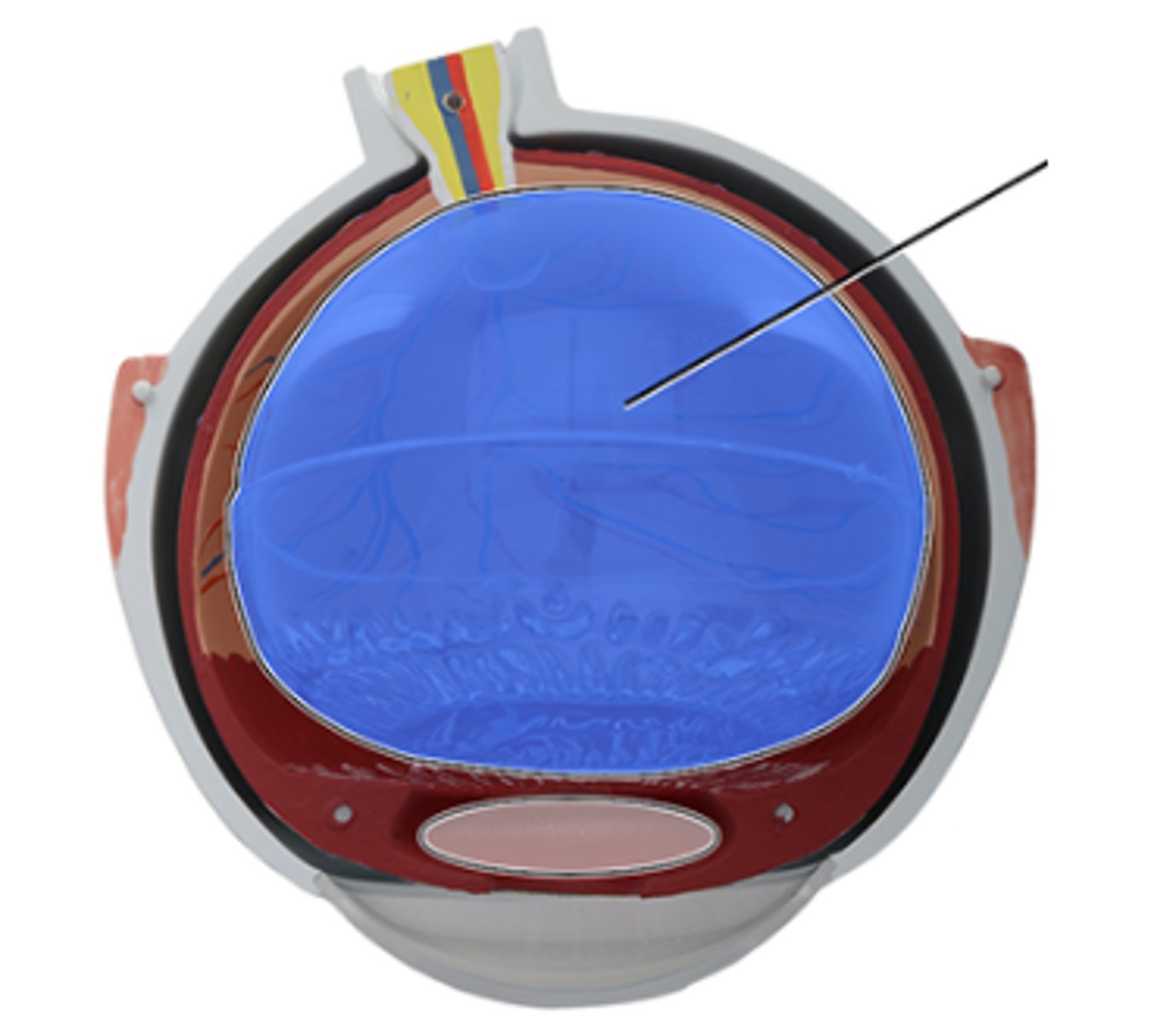
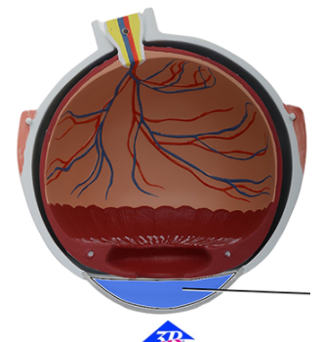
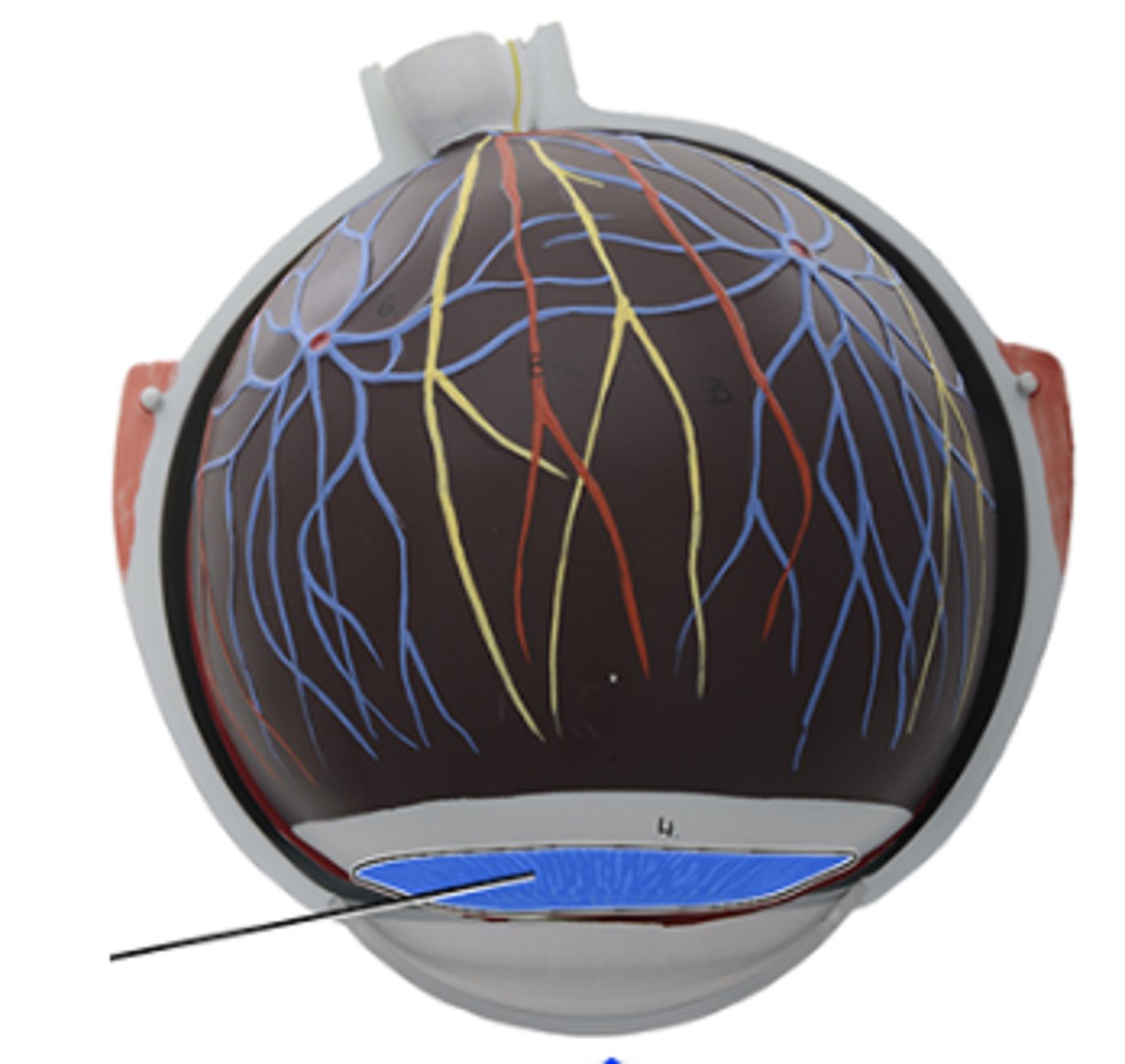
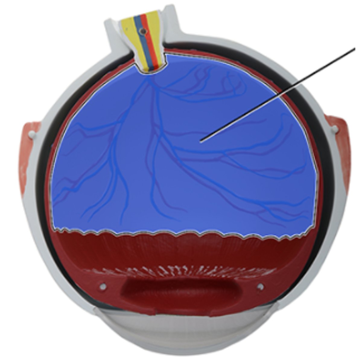
e. Posterior cavity
Which structure is highlighted and indicated by the leader line?
a. Anterior chamber
b. Retina
c. Lens
d. Choroid
e. Posterior cavity
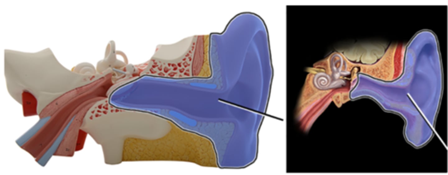
b. External ear
Which structure is highlighted and indicated by the leader line?
a. Inner ear
b. External ear
c. Middle ear
d. Cochlear
e. Occipital bone
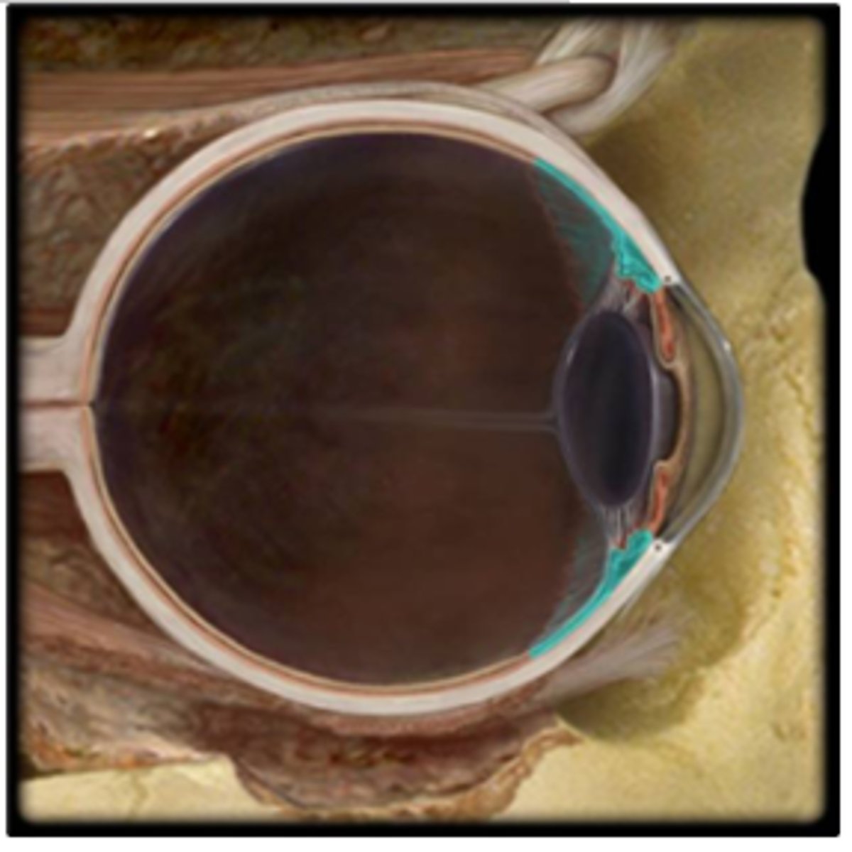
c. Anterior chamber
Which structure is highlighted and indicated by the leader line?
a. Sclera
b. Ciliary body
c. Anterior chamber
d. Suspensory ligaments
e. Ora serrata
c. External acoustic meatus
Which structure is highlighted and indicated by the leader line?
a. Auricle
b. Tympanic membrane
c. External acoustic meatus
d. Temporal bone
e. Auditory tube

a. ciliary body
Which structure is highlighted?
a. ciliary body
b. pupil
c. cornea
d. iris
e. lens
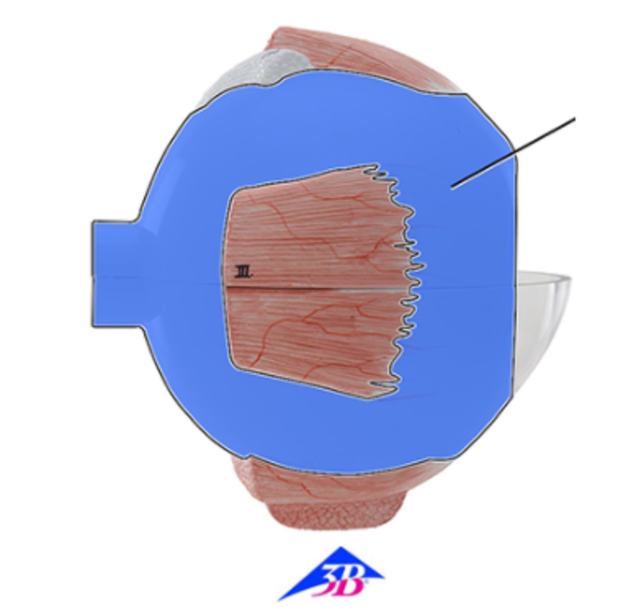
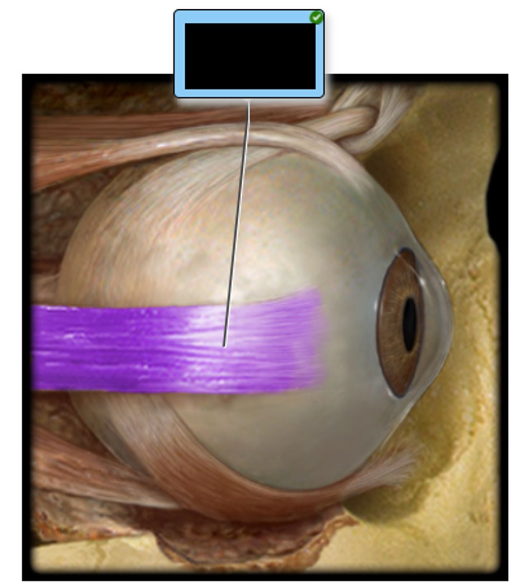
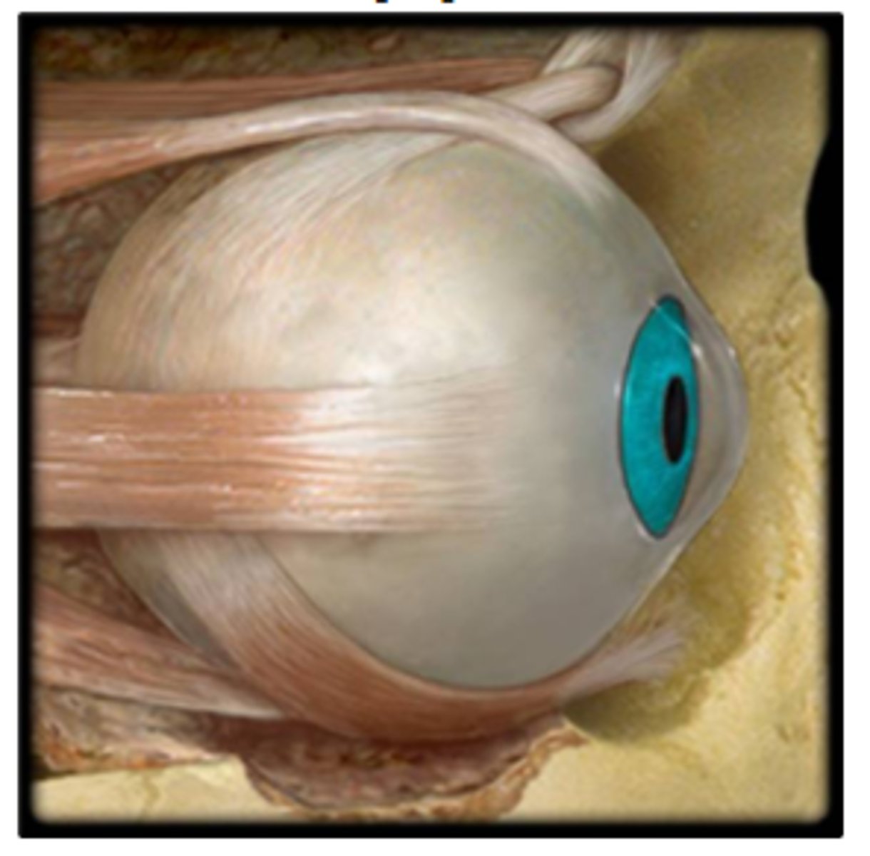
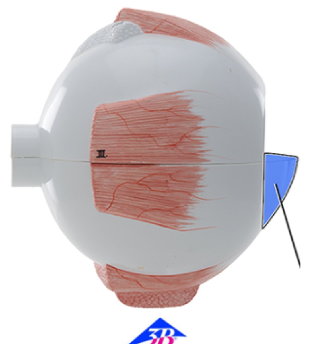
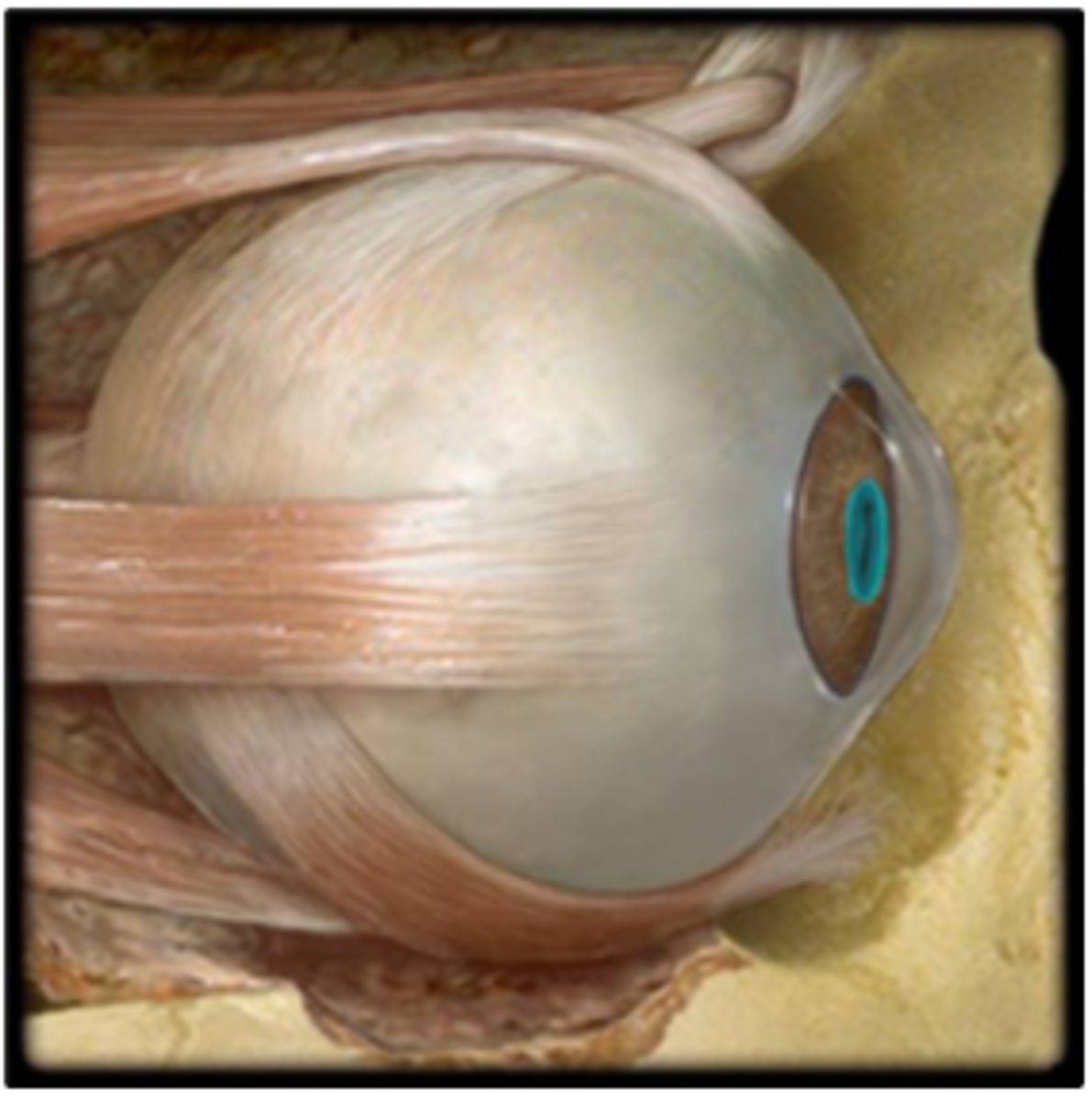
a. Abduction of eyeball
Indicate the action of the highlighted muscle of the eye.
a. Abduction of eyeball
b. Dilation of pupil
c. Elevation of eyeball
d. Adduction of eyeball
e. Depression of eyeball
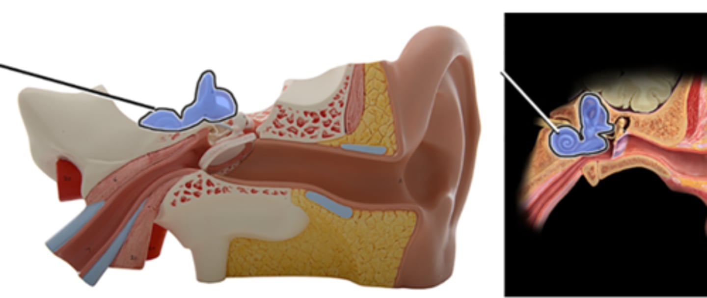
b. Inner ear
Which structure is highlighted and indicated by the leader line?
a. Middle ear
b. Inner ear
c. Tympanic membrane
d. External ear
e. Occipital bone
c. Pupil
Which structure is highlighted and indicated by the leader line?
a. Cornea
b. Iris
c. Pupil
d. Sclera
e. Ciliary body
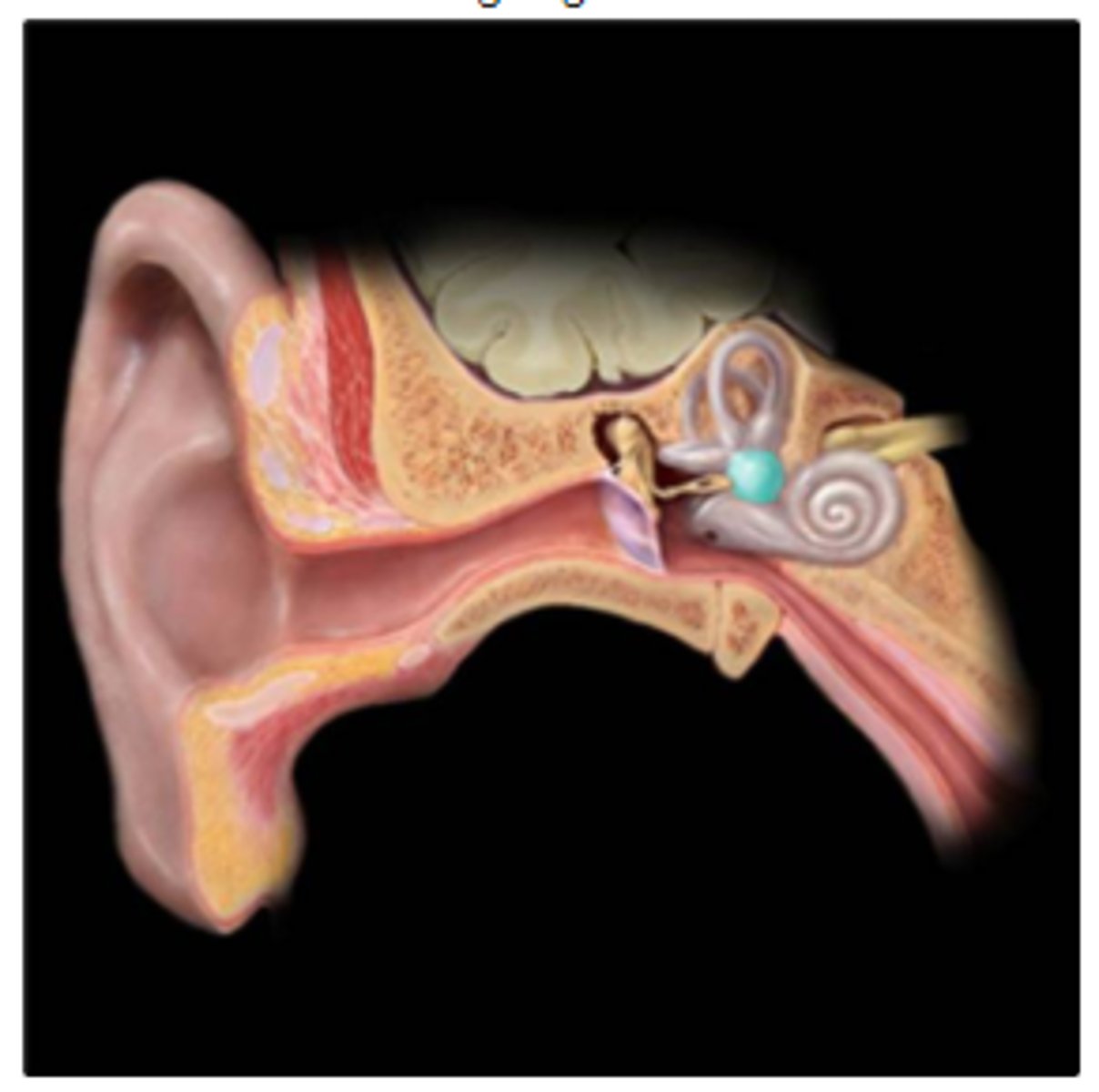
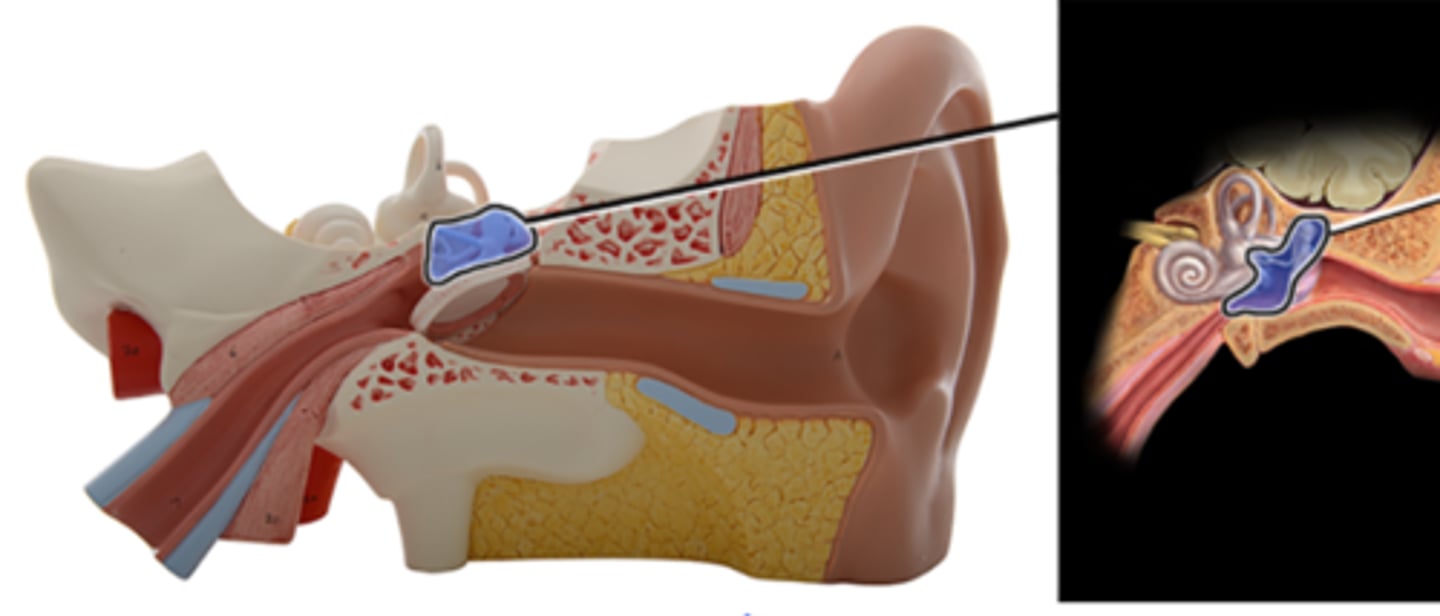
c. Vestibule
Which structure is highlighted and indicated by the leader line?
a. Semicircular ducts
b. Cochlea
c. Vestibule
d. Stapes
e. Malleus
f. Incus
f. Stapes
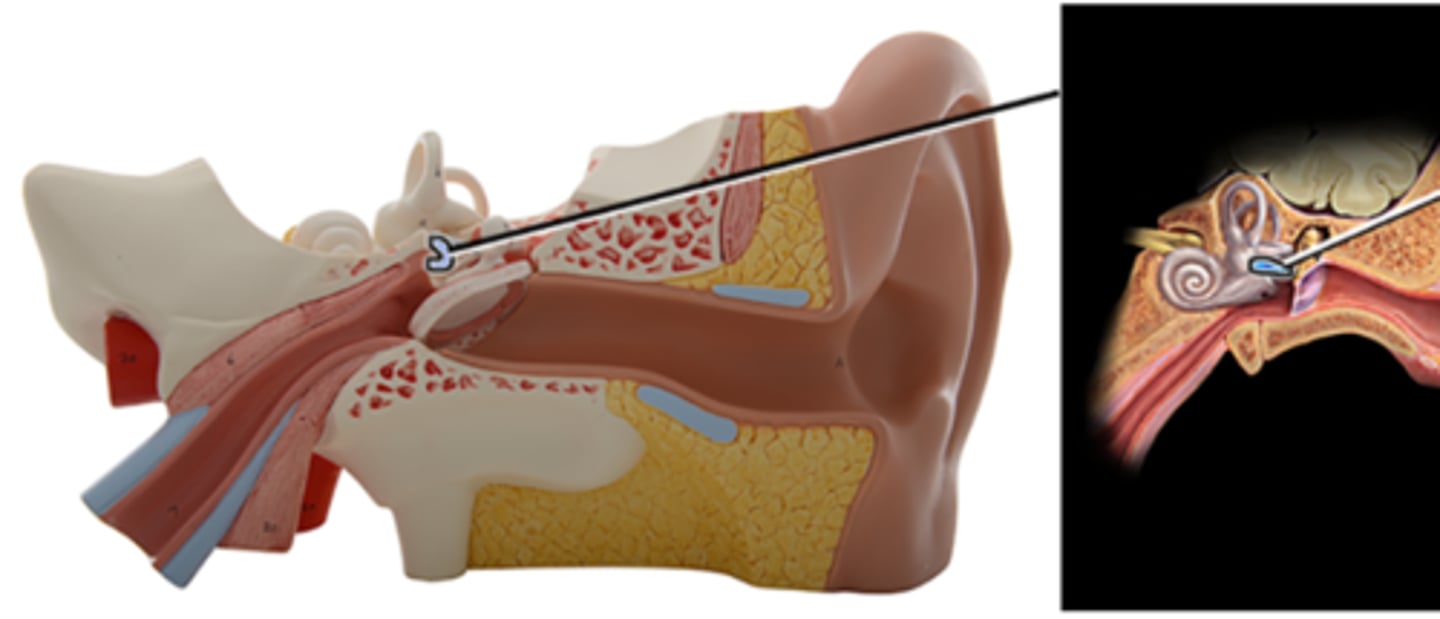
Which structure is highlighted and indicated by the leader line?
a. Malleus
b. Semicircular ducts
c. Incus
d. Vestibule
e. Cochlea
f. Stapes
e. Iris
Which structure is highlighted and indicated by the leader line?
a. Cornea
b. Ciliary body
c. Sclera
d. Choroid
e. Iris
f. Retina
Which structure is highlighted and indicated by the leader line?
a. Choroid
b. Optic nerve (CN II)
c. Optic disc
d. Central vein of retina
e. Central artery of retina
f. Retina
d. iris
a. choroid
b. cornea
c. pupil
d. iris
e. lens
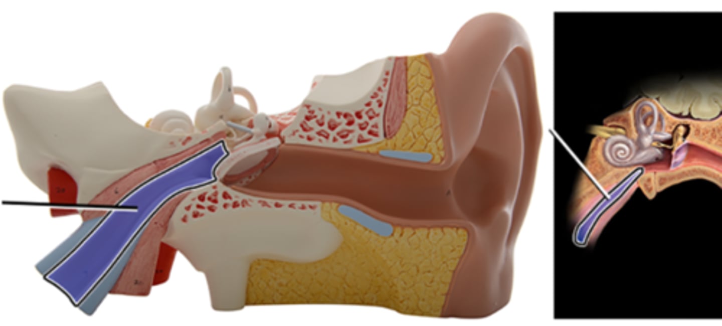
a. Auditory tube
a. Auditory tube
b. Auricle
c. Tympanic membrane
d. Temporal bone
e. External acoustic meatus
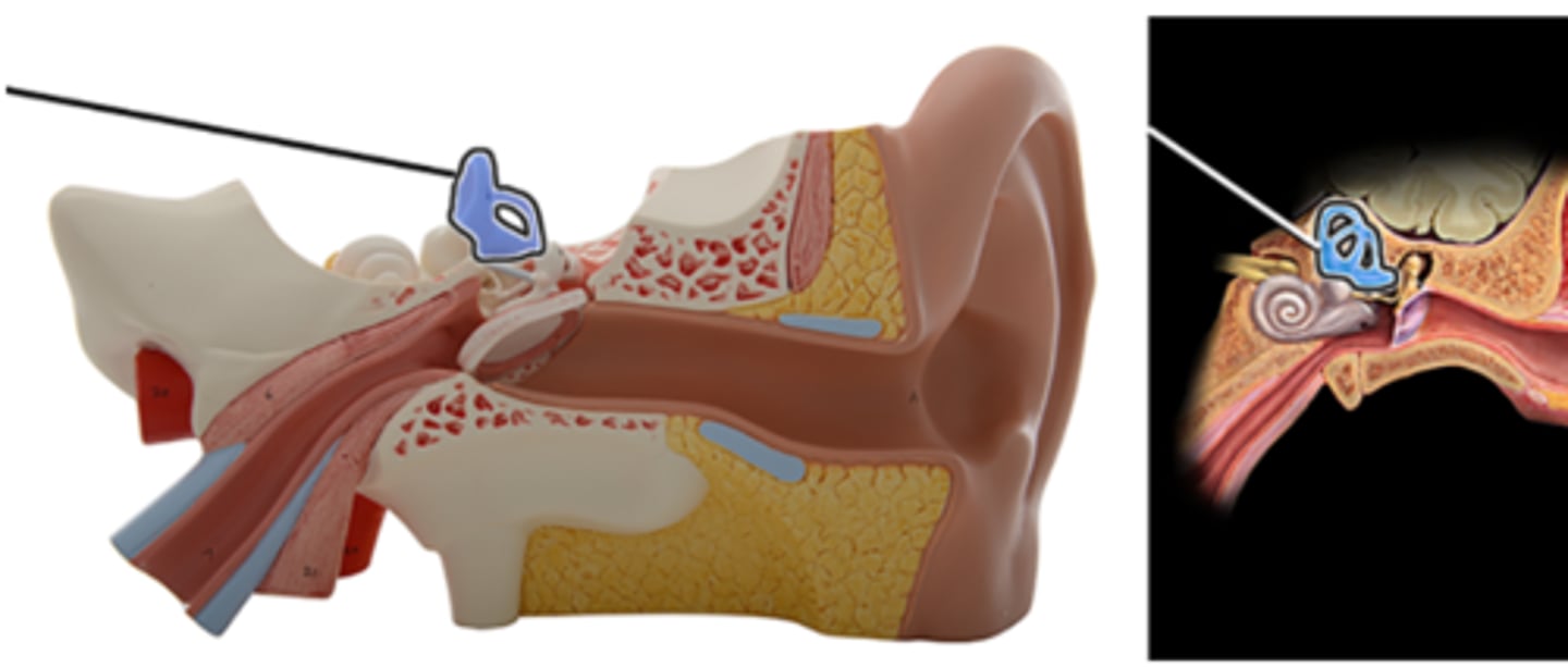
d. Semicircular canals
a. Vestibule
b. Incus
c. Malleus
d. Semicircular ducts
e. Stapes
f. Cochlea
a. Malleus
a. Malleus
b. Semicircular ducts
c. Cochlea
d. Stapes
e. Vestibule
f. Incus
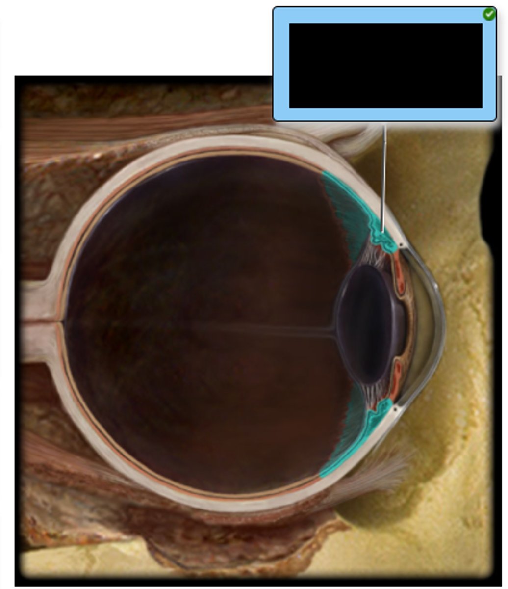
d. Ciliary body
Which structure controls the tension of the suspensory ligaments of the lens?
a. Iris
b. Choroid
c. Ora Serrata
d. Ciliary body
e. Optic disc
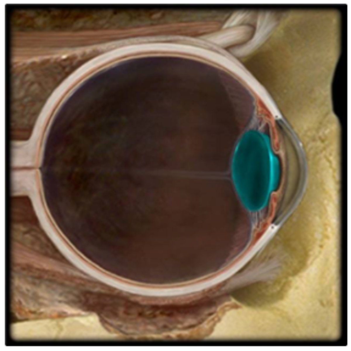
e. lens
a. cornea
b. sclera
c. choroid
d. iris
e. lens
a. Cornea
a. Cornea
b. Lens
c. Choroid
d. Sclera
e. Retina
b. pupil
a. cornea
b. pupil
c. choroid
d. iris
e. lens
a. Cochlea
a. Cochlea
b. Stapes
c. Semicircular ducts
d. Vestibule
e. Incus
f. Malleus
e. cornea
a. sclera
b. choroid
c. lens
d. iris
e. cornea
d. Middle ear
a. Auricle
b. Inner ear
c. Cochlear
d. Middle ear
e. External ear
b. Sclera
a. Choroid
b. Sclera
c. Retina
d. Ciliary body
e. Suspensory ligaments
f. Cornea
e. Tympanic membrane
a. Temporal bone
b. Auditory tube
c. External acoustic meatus
d. Auricle
e. Tympanic membrane