Macroscopic anatomy of the urinary tract
 Call Kai
 Learn
Learn Practice Test
 Spaced Repetition
 Match
Match Flashcards
 Knowt Play
1/123

There's no tags or description
Looks like no tags are added yet.
Name | Mastery | Learn | Test | Matching | Spaced | Call with Kai | Chat |
|---|
No analytics yet
Send a link to your students to track their progress
124 Terms

Identify what has been lebelled here
RK = right kidney
LK = left idney
B = bladder
P = prostate
Sp = spleen
U = ureter
Ur = urinary bladder
what is the product from the kidneys?
urine
Describe the location of the kidneys within the abdomen
retro-peritoneal, dorsal abdomen
in ventral lumbar region with limited mobility
right kidney is most cranial
left kidney is most mobile
in which species is the kidney more mobile?
cats
In which species is the right kidney not the most cranial kidney?
pig
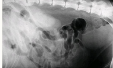
Identify the kidenys
Remember right is more cranial than left

what is the cranial landmark of the left kidney?
greater curvature of the stomach
what does the left kidney attach to dorsomedially
spleen
what may the left kidney be in contact with cranially?
left limb of pancreas
what is attached to the left kidney medially?
left adrenal gland
what does the left kidney attach to caudally?
small intestine and descending colon
in females what may the left kidney be in contact with?
ovary
what does the right kidney attach to cranially?
renal fossa of caudate lobe of the liver
what is the right kidney attached to caudally?
caudal vena cava
what is the right kidney attached to dorsolaterally?
right adrenal gland
what is ventral to the right kidney?
descending duodenum
what is ventrally and medially to the right kidney?
right pancreatic limb
in females, what is attached to the right kidney?
ovary
what do we need to retract surgically to see the left kidney?
mesocolon
what do we ened to retract on the RHS of the abdomen surgically to visualise the right kidney?
mesoduodenum
state the 3 basic components of the kidney
outer fibrous capsule
renal cortex
renal medulla
what is the renal hilus?
area where the artery, vein and ureter enter/exit the kidney
what makes up a renal lobe?
cortex + pyramid + papilla
where do blood vessels travel in the kidney/
in the renal columns
what is each minor calyx fixed to?
one papilla
in which species is the kidney ‘bean shaped’
dog
cat
sheep
goat
when cutting sagitally through a kidney, what structure may be identifiable?
renal crest
describe the shape of renal kidneys in the horse
more triangular in shape
Right = ‘heart’ shaped
left = ‘pyramidal’ - J shaped (VD view)

describe the shape of porcine kidneys
long and flat
more rectangular
describe the structure of the bovine kidney
reniculate
which other species share a structure similar to that of a bovine kidney?
acquatic mammals
bears
elephants
what are the 2 regions of the cortex?
external
internal - juxtamedullary
what is found in the cortex
glomeruli
convoluted tubules
what do we find in the medulla?
loops of henle and collecting ducts
what are the parts of the renal medulla?
outer part = base of pyramid
inner part = renal papilla - only collecting ducts
describe unilobar kidneys
single renal lobe
single pyramid
single papilla
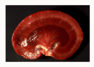
which species have a unilobar kidney?
rodents and lagomorphs
what kind of kidney do other animals, not rodents and lagomorphs have?
multilobar kidney
describe the structure of the kidney in cows, elephants, bears and acquatic mammals;
cortex?
medulla?
lobes?
renal papillae?
calyx?
pelvis?
surface appearance?
separate
separate
multiple
multiple
multiple
no
lobulated - reniculate

describe the appearance of the pig and primate kidney:
cortex?
medulla?
lobes/pyramids?
renal papillae?
calyx?
pelvis?
surface appearance?
fused
separate
obvious ones
multiple
multiple
branched
smooth
Describe the anatomy of the kidney of the dog, cat, sheep and horse:
cortex?
medulla?
lobes?
pyramids?
renal papillae?
calyx?
pelvis?
surface appearance?
fused
fused
internally, yes
partially merged
merge to form renal crest
no
single one
smooth

Fill in
globate
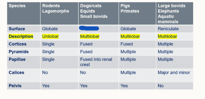
Fill in
globate
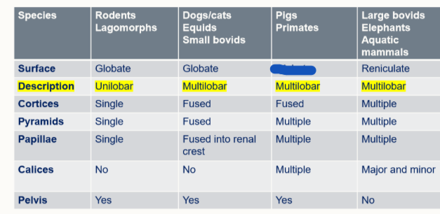
Fill in
globate

Fill in
reniculate

Fill in
unilobar

Fill in
multilobar
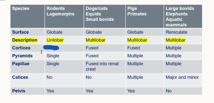
Fill in
single

Fill in
fused

Fill in
multiple

Fill in
single
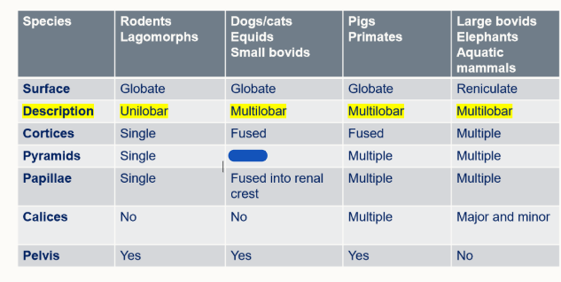
Fill in
fused

Fill in
multiple

Fill in
single
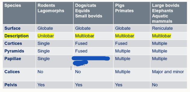
Fill in
fused into renal crest
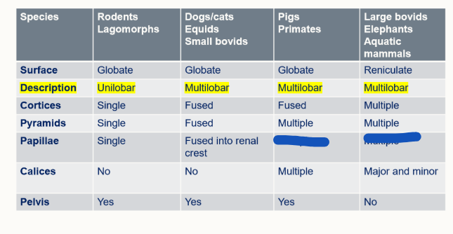
Fill in
multiple

Fill in
no
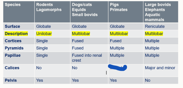
Fill in
multiple

Fill in
major and minor

Fill in
yes

Fill in
no
what are minor calyx?
where the urine first drains into from the medullary pyramids
what is a major calyx?
where 2-3 minor calyx drain into a bigger calyx = major calyx
what do pigs and primates have, which aren’t major/minor calyx?
renal pelvis - where the major calices drain into → ureter
in dogs, cat, sheep and horses, what structure do they have instead of calyx?
renale crest - as the medullary cones are fused
renal crest → renal pelvis
what do we call it in dogs and cats where the medullary pyramids are fused?
renal crest
what do we call the space the urine drains into in dogs and cats
renal pelvis
where is the renal pelvis located in dogs and cats
in the renal sinus
what intersting feature does the renal pelvis have ina dog and a cat we may visualise on radiography?
folds in its renal pelvis - called recesses
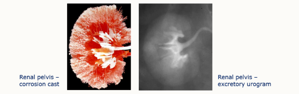
what are recesses
large, irregular structure with broad processes on the renal pelvis
in which species do we find recesses/
single lobe speices
how many recesses are there in the horse?
two
what are calyces?
small, irregular structure with short stems ending in dilated regions - pigs and primates
do cattle have a pelvis?
no
what do cattle have instead of a pelvis?
proximal ureter leading to multiple calyces
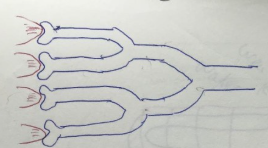
What species is this?
cattle

What species is this
pigs and people

What animals have this structure?
carnivores
sheep
goats
describe the basic flow of blood in the kidney
hilus
cortex
medulla
cortex
hilus
what additional system do we find around the nephrons?
intra-renal portal system

Name the vessels working from largest to smallest
aorta → renal a.
interlobar artery and vein
arcuate artery and vein
interlobulary artery and vein
renal vein → CVC
state the renal blood flow circuit
aorta
renal artery
inter-lobar artery
arcuate artery
inter-lobular artery
afferent glomerular arteriole
glomerulus
efferent glomerular arteriole
inter-lobular vein
arcuate vein
inter-lobar vein
renal vein
caudal vena cava
describ the blood supply to the:
cortical glomeruli
juxtamedullary glomeruli
peritubular capillaries
peritubular capillaries (cortex) and vasa recta (medulla)
describe the venous outflow from a nephron
venules drain peritubular capillaries and vasa recta → arcuate or interlobulary veins
DO NOT RE-ENTER BOWMAN’S CAPSUL
what supplies the blood to the PCT+DCT?
peritubular capillaries
what supplies the loop of henle blood?
vasa recta
what is the role of the peritubular capillaries and vasa recta?
resorb many solutes
why do the medulla and cortex need different blood supplies?
the solutes resorbed in the PCT and DCT would negatively impact the medullary tissue
what are end arteries?
arteries which don’t have a back-up supply/anastamoses with other vessels
what is the consequence of an end artery if it’s damaged?
the area of tissue it supplies will die and necrose off
what do all carnivores have an additional of in terms of blood supply?
cortical venous drainage
what is a cortical venous drainage in carnivores?
drain twoards hilum of kidney without going towards renal medulla
describe the cortical venous drainage in dogs
veins flow through cortex to hilum and join renal vein - anastamose with deep cortical veins
describe the cortical venous drainage in cats
subcapsulr veins run over cortex - distinctive feature - do not anastamose, unlike dog ones

What species kideny is this
cat
describe the wall of the bladder
mucous membrane
urolethium
tight junctions
lamina propria
muscular layer
adventitia
why does the baldder wall have tight junctions between its cells
to protect against urine
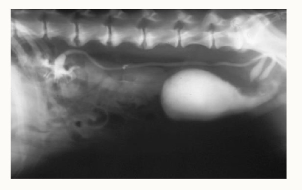
What has been demonstrated here by contrast?
abnormal ureters near bladder - likely due to a prostate tumour (near the neck of the bladder!)
what are the two portions of the ureters?
abdominal
pelvic