Exam 1: Values + condensed version
 Call Kai
 Learn
Learn Practice Test
 Spaced Repetition
 Match
Match Flashcards
 Knowt Play
1/141

There's no tags or description
Looks like no tags are added yet.
Name | Mastery | Learn | Test | Matching | Spaced | Call with Kai | Chat |
|---|
No analytics yet
Send a link to your students to track their progress
142 Terms
Sensitivity Formula
Number of true positive tests
Number of all positive tests by gold standard
Specificity Formula
Number of true negative tests
Number of all negative tests by gold standard
Positive Predictive Value (PPV) Formula
Number of true positive tests
Number of all positive noninvasive tests
Negative Predictive Value (NPV) Formula
Number of true negative tests
Number of all negative noninvasive tests
Accuracy Formula
Total number of correct tests
Total number of all tests
Celiac Artery PSV
101 cm/sec
Celiac Artery RI
0.7
70% Celiac Artery Stenosis PSV
≥ 200 cm/sec
Celiac Artery PSV with MALS
> 250 cm/sec during expiration that normalizes with inspiration
SMA PSV
113 cm/sec
70% SMA Stenosis PSV
≥ 275 cm/sec
IMA PSV
141 cm/sec
IMA Stenosis PSV
> 200 cm/sec
Left Gastric Vein (coronary vein) Diameter
> 6 mm
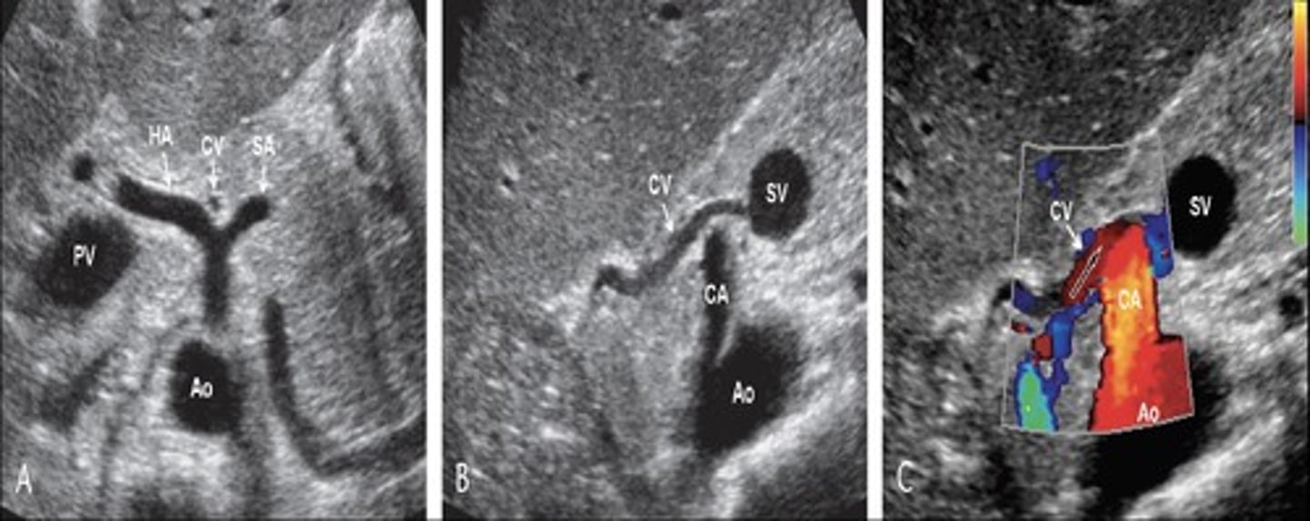
Main Portal Vein Diameter
≤ 13mm (resting)
≤ 16 mm (deep inhalation/valsalva)
Main Portal Vein PSV
16-31 cm/sec
Hepatic Vein Diameter
6 mm
≤ 9 mm (when CHF is present)
Hepatic Vein PSV
20-39 cm/sec
Hepatic Artery PSV
70-120 cm/sec
Hepatic Artery RI
0.5 - 0.7
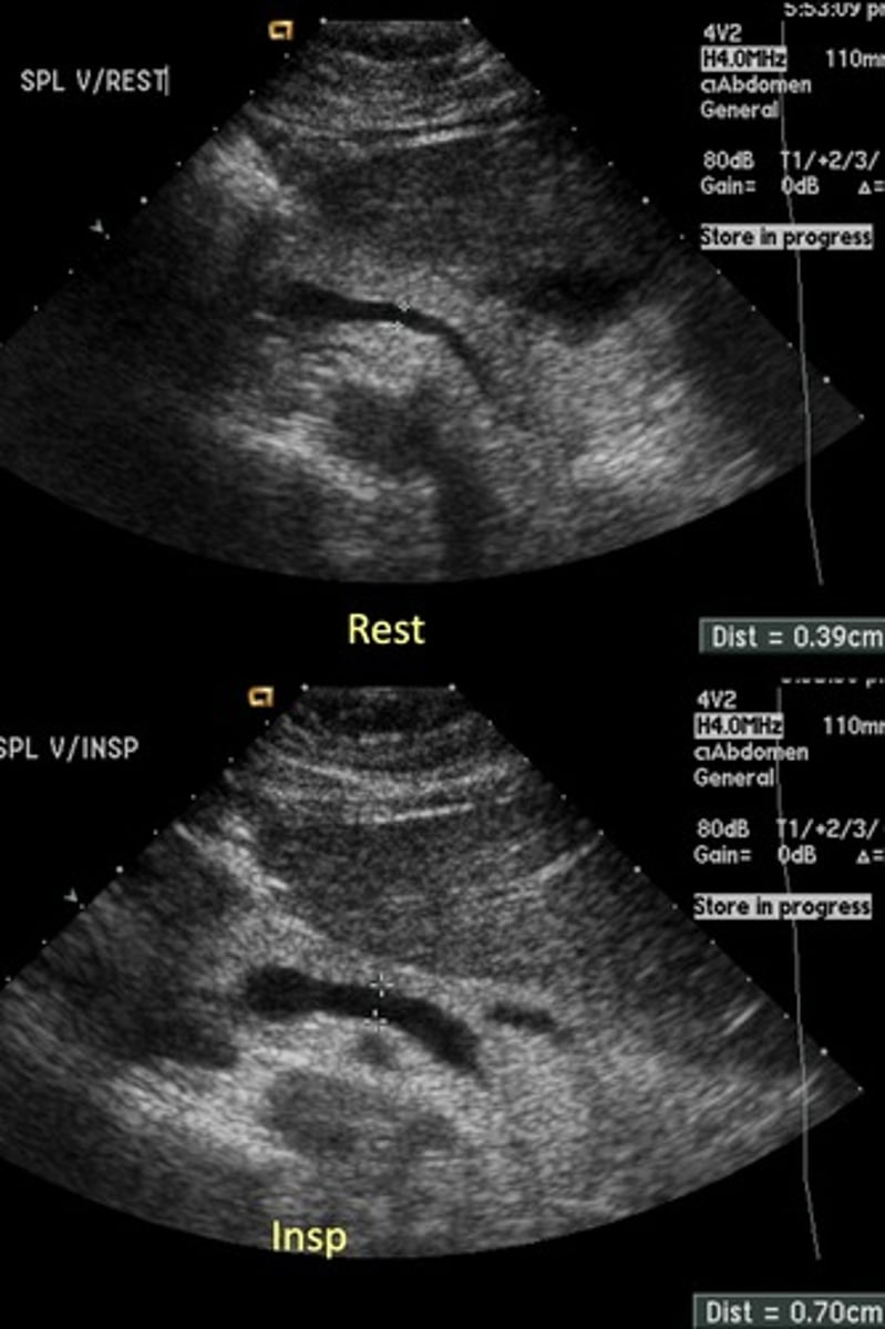
Splenic Vein Diameter
10 mm
Splenic Vein PSV
9-30 cm/sec
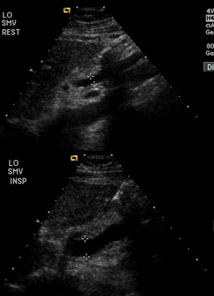
SMV Diameter
10 mm
SMV PSV
8-40 cm/sec
IVC Diameter
15-25 mm
valsalva = max diameter
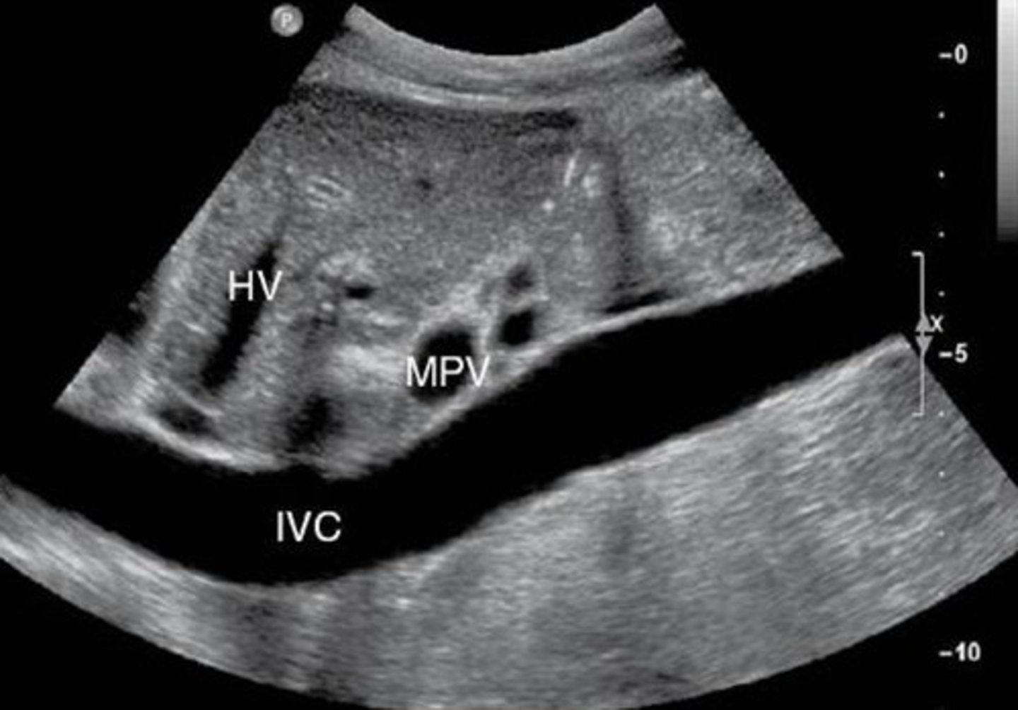
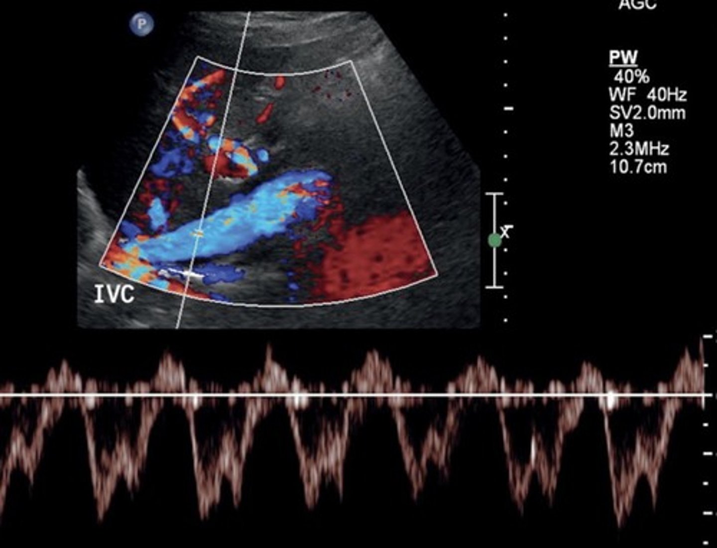
IVC PSV
44-118 cm/sec
Increases with inspiration
Portal Pressure Formula
IVC pressure - portal vein pressure
Portal Vein Pressure
5-10 mmHg
≥ 15 mmHg = clinically significant
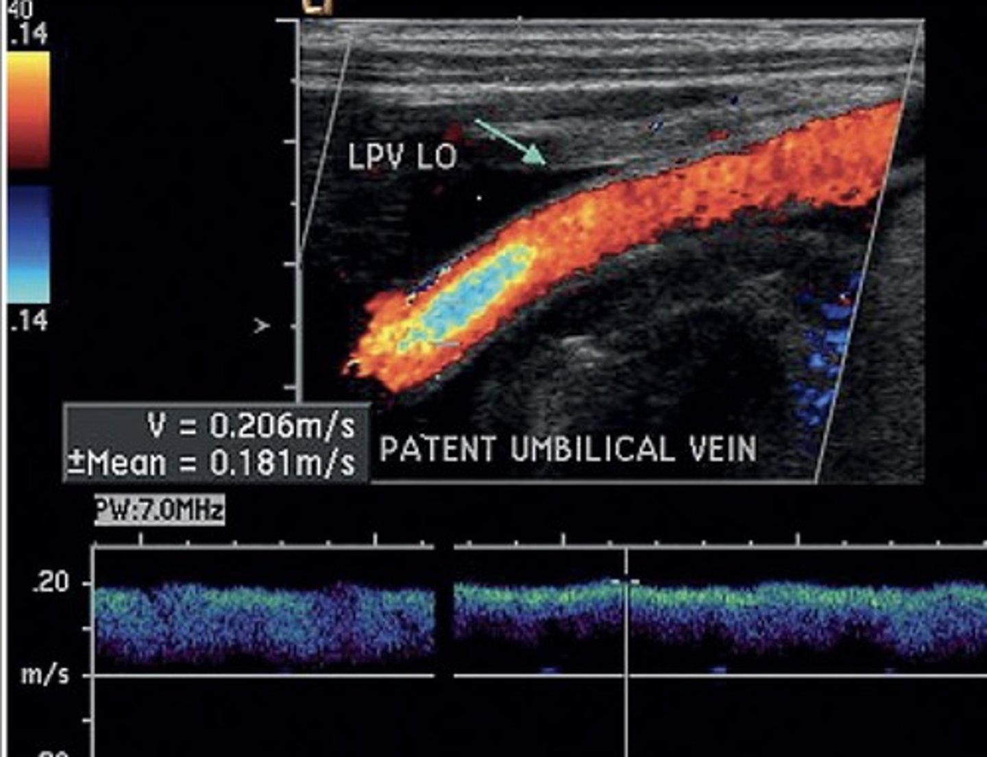
Recanalized Paraumbilical Vein Diameter
> 3 mm
Recanalized Paraumbilical Vein PSV
> 5 cm/sec
Gallbladder Varices Diameter
3-8 mm
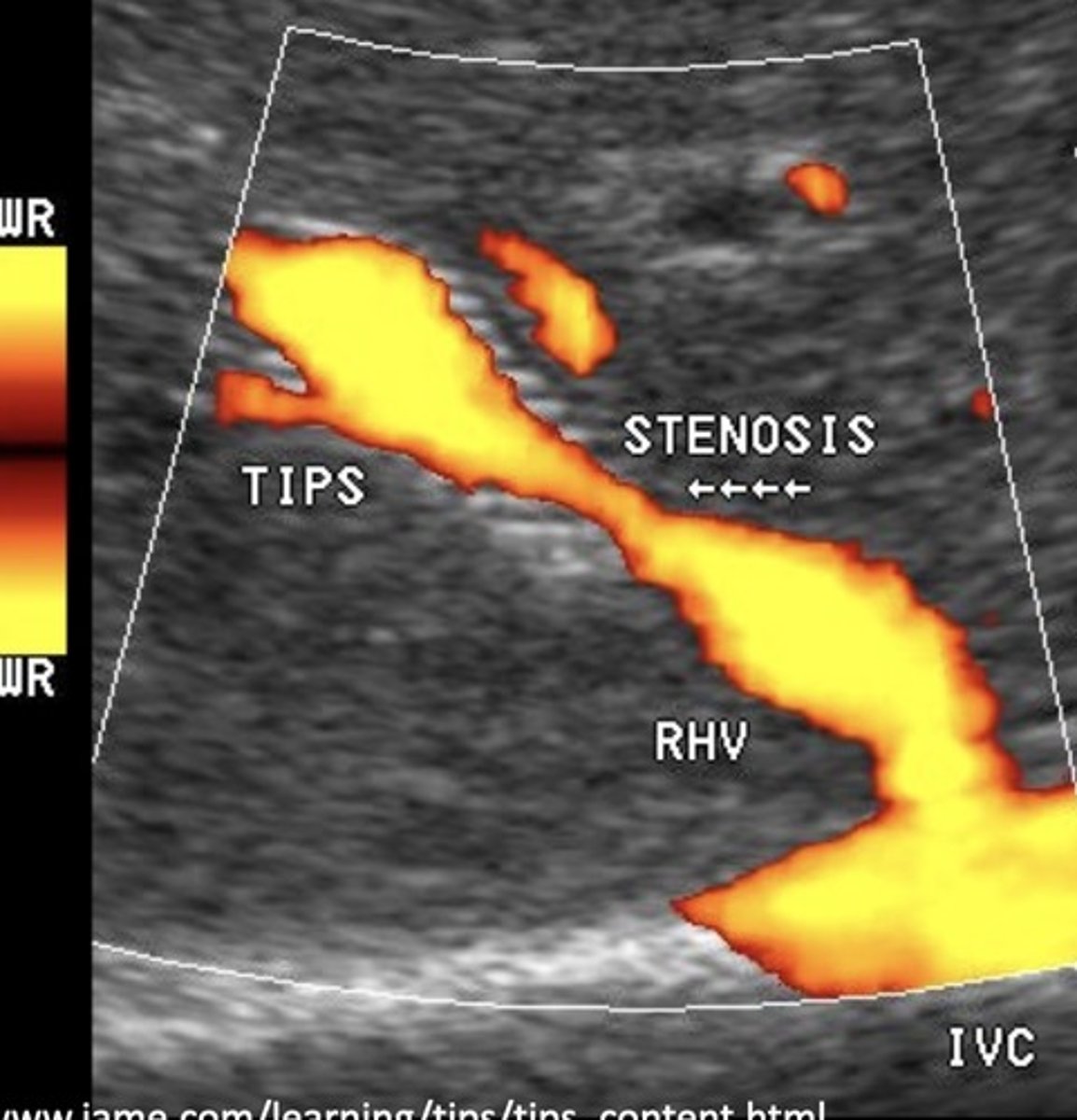
TIPS Shunt PSV
90-190 cm/sec
TIPS Shunt Stenosis Findings
< 50 cm/sec velocity within stent
Focal area velocity increase > 200 cm/sec
> 50 cm/sec velocity change in same portion of stent compared to past exams
Normal Kidney Size
9-13 cm
No more than 2 cm difference between left & right
Aorta PSV at Renal A. Level
60-100 cm/sec
Low resistant
Renal A. Origin PSV
74-127 cm/sec
Distal Renal A. PSV
70-90 cm/sec
Renal A. Sinus PSV
30-50 cm/sec
Renal A. Cortex PSV
10-20 cm/sec
Renal A. PSV Criteria for ≥ 60% Stenosis
> 180 cm/sec (without angle correct)
> 150 cm/sec (with angle correct & turbulence)
Renal Aortic Ratio (RAR) Formula
PSV renal artery
PSV aorta
Angle independent
Normal RAR
< 3.5
Normal Acceleration Time
< 0.1 seconds
Normal Acceleration Index
> 3.78 kHz/sec/MHz
Normal Acceleration
> 291 cm/sec^2
Gold Standard for Vascular Imaging
Angiography
Validity
Ability of a test to distinguish between who has the disease and who does not
Accuracy
Degree of closeness of a test result to the actual value
Percentage of overall correct results
Must lie between sensitivity & specificity and PPV & NPV
Reliability
Consistency of obtaining similar results under similar conditions - reflects accuracy over time
Increasing Cut-Off Values
Improves specificity
More true negatives
Decreasing Cut-Off Values
Improves sensitivity
More true positives
Celiac Artery Occlusion
Results in SMA collaterals diverting blood through gastroduodenal artery toward the liver and spleen
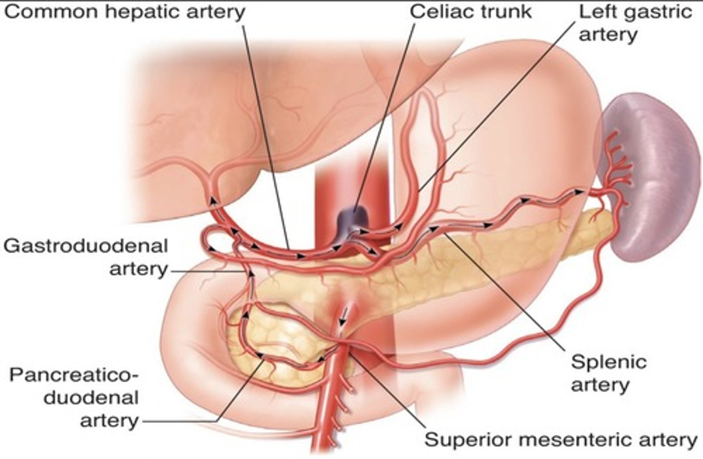
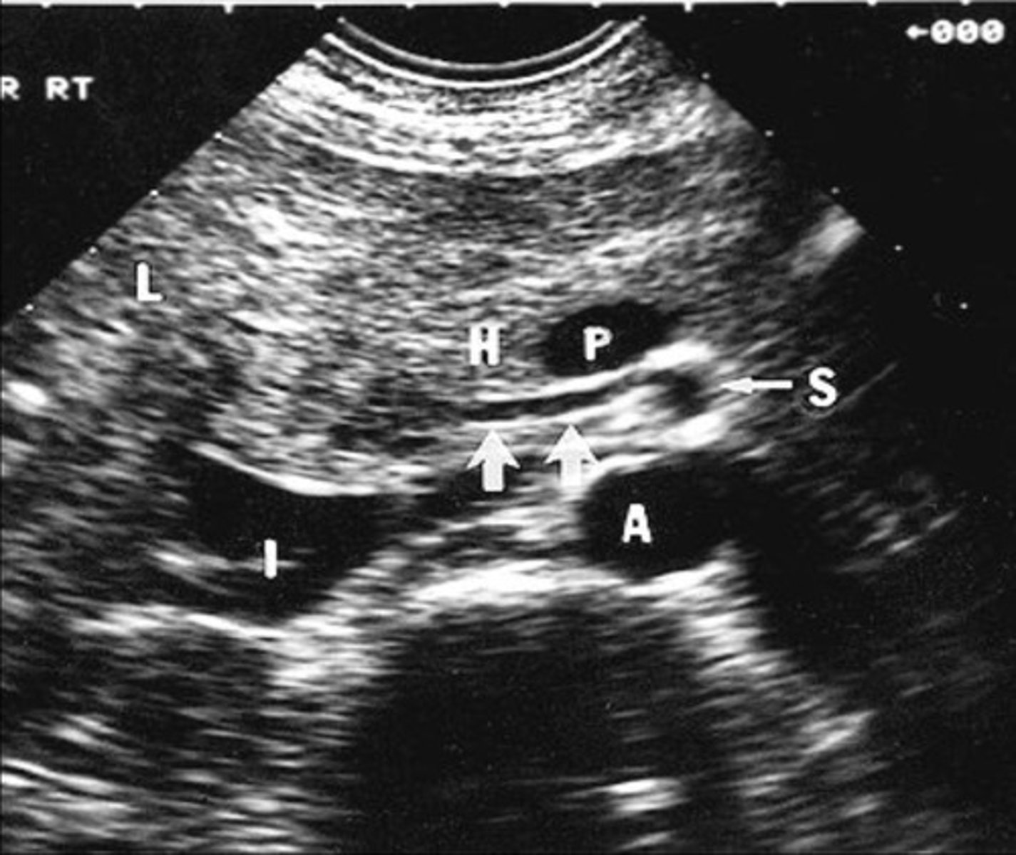
Replaced Right Hepatic Artery
Right hepatic artery branches off something else besides celiac artery - usually SMA
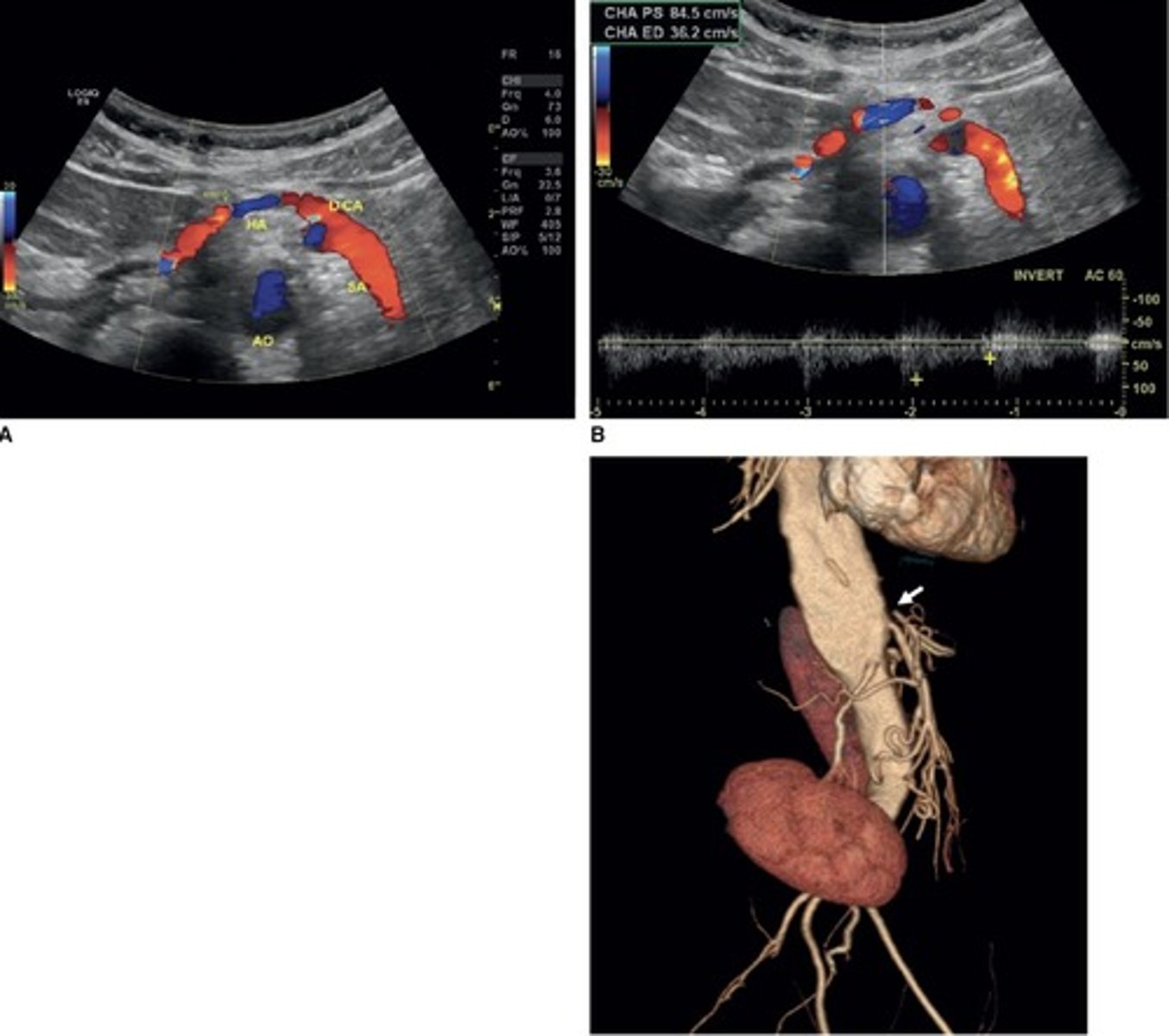
Hepatic Artery Retrograde Flow
Due to celiac artery occlusion
Blood flows towards splenic artery - RABT color pattern
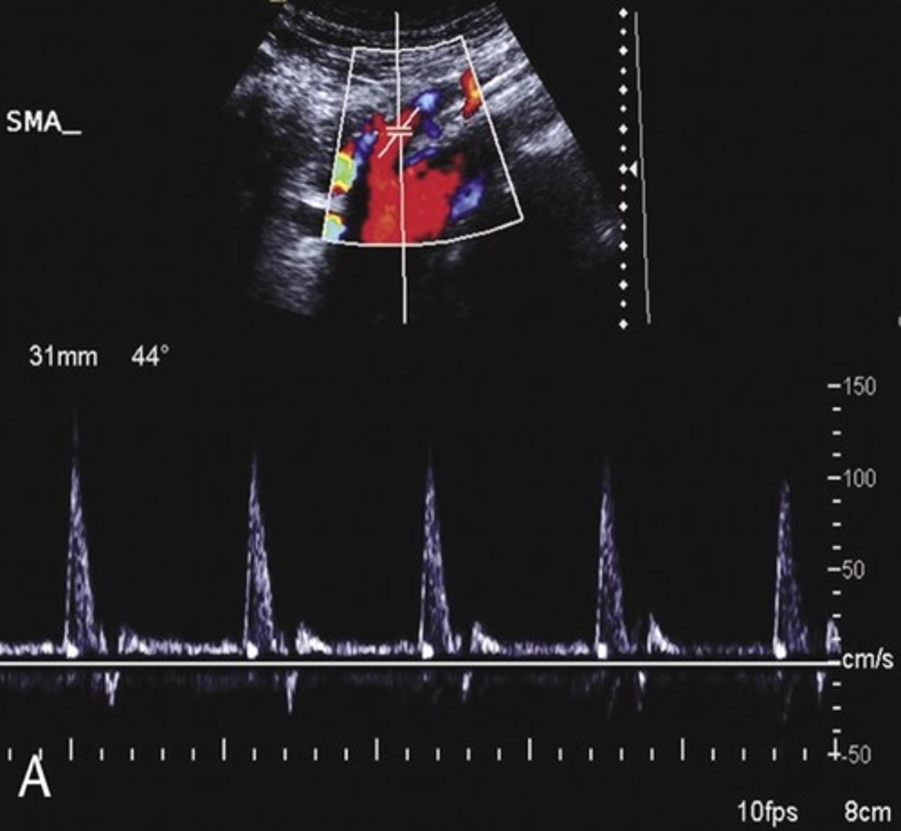
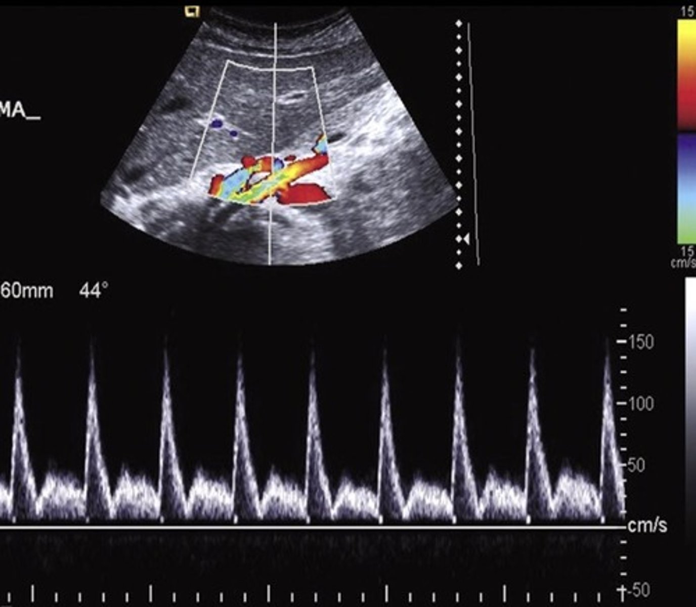
Pre-Prandial SMA Doppler
Post-Prandial SMA Doppler
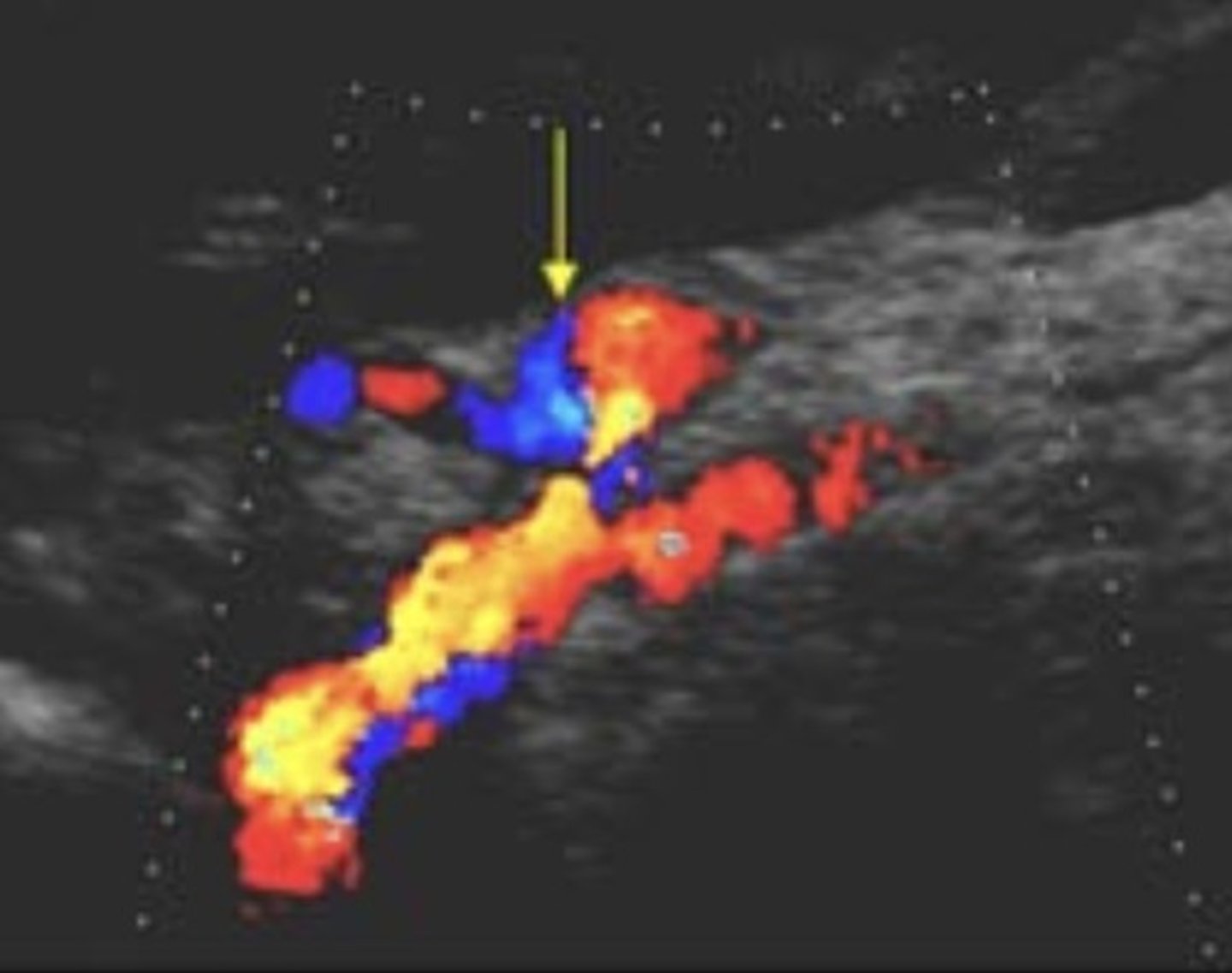
Common Trunk Variant
Celiac and SMA come off common trunk
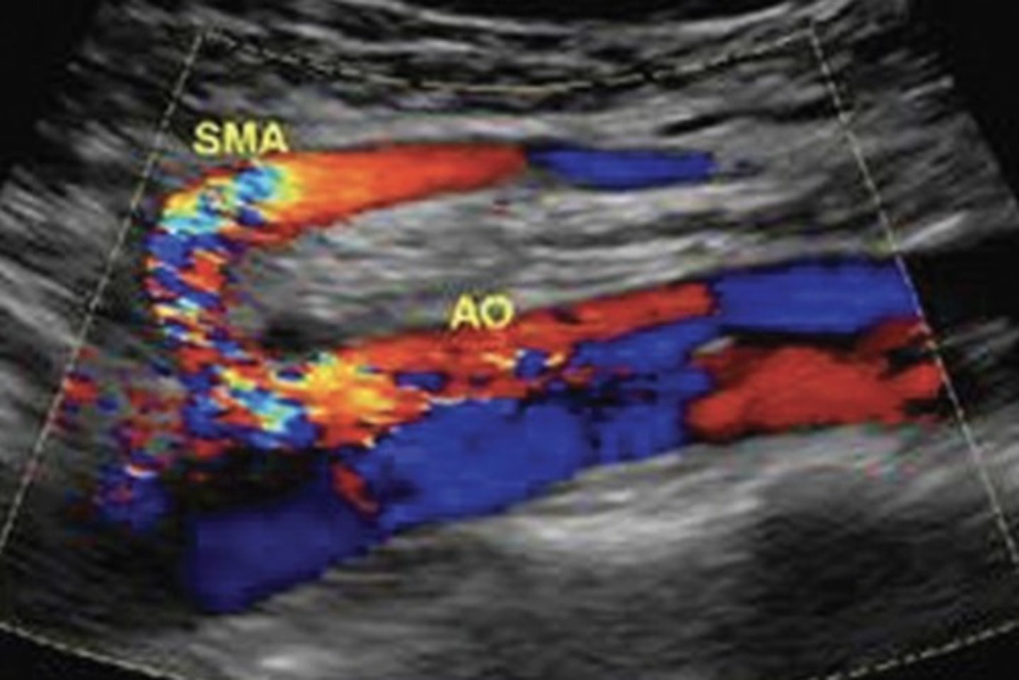
Prominent IMA
Due to SMA occlusion
Acute Mesenteric Ischemia
Thrombosis of one or more mesenteric vessels
Life threatening - requires immediate intervention
Severe cramping/pain - disproportional pain
Chronic Mesenteric Ischemia
Low resistant pre-prandial doppler signals
70% occlusion of 2/3 splanchnic arteries required for diagnosis (celiac, SMA, IMA)
Epigastric pain after eating - fear of food, weight loss, decreased nutrition
Compensatory Flow
Elevated velocities in normal collateral vessels
No stenotic profile seen
Dissection
Separation of channels - flap line
To and fro flow
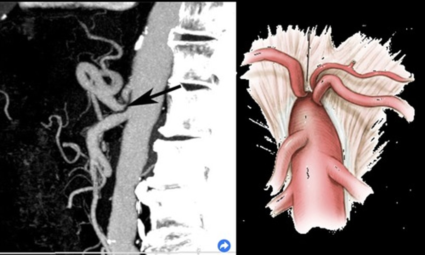
Median Arcuate Ligament Syndrome (MALS)
Compression of celiac axis during exhalation by median arcuate ligament
Pain relieved by inhalation
Evaluate in supine & upright positions and with inspiration & expiration
Pre-Hepatic/Inflow Vessels
Portal Vein
Hepatic Artery
Intrahepatic/Sinusoidal Vessels
Sinusoids/capillaries
Hepatocytes
Post-Hepatic/Outflow Vessels
Central Veins
Sublobular Veins
Hepatic Veins
IVC
Left Gastric Vein (coronary vein) Doppler Flow
Hepatofugal
Main Portal Vein Doppler Waveform
Monophasic continuous waveform
Hepatopetal flow (antegrade flow)
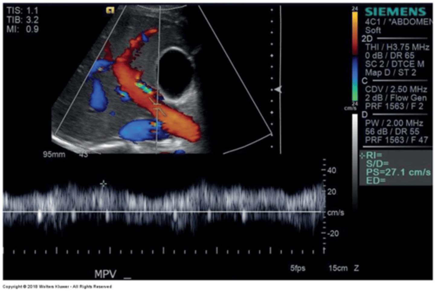
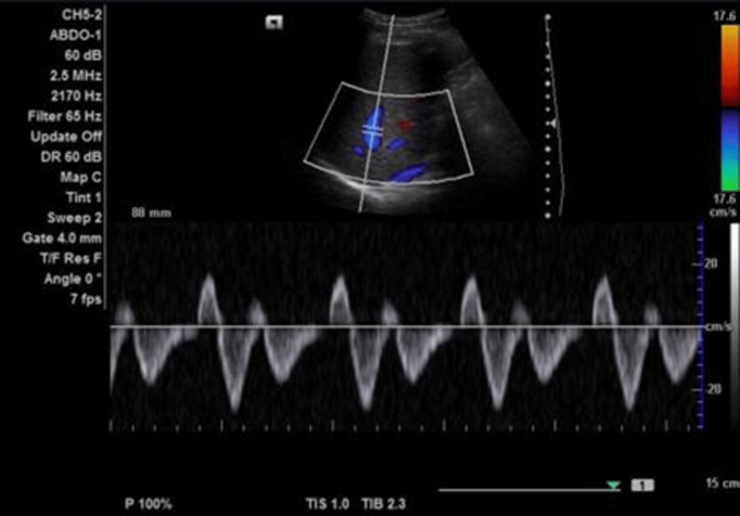
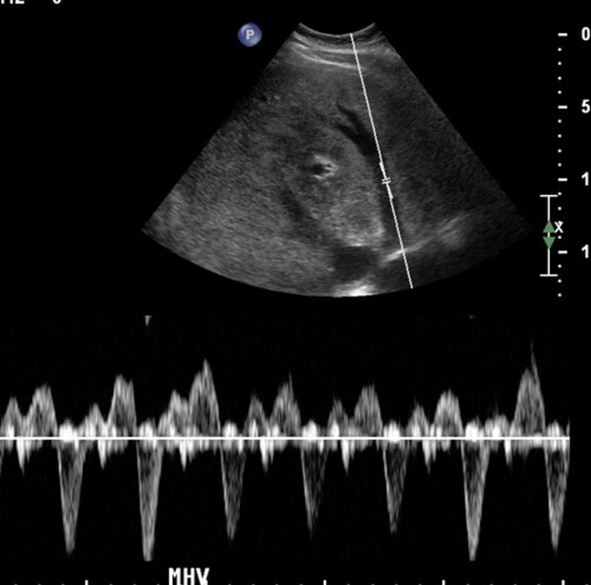
Hepatic Vein Doppler Waveform
Triphasic
Antegrade & retrograde flow - cardiac pressure changes
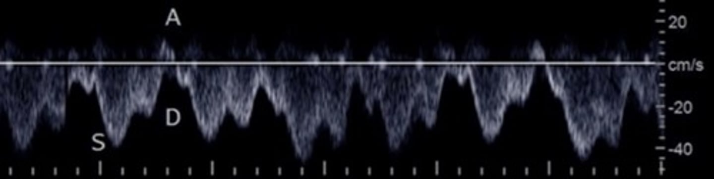
Hepatic Vein Doppler
S wave: ventricular systole
D wave: atrial filling
A wave: atrial contraction
Inspiration depresses systolic wave
Exhalation augments systolic wave
Valsalva diminishes pulsatility
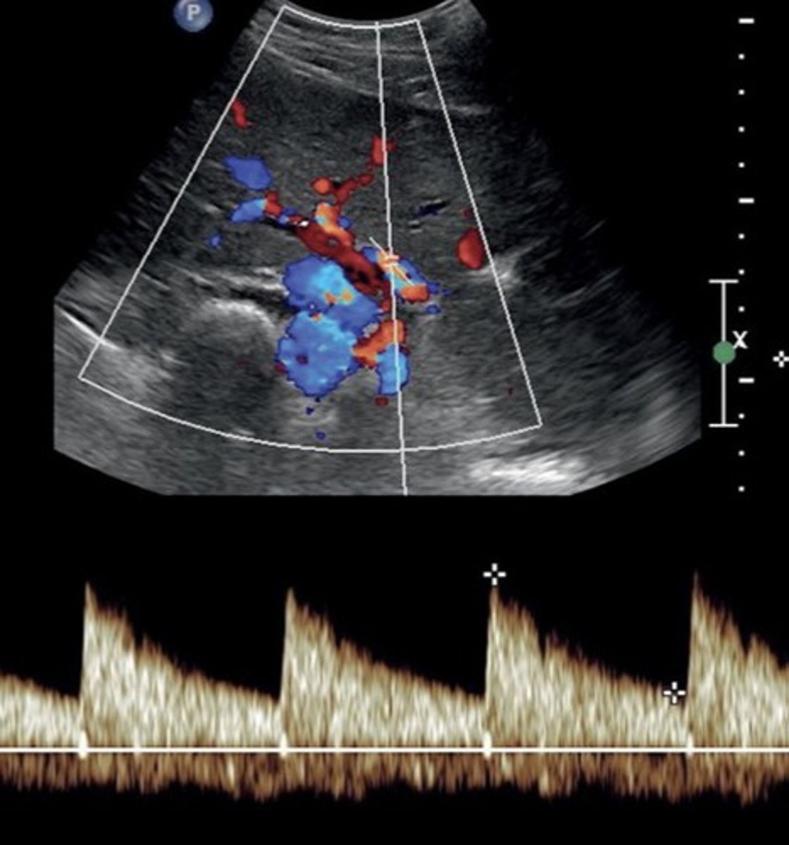
Hepatic Artery Doppler Waveform
Hepatopetal
Low resistant monophasic pan-diastolic forward flow
Hepatic Buffer Response
When PV flow increases, HA flow decreases (post-prandial)
Splenic Vein Doppler Waveform
Hepatopetal flow
Continuous monophasic with slight respiratory variation
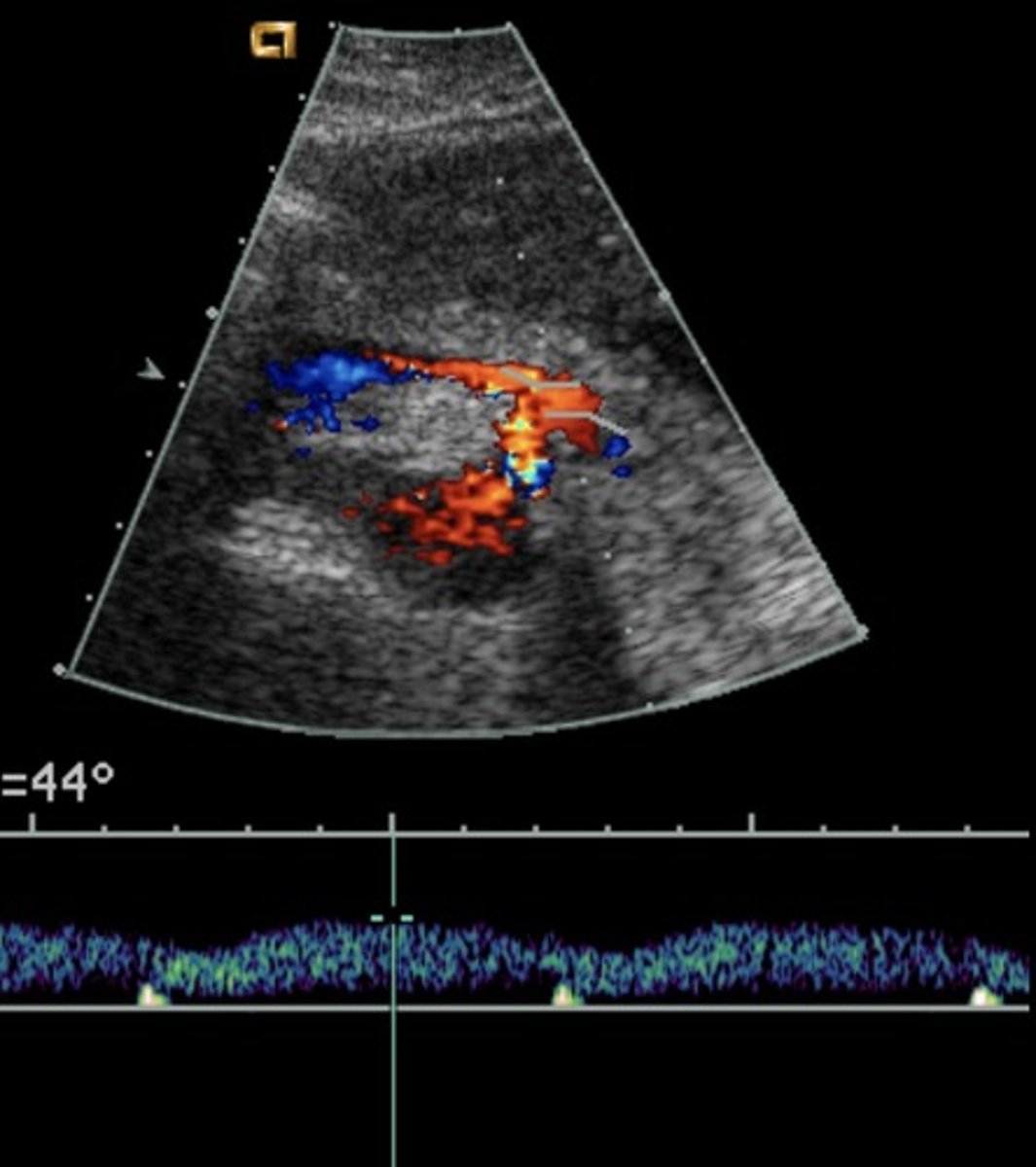
SMV Doppler Waveform
Hepatopetal flow
Continuous monophasic with slight respiratory variation
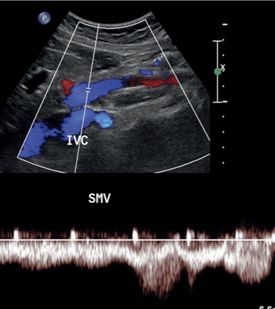
Portal HTN
Increased pressure in portal venous system
Not diagnosed with spectral Doppler -> diagnosed with gray scale & color
Causes of Portal HTN
Hepatitis C
Hepatitis B
Alcoholic cirrhosis
Primary Biliary Cirrhosis
Autoimmune Hepatitis
Hereditary Hematochromatosis
Schistosomiasis
Portal HTN 2D Findings
Large pulmonary vein
Collaterals
Splenomegaly
Ascites
Enlarged hepatic artery
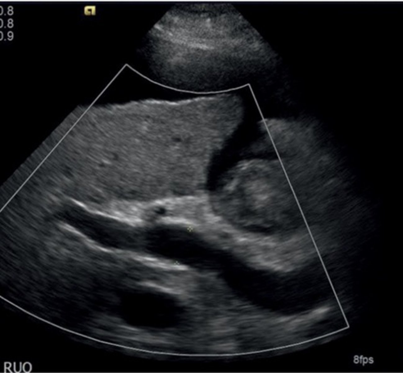
Portal HTN Doppler Findings
Slow, hepatofugal flow in portal vein
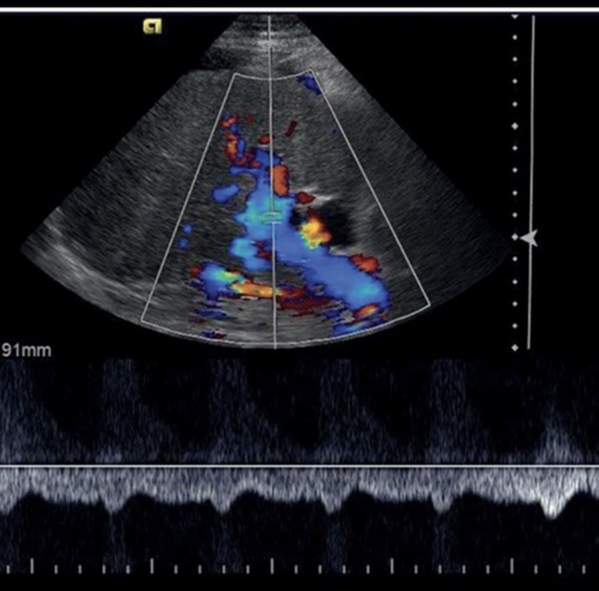
Cirrhosis
End-stage liver disease
Cirrhosis Findings
Portalization of hepatic veins
Collaterals
Hepatofugal flow in portal vein & splenic vein
Coronary Vein Collateral
Can lead to esophageal varices
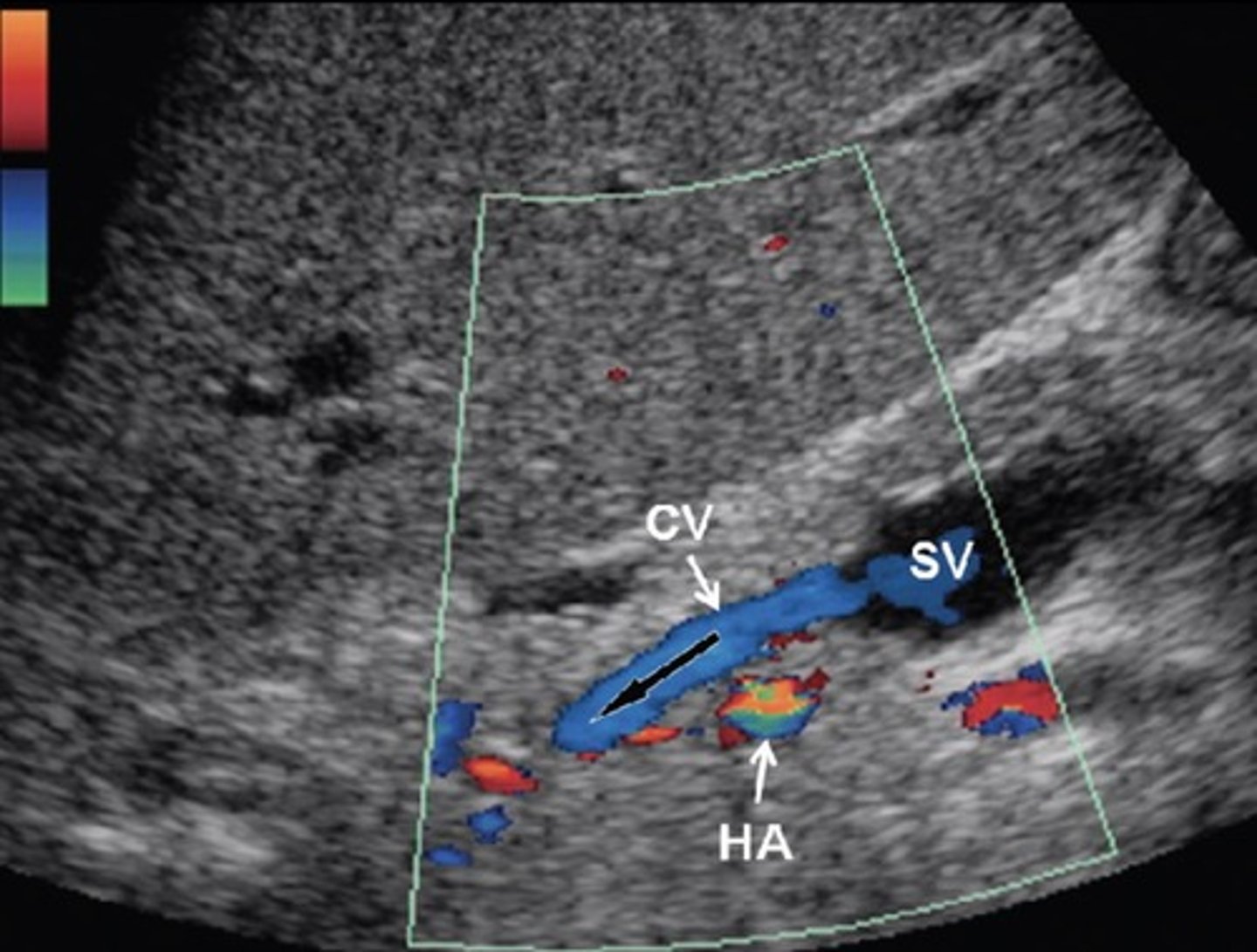
Recanalized Paraumbilical Vein
Ligamentum teres recanalizes
Courses from left portal vein to anterior abdominal wall
Hepatofugal flow
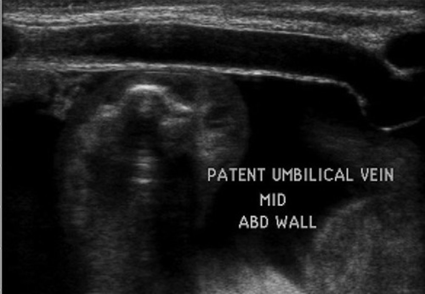
Splenorenal Shunt
Prominent veins at splenic hilum
Hepatofugal flow in splenic vein
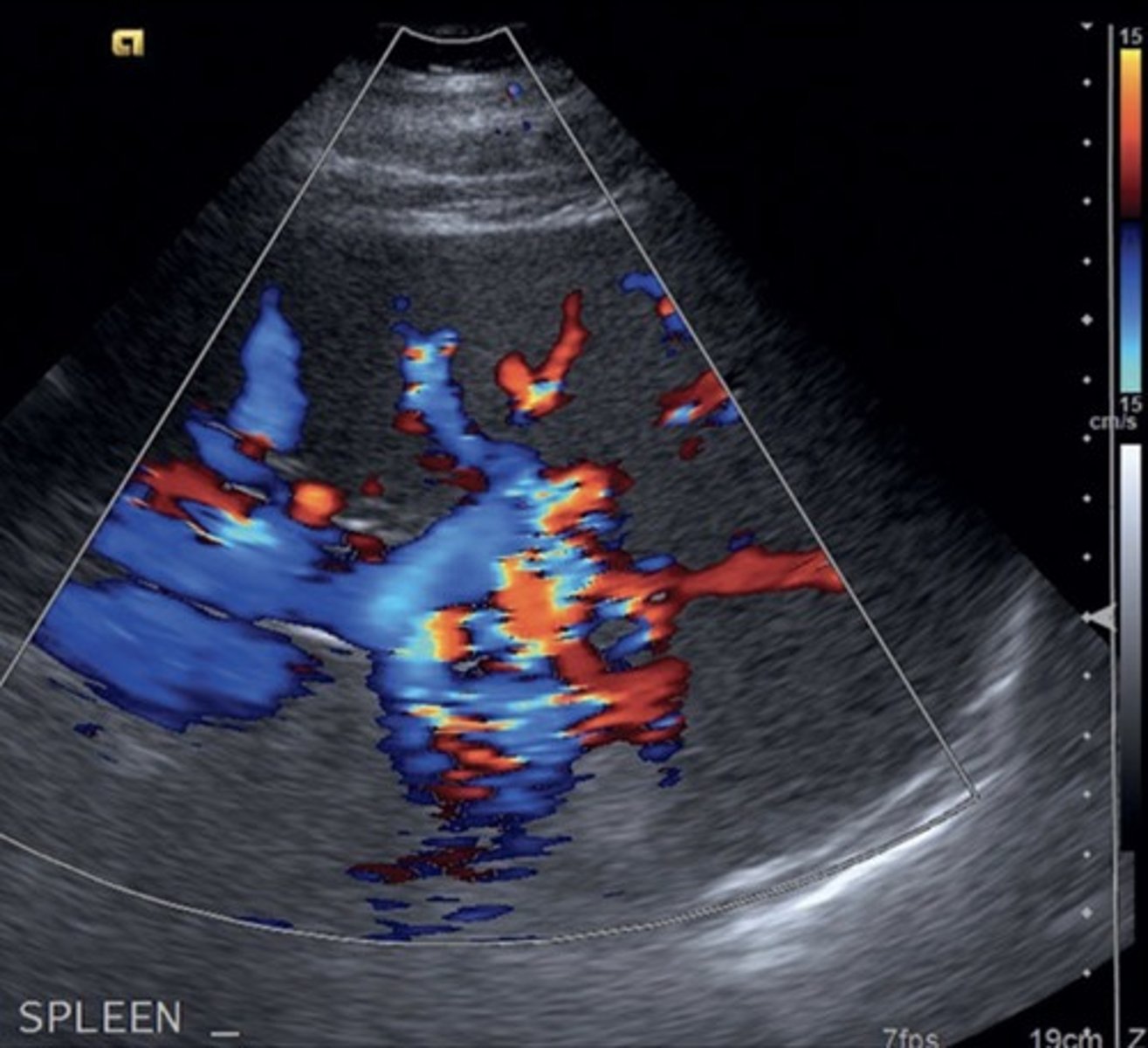
Gastroesophageal Veins/Esophageal Varices
Originate from gastroesophageal junction posterior to left liver lobe
Due to hepatofugal flow in coronary vein shunt
High risk of rupture
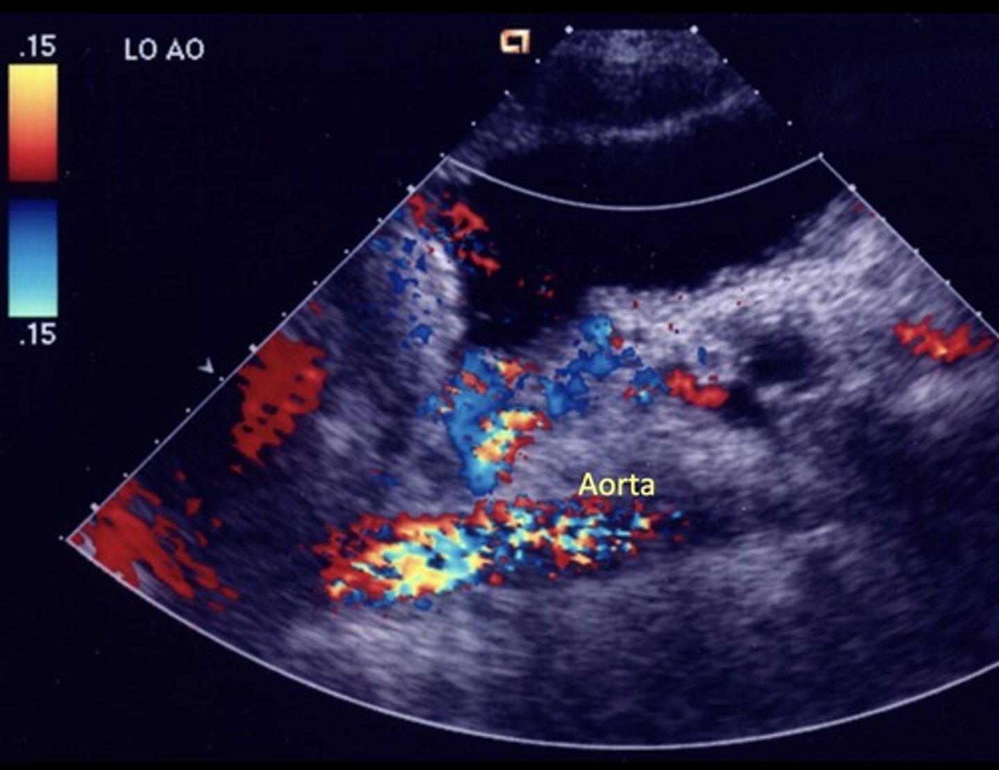
AV Fistula
Arterialized portal vein flow - hepatic artery to portal vein shunting
Leads to portal HTN
AV Fistula Findings
Large anechoic spaces
Increased portal vein pulsatility & velocities
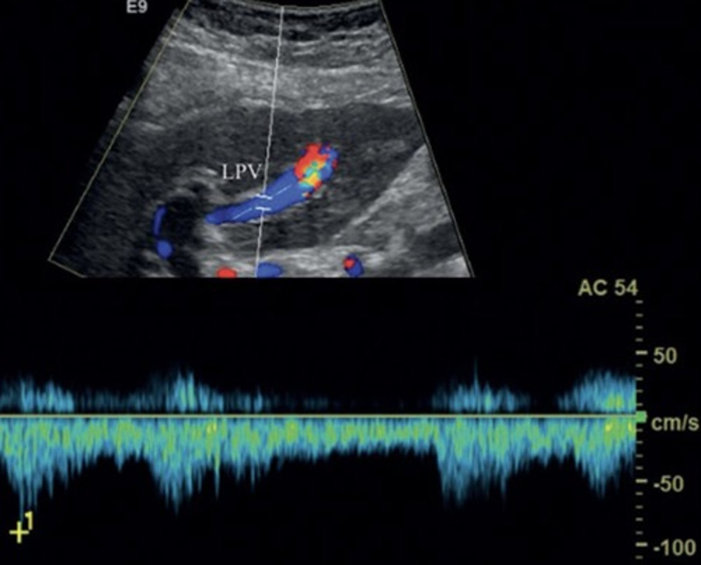
Aterialization of the Liver
Max portal vein pressure leads to increased hepatic artery flow
Cavernous Transformation
Due to chronic portal vein thrombosis
Occurs within 6 days after thrombotic event
Absent portal vein flow
Formation of new peri-portal vessels around portal vein
Hepatopetal flow within serpentine vessels
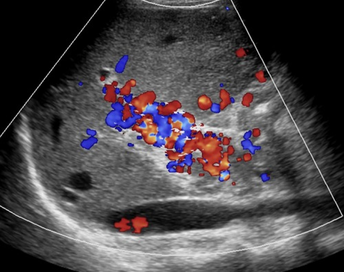
Budd-Chiari Syndrome
Hepatic vein/liver outflow obstruction
Causes increased sinusoidal pressure
Budd-Chiari Syndrome Findings
Hepatic vein thrombosis/post-sinusoidal thrombosis
Monophasic, reduced/reversed flow in portal vein
Enlarged caudate lobe
Ascites
Splenomegaly
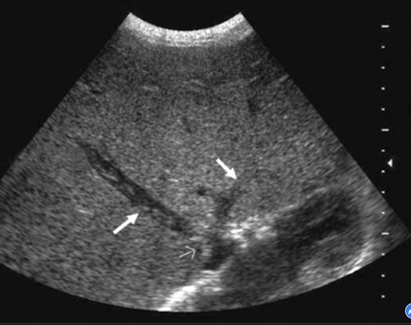
Cardiac Cirrhosis
Liver edema caused by congestive heart failure
Portal Vein with Cardiac Cirrhosis
Pulsatile
Biphasic
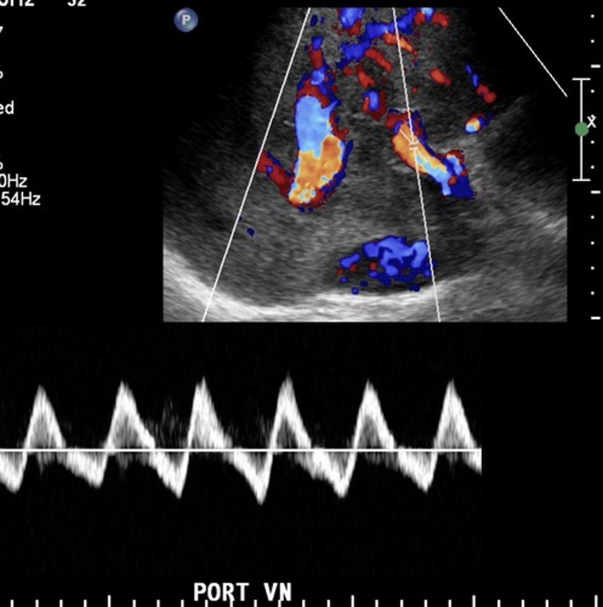
Hepatic Veins with Cardiac Cirrhosis
Compromised flow - over congestion
"w" pattern
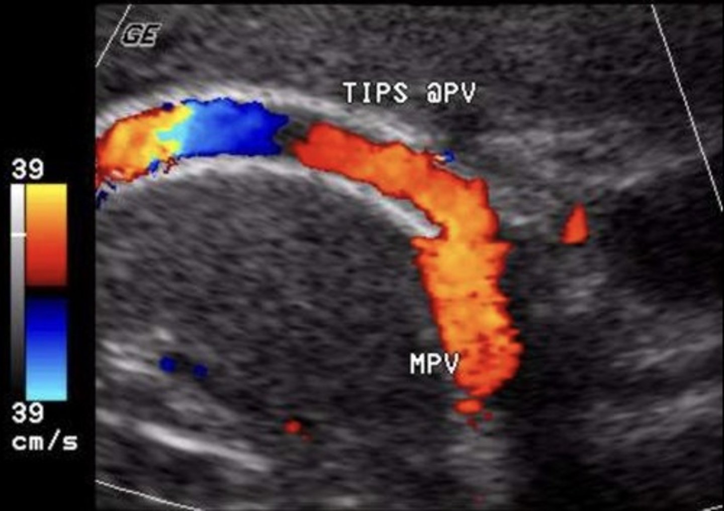
TIPS Shunt
Stent between portal vein & hepatic vein
(commonly right portal vein and right hepatic vein)
TIPS Shunt Normal Findings
No focal aliasing
Hepatofugal flow in portal veins beyond stent
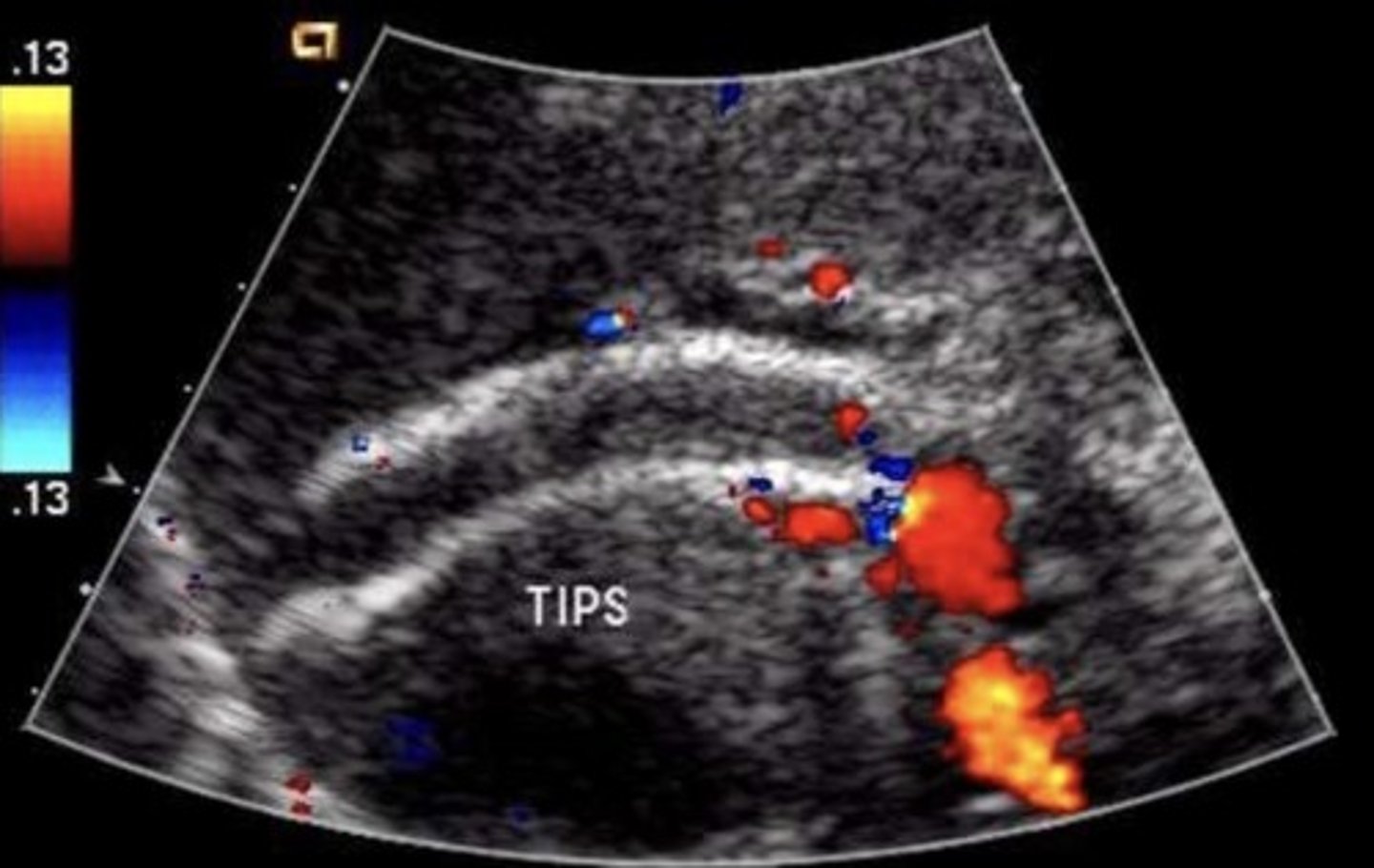
TIPS Shunt Abnormal Findings
Focal aliasing
Velocity changes
Antegrade flow in right & left portal veins
Retrograde flow in hepatic vein
Developing ascites or collaterals
TIPS Shunt Occlusion Findings
Hepatopetal flow in right & left portal veins
Hepatofugal flow in main portal vein
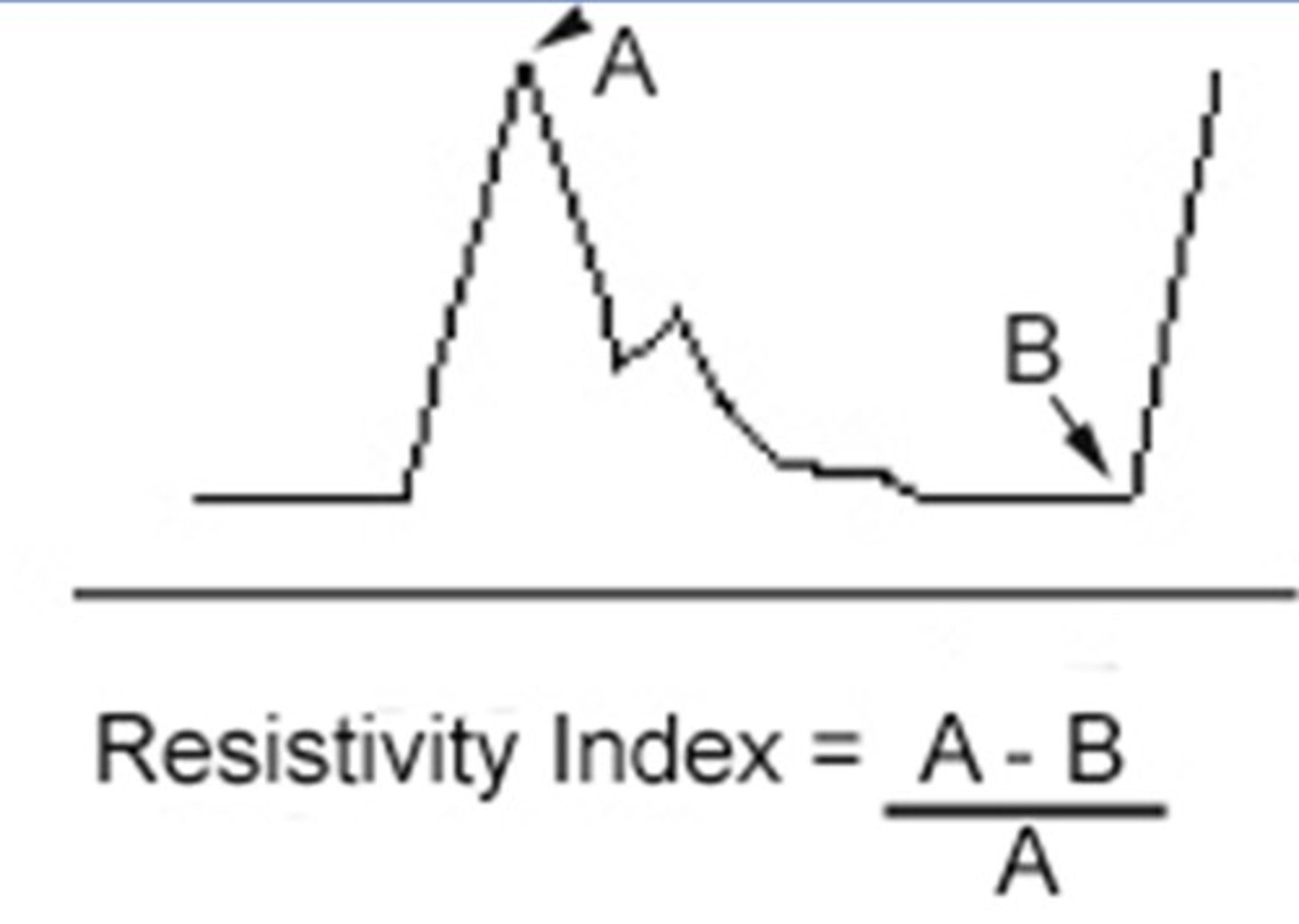
Resistive Index
Resistance of the end/target organ
Orthotopic Liver Transplant
Whole liver is transplanted from a deceased donor
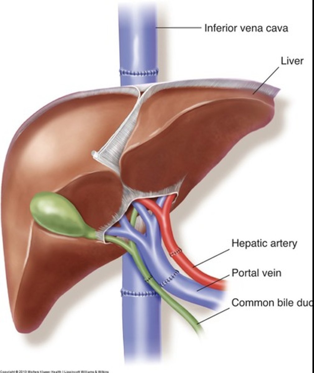
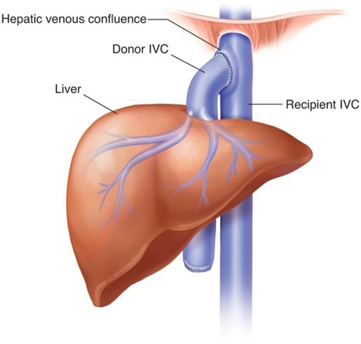
Piggyback IVC Anastomosis
Donor supra hepatic IVC attached to recipients hepatic venous confluence