NEUR1020
 Call Kai
 Learn
Learn Practice Test
 Spaced Repetition
 Match
Match Flashcards
 Knowt Play
1/103

Earn XP
Description and Tags
Final Exam
Name | Mastery | Learn | Test | Matching | Spaced | Call with Kai | Chat |
|---|
No analytics yet
Send a link to your students to track their progress
104 Terms
Testability
Does the theory generate hypotheses that can be evaluated against data?
Falsifiability
Are there potential observations, or patterns of data, that are disallowed by the theory?
Paradigm
A framework for understanding and investigating phenomena with a discipline.
Defines concepts that are used in theories, research questions that are addressed by a discipline, and methodologies used to investigate these questions.
Origins of the Behaviourist Paradigm
Freud urged us to ‘look inward’ and consider how the unconscious mind influenced our thoughts and behaviour. Behaviouristst encouraged us to consider how the external environment shapes our thoughts and behaviours.
Watson’s Methodological Behaviourism
Rejected the study of unobserved phenomena (such as overt behaviours) could be studied scientifically.
Prompted a shift in focusing on relationships between publicly observable stimuli and the behavioural consequences.
Skinner’s Radical Behaviourism
Broadened the definition of ‘behaviour’ to include ‘private’ events (e.g., thoughts and feelings) as legitimate scientfic topics of study.
Argued that the external environment was the determinant of both observable and unobservable behaviours.
Mental events, therefore, are not causes of behaviour, but are themselves behaviours caused by the environment.
Cognitive Paradigm
Framework focusing on internal mental processes
Biological Paradigm
A theoretical framework in psychology that explains human behaviour, personality, and mental disorders through physical factors (including genetics, neurochemistry and brain structure.
Features of a Science
Grounded in observation. Science is cumulative. Self-correcting. Achieves explanation and understanding.
Behavioural Paradigm
A psychological framework focusing on observable, measurable behaviours rather than internal mental processes.
Control (Least to Most)
Introspection, Natural Observation, Case History, Surveys, Correlational Designs, Experiments.
The Stroop Effect
Refers to the phenomenon where individuals take longer to name the colour of ink that the names of colours are written in than it does to read the colour names.
Cerebrum (Cerebral Hemispheres)
Two hemispheres divided by longitudinal fissure or inter-hemispheric fissure
Cerebral Cortex
Outermost surface layer of the cerebrum.
Grey Matter (Surface of the brain 2-4mm thick, highly folded to maximise surface area)
White Matter (underneath grey matter is all “wiring” such as axons of teh neurons connecting to the spinal cord and to other areas of the cortext)
Frontal Lobe
Primary Motor Cortex
Premoteor Area
Motor Speech Area
Prefrontal Area
Executive functions (reasoning, planning, problem-solving, inhibitory control, working memory)
Motor functions (premotor - motor planning, cortex - execution)
Speech Production (Broca’s area)
Parietal Lobe
Primary Somatosensory Cortex (perception of touch)
Sense of space and locations (gives sense of stable world around us relative to body position)
Spatial attention (directing attention and eye movements to explore the visual world)
Linking vision to action (represents spatial location of objects around us for guiding actions)
Occipital Lobe
Primary Visual Cortex (V1) - all visual perception
Higher visual areas - different regions process shape, colour, orientation and motion
Temporal Lobe
Primary auditory cortex (perception of sound)
Language comprehension, Wernicke’s area)
Medial Temporal Lobe (Limbic system, amygdala and hippocampus)
Limbic System
Amygdala - Fear and arousal, responds to threats/danger, fear/learning phobias
Hippocampus - learning and memory, forming new episodic memories, damage causes anterograde amnesia (can’t form new memories)
Corpus Callosum
Neuron connections between the left and right hemispheres and allows communication between hemispheres
Broca’s Area
Speech production
1861 Paul Broca described a patient who was unable to speak after damage to the left frontal lobe
Speech is slow and non-fluent
Difficulty finding appropriate words (anomia)
Speech still caries meaning, comprehension is mostly unaffected
Wernicke’s Area
Language comprehension
1874 Carl Wenicke suggested that lesions to the left posterior temporal lobe led to deficits in language comprehension
Unable to understand language, deficit in comprehension
Speech is fluent with normal prosody (rhythm, intonation)
Speech has no meaning
Wilder Penfield
Stimulated the brain with electrical probes while the patients were conscious, during surgery for epilepsy
1951 published maps of motor and sensory cortices of the human brain
Homunculus
Primary sensory cortex and Primary motor cortex
Brain function “mapped” by electrical stimulation, brain stimulation leads to sensation or movement
Size of area on cortex determines sensitivity or five motor control
Nervous System Break Down
CNS (Brain and spinal cord) and PNS
PNS - Autonomic (involuntary) Somatic (voluntary, sensory and motor)
Autonomic - Sympathetic (emotional arousal, stress, fear, fight or flight response, increases heat-rate, respiration, perspiration, pupils dilate) and Parasympatheic (rest and digest, lowers heart-rate, respiration, increases stomach and intenstine activity)
Brain Stem
Medulla
Autonomic nervous system functions
Controls heart rate, respiration, regulation of blood pressure, body temperature
Reflex centres for coughing, sneezing, swallowing, vomiting
Persistent Vegetative State
Severe damage to upper brain
If brain stem is not damaged, autonomic nervous system functions can remain
Sometimes normal respiration, control of heart rate, and some face and eye movements remain
Locked-In Syndrome
Amyotrophic Lateral Sclerosis (ALS) or Motor Neuron Disease (MND)
Brain Injury
Intact cerebrum and brain stem but disconnected from spinal cord
Normal cognitove function, vision, and hearing but mostly cannot move
Patients may be fully conscious and aware but totally unresponsive
Pateints apprea the same as those with persistent vegetative state
High to Low level function
Cerebral Hemispheres - Cortex, planning, reasoning, problem-solving (frontal lobe), language and perception
Brainstem - Autonomic nervous system functions (heart-rate, respiration, blood pressure)
Cerebellum
Hindbrain
Sense of balance and co-ordination of complex movement
Motor-learning - fine adjustment of movement based on feedback
Glial Cells
Oligodendrocytes - Produce the myelin sheath that wraps around axons
Astrocytes - Supply nutrients from blood to the neurons, maintain “blood-brain barrier”
Microglia - brain’s immune system, clean up foreign of toxic substances
The Myelin of Axons
Oligodendrocytes from myelin sheath by wrapping around the axon
Essential for efficient communication, for propagation of signals along axon
Multiple sclerosis involves loss of myelin, disrpution of efficient neurl communication throughout the body
Neural signals go one way
Pre-synaptic: before the synapse, from cell body to axon terminal
Post-synaptic: after the synapses, from dendrite to the cell body
Neurons Electrical Signals
Electrical signal “pulse” travels along the axon
Fixed size - either on or off, signal or no-signal (not large or small)
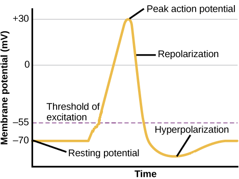
Membrane Potential
The electrical voltage difference between the inside and outside of a biological cell
Resting Potential
The stable electrical charge difference across the cell membrane of an excitable cell (neuron) when it is not actively transmitting signals
Action potential
A rapid, temporary reversal of electrical charge across the membrane of an excitable cell
Depolarisation/repolarisation is fact (occurs in less than 0.002 seconds)
Repolarisation undershoots - (refractory period - more difficult for another action potential to occur), further to threshold to trigger another action potential
Cell Membrane Wall
70% of the brain is water with water surrounding (extra-cellular) and filling (intra-cellular) cells
Cell membrane forms barrier between extra-cellular and intra-cellular fluid
Ions and Electrical Potential across Cell Membrane
Sodium (Na+) and Potassium (K+) positively charged ions
Different concentrations outside and inside cell, across cell membrane
Gives difference in electrical charge (potential) across cell membrane
Ion Channels in Cell Membrane
Open and close to pass or block movement of ions across cell membrane
Ions move between intra and extra-cellular fluid
Movement of ions changes electrical potential
Sodium Potassium Pump
Actively pumps Na+ and K+ across cell membrane
Overly pumps positive charge out of cell (3 Na+ out for every 2 K+ in)
Positive charge will naturally move towards negative area (opposites attract)
Maintains negative resting membrane potential (approx ~70mv)
Uses about 25% of body total energy (70% of brain energy)
Voltage-Dependent Ion Channels
Voltage-dependent ion channel, closed at resting potential
Open when membrane potential reaches threshold voltage
Allows flow of ions across membrane
Positive ions (Na+) can flow from outside into the cell (because positive charge will naturally move towards negative area)
Causes depolarisation of cell (voltage less negative = closer to 0)
Depolarisation
Na+ channels open when voltage exceeds threshold
Na+ flows into the cell
Less negative potential
Repolarisation
Na+ channels close and K+ channels open after depolarisation
K+ flows out of cell
Plus Na/K pump
More negative potential
Synapses and Neural signals
Neurotransmitter release - depolarisation of axon terminal (action potential) triggers release of neurotransmitter which acts on receptor on post-synaptic neuron to open ion channels and pass signal
Synaptic vesicles - stores neurotransmitter in pre-synaptic terminal (recycled neurotransmitter taken back into pre-synaptic terminal is re-packaged into vesicles)
Re-uptake pump - clears neurotransmitter form synaptic cleft back into pre-synaptic terminal
Dopamine - Parkinson’s Disease
Loss of dopamine in the basal ganglia deep in the brain
Primarily affects movement
Treatment with L-DOPA replaces the dopamine in the brain
SSRI
Anti-depressant drug for serotonin
Selective Serotonin Re-uptake Inhibitors
MAOIs
Anti-Depressant Drugs for serotonin
Monoamine Oxidase Inhibitors
Sending Signals Neurotransmitter Release
Depolarisation of axon terminal triggers release of neurotransmitter
Neurotransmitter acts on receptor and post-synaptic neuron to open ion channels and pass signal
Ligand Gates
Neurotransmitter receptors open ion channels when neurotransmitter binds
Different neurotransmitters bind to and open different ion channels (Na+, K+, Cl-) to change membrane potential in different ways
Receptor Binding - can cause depolarisation and hyperpolarisation
Excitatory Post-Synaptic Potential
Receptor opens channels that cause depolarisation, closer to threshold for action potential
Inhibitory Post-Synaptic Potential
Receptor opens channels that cause hyperpolarisation, further from threshold for action potential
Graded Potentials
Excitatory and inhibitory inputs combine together which change the membrane potential on post synaptic cell
Depends on the strength of synapse connection (strong connection causes large change in membrane potential, weak connection causes small change)
When do inputs trigger an action potential?
Membrane potential at axon hillock depends on sum and timing of inputs through dendrites
If enough excitatory inputs occur together close enough in time, membrane potential will exceed threshold level for action potential
If membrane potential exceeds threshold level (at axon hillock) it triggers action potentials and neuron fires
Neuropsychology - Brain Lesions
Explains normal brain function by examining what changes when part of the brain is damaged (stroke or brain injury inhumans, induced lesions in animals from electrical or chemical stimuli)
Assumption is whatever changes in behaviour/cognition must rely on that part of the brain that is damaged
Single Neuron Recording
Place a single thin electrode to an animals brain and record action potentions firing form a single neurons
Measures what the neuron encodes or detects
Best localisation and timing of brain function
EEG - Electroencephalography
Summed activity from action potentials of neurons in the cortex cause electrical activity change on the scalp
Measure voltage changes from electrodes placed on the scalp
Wave forms vary with brain states
Constant oscillations and frequencies change with alertness and sleep
Clinical use - detecting stages of sleep, monitoring for epileptic seizures
ERPs - Event-Related Potentials
Brain activity related to a specific event or stimulus
Average together >100 trials of EEg response to the stimulus
Peaks represent different stages of processing of the stimulus
Can show precise timing of information processing in the brain
Face Processing N170
ERPs can show precise time of information processing in the brain
100ms viewing any stimuli (peak brain activity 100ms after seeing visual stimulus, early brain processing of general visual features)
170ms viewing faces (peak of brain activity 170ms after seeing face, brain processing for face-recognition in visual cortex)
PET
Positron Emission Tomography
1980-late 90s
Uses radioactive substances injected into the blood stream
Used now to map neurotransmitters or receptors in the brain
fMRI
Functional Magnetic Resonance Imaging
1992 to current
Masures changes in blood oxygen level (BOLD signal)
Studies brain function
Oxygen is carried in blood and delivered to active neurons
Change in blood oxygen level = change in brain activity
Spreading Activation Model Theory
Neurons represent a specific concept
Share connections with neurons that represent related concepts
Activation of one neuron leads to spreading activation to related or connected neurons
Neuroplasticity - With Experience
Sensory and motor cortex areas expand with use and experience
Neuroplasticity - After Damage
No rehab - no movement, motor cortex gets small maladaptive plasticity
With rehab - movement training, motor cortex area for hand expanded and movement improved
Neuroplasticity in Blind People
Brain activity in visual cortext while reading braille
Visual cortex change only in blind people, not sighted people reading braille
Physiological Blind Spot
Point at which the optic nerve leaves your eye is called the optic dics
At the top of the disc there can be no photoreceptors so you are blind to images that project to that position on your retinae
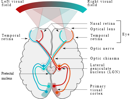
Visual Signals
Left Hemisphere Lateralised Function
Language and Speech
Right Hemisphere Lateralised Function
Tone of voice, face perception, perceptual grouping
Contralateral Functions
Movement, sensation and visoin
Left Hemisphere - right body movement, sensation and vision
Right Hemisphere - left body movement, sensation and vision
Language Left Hemisphere
Language comprehension, speech, reading and speech production
Language and hand dominance, no overall dominant hemisphere
Corpus Callosum
Connects left and right hemisphere
Axons of neurons crossing to the opposite hemisphere
Inter-Hemisphere Communication
Vision goes to contralateral hemisphere (left of screen → right hemisphere)
Split Brain (Severed Corpus Callosum)
Last resort surgical treatment for very severe epilepsy to stop seizure activity from spreading to the other hemisphere
Transduction
The conversion of electromagnetic radiation into neural events
Coloured Aftereffects
Brain causes you to see things that are not present on the retinae
After protracted viewing of certain colours, you can see opppositely coloured after images
Face Aftereffects
Perceptual illusions where prolonged viewing a face causes subsequent faces to appear distorted in the opposite direction
What causes after effects
Occur because the firing rates of neurons that are responsive to an input adapt over time which means they become less responsive
Response Selectivity in the Human Retinae
The ability of retinal ganglion cells to respond preferentially to specific visual features (such as direction of motion, edge orientation, or colour) rather than simply registering light intensity
V1 Response Selectivity
Simple V1 cells are responsive to oriented stripes of contrast located in a specific subfield of its receptive field
End-stopped V1 cells are responsive to oriented stripes of contrast of a specific (limited) length
Retinotopic Map
Adjacent neurons in visual brain regions encode information from adjacent regions on the retinal surface
Retinotopic mapping is most apparent in lower-level visual brain structures where neurons are associated with small receptive fields
Lateral Geniculate Nucleus (LGN)
A key thalamic structure acting as the primary relay station for visual information between the retina and the primary visual cortex
Primary Visual Pathway
Main route for conscious vision, transmitting retinal information via the LGN to the primary visual cortex (V1) for detail and colour processing
Secondary Visual Pathway
Involves areas like V2-V5 and subcortical structures handling higher-order processing like motion, depth, and spatial awareness
V1-V5
V1 - Primary visual cortex
V2 - Secondary visual cortex
V3 - Third visual area (processing dynamic form)
V4 - Fourth visual area (involved in color perception and shape recognition)
V5 - Middle Temporal Area (specialised motor detection, react to the direction and speed of movement)
Cerebral Achromatopisa
Rare, acquired form of colour blindness due to damage of the V4 cortex (black and white vision)
Prosopagnosia
Face blindness (Occiputal face area)
Adjacent to V1 and V4
Cerebral Akinetopsia
Rare neuropsychological disorder where patients lose the ability to perceive visual motion, even though their eyesight and ability to see stationary objects remain normal
Damage to V5
Limbic System
Hippocampus - medial temporal lobe, memory (forming new episodic memory and damage causes memory loss in the form of not being able to retain new memories), spatial navigation
Top-Down
Cognitive control or volitional choice
Modulation by prior knowledge and experience
Bottom-Up
Driven by external stimuli or unconscious states
Spatial Neglect
Lesion to one hemisphere (frontal or parietal)
Commonly caused by a stroke
Deficit in directing to one side of space
Fronto-Temporal Dementia (FTD)
Degeneration of neurons in frontal and temporal lobe
Symptoms
inappropriate actions (aggressive, sexual, or lack of care)
Apathy
Loss of empathy
Deficits in executive function
Speech, language and motor deficits
Extinction (Classical Conditioning)
CR response will reduce in strength and disappear if the CS is repeatedly presented without the UCS
Spontaneous recovery
Sudden reemergence of a previously extinguished conditioned response after a period of rest or delay
Generalisation
Tendency for CR to appear with similar stimuli to CS
Discrimination
Learned ability to differentiate between a CS and US
Multi-Store Model of Memory

Capacity of Sensory Memory
Nearly unlimited capacity but brief duration if not encoded into short-term memory
Capacity of Short-Term Memory
7 +_ 2