IPC Kidney Week 1
 Call Kai
 Learn
Learn Practice Test
 Spaced Repetition
 Match
Match Flashcards
 Knowt Play
1/42

Earn XP
Description and Tags
AKI, Acid-Base Disorders, Drug Induced AKI, Mechanisms of Kidney Disease
Name | Mastery | Learn | Test | Matching | Spaced | Call with Kai | Chat |
|---|
No analytics yet
Send a link to your students to track their progress
43 Terms
Acute Glomerulonephritis (GN)
Autoimmune
Infection
Lupus
Ischemia
Drugs / toxins
Chronic Glomerulonephritis (GN)
Systemic Disease
Diabetes Mellitus, HTN
most common cause of CKD
Glomerulus Microanatomy
Endothelial fenestrations
Initial filter
Basement membrane
prevents passage of large molecules through split pores and negative charge
Epithelial Podocytes
foot processes
negative charge
repels charged molecules (proteins)

GN Pathophysiology
Immune Filtration
Innate immune system → WBC drive inflammation
Immune Complex deposition
Ag-Ab Complex → blocked by filter, deposit in basement membrane → WBC drive inflammation (cytokine & complement release) → leaky filter
Foot process malformation
Flattened out → leaky filter
Mesangial Cell Hyperplasia
MCell sense inflammation → rapidly multiply → squeeze capillaries, take up filtration space
All lead to leaky filter
Nephrotic vs. Nephritic Syndrome
Nephrotic
Total proteinuria >3.5g/day → hypoproteinemia
Edema (loss of oncotic pressure)
commonly presents as periorbital edema
Hyperlipidemia
liver creates VLDL to make up for decrease in oncotic pressure
Hypercoagulability → loss of protein C and S, antithrombin
Non-inflammation
Podocyte injury
Nephritic
Inflammatory
some proteinuria
granular casts in urine
endothelial cell injury
Acute Tubular Necrosis (ATN) Pathophysiology
Most common in-hospital AKI
Necrosis = misnomer
ATN has majority of tubular injury but not necessarily necrosis
Tubular obstruction: sloughing of cells → plug and obstruct tubules → blocks ultrafiltrate → decrease in GFR
Caused by pro-longed ischemia, endogenous toxins (myoglobin), exogenous toxins (drugs)
ATN Pathophysiology → Oxygen Supply
Proximal tubule and thick ascending limb at highest risk
high metabolic demand, low oxygen environment
medulla has little oxygen
ischemia quickly depletes ATP stores
Heme Pigment Nephropathy
ATN
Rhabdomyolysis → Kidney vasoconstriction and filtering of myoglobin
Myoglobin precipitates in tubules → obstruction and AKI
ATN Clinical Presentation
AKI
increase SCr, BUN, decrease GFR, urine output
Urinalysis
Muddy brown casts
Cellular debris
normal/high FeNa → loss of tubular function = lack of Na reabsorption
Tubulointerstitial Nephritis (AIN) Pathophysiology
Inflammatory cell infiltration of the interstitium
hypersensitivity reactions to an Ag (drug or infectious agent)
Type 4 Hypersensitivity reaction (slow onset)
Caused by
drugs, hypercalcemia, infections, idiopathic
dose independent
AIN Clinical Presentation
Renal biopsy for Dx
Triad → Eosinophilia, Fever, Rash
Arthralgia
Urinalysis → pyuria, WBC casts, hematuria
Vasculitis
Systemic vasculitis condition → inflammation of the blood vessels within the kidney
Kidney Disease State Timelines
AKI → decline of kidney function within 7 days
may represent injury, risk for damage, or actual damage
AKD → decline of kidney function within 7-90 days
CKD → >90 days of kidney function decline
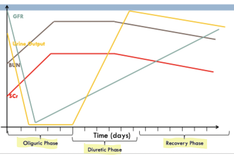
AKI Timeline
Oliguric Phase
decrease in GFR, Urine output (first to drop)
increase in BUN, SCr
Diuretic Phase
Improvement → urine output unproportionately increase compared to GFR increase
Recovery Phase
return to normal function over days
AKI Stage Classification
Stage 1
1.5-1.9 times baseline or
>0.3mg/dl increase within 48 hrs of normal baseline
Stage 2
2.0-2.9 times baseline
Stage 3
SCr >4 or
3 times baseline or
initiation of renal replacement therapy
Pre-Kidney AKI
Decrease kidney perfusion → severe dehydration
Decrease effective circulating volume → HF
Renin-Angiotensin-Aldosterone System (RAAS)
Process:
Decrease in renal perfusion → renin release from kidneys → conversion of angiotensinogen to angiotensin I → ACE converts to angiotensin II
Angiotensin II Effects:
Increase in sympathetic system
Aldosterone secretion → Na & Cl absorption, K excretion
Vasoconstriction → increase in BP
ADH secretion → water retention
Goal
water & salt retention increase effective circulating volume → increase in kidney perfusion → decrease in renin release
Ischemic ATN Pathophysiology
Auto-regulation of afferent arterioles vasoconstrict to decrease oxygen demand in medullary nephrons to prevent damage
Cost is decrease in GFR
Ameliorating Effects
Decrease in O2 demand → decreased tubular transport & GFR
Vasodilators → PG, Adenosine, bradykinin, NO
Exacerbating Effects
Nephrotic medications
NSAIDS
Angiotensin II (potent vasoconstrictor = decrease O2 supply
Calcium
Myoglobin
Post-Kidney AKI
Nephrolithiasis → block renal pelvis or ureter
hydronephrosis (expansion of kidney pelvis)
BPH → block urethra in men
Malignancy
Complications of AKI
Edema
Uremia
Electrolyte imbalance
CKD risk
Acid & Base imbalance
CVD risk
Risk Factors for Drug-Induced Kidney Disease
Age >65
CKD, DM, HTN
concomitant nephrotoxins
Renin-dependent state
HF, cirrhosis
Drug allergy
Duration of therapy
Prevention
Direct prevention strategies
Avoid nephrotoxins in high risk patients
TDM
Maintain hydration
Drug-Induced Pre-Renal AKI
ACEi/ARBs → efferent arteriole dilation
NSAID & Calcineurin inhibitor → afferent arteriole constriction
Loops → decrease circulating volume
Loss of autoregulation = risk of decrease in GFR due to decrease in hydrostatic glomerular pressure
Drug-Induced Pre-Renal AKI Prevention and Treatment
Prevention
Maintain fluid intake
Avoid concomitant nephrotoxins
Monitor SCr, BUN, K+, weight
start low dose, titrate up
Avoid NSAID + ACE/ARB combo in patients with CKD, HF, liver disease
Treatment
Discontinue offending agent (NSAID)
Provide fluids (0.9% NS) to maintain effective circulating volume
Acute Tubular Necrosis (ATN) → Causative Agents
Causative Agents
Aminoglycosides (systemic)
Tobramycin, Amikacin, Gentamycin
Amphotericin B
IV iodinated contrast media
ATN & Aminoglycosides
Nephrotoxicity related to trough concentrations (TDM)
Goal Trough concentrations:
Normal Dosing
Gentamycin & Tobramycin → < 2 mg/L
Amikacin → <8 mg/L
Extended Dosing
undetectable (~0.5mg/L)
optimizes PKPD compared to traditional dosing
CIN Risk Factors
CKD
Diabetes mellitus
concomitant nephrotoxins
Contrast Media
large iodinated contrast dose
high osmolality contrast
Ionic contrast
short interval between 2 administrations
CIN Prevention
Saline Hydration
0.9% NS 12 hours prior and after
“flush out the kidneys”
Sodium Bicarb
conflicting evidence, may cause harm, usually avoided
Acetylcysteine
maybe beneficial, but not harmful
Additional to fluids with patients who are high risk
1200 mg PO BID
Drug-Induced AIN + Treatment
Beta lactams
Oxacillin, methicillin, etc.. (penicillins)
NSAIDS
Sulfa-containing drugs
PPI
Treatment → aggressive high dose steroids as soon as possible once diagnosed
Vancomycin AKI
Nanospheric obstructive vancomycin → AIN or ATN
TDM
Trough of 10-20 mg/dL
AUC of < 600 mcg*h/mL
Avoid concomitant nephrotoxins
avoid in weight >101.4 due to weight based dosing toxicity
avoid duration > 7 days
Rhabdomyolysis Intra-Tubular Obstruction
precipitation in tubules from muscle breakdown
statins & statin-fibrate combos
Counsel on muscular symptoms and changes in urine color
Management → aggressive fluid administration
Lithium induced CKD
Nephrogenic diabetes insipidus
Risk factors
duration of therapy
episodes of acute lithium toxicity
cumulative lithium exposure
Treatment
d/c Lithium
K+ sparing diuretic
triamterene or amiloride
Only treats Symptoms (polyuria, polydipsia)
Acid / Base Laboratory Values
pH → 7.4
pCO2 → 40 mmHg
pO2 → 80 mmHg
HCO3- → 24 mEg/L
SaO2 → 97%
Assessment for Acid/Base Disorders
pH < 7.35 → Acidemia
↑ pCO2 → respiratory
↓ Bicarb → metabolic
pH > 7.45 → Alkalemia
↑ Bicarb → metabolic
↓ pCO2 → respiratory
Metabolic Acidosis Overview
pH < 7.35, Bicarb < 22
Causes:
loss of bicarb (diarrhea, vomiting)
increase in organic acids
acid accumulation (renal failure)
Compensation
Increased ventilation = ↓ pCO2
tachypnea (RR > 22)
Anion Gap Interpretation
Normal 8-12 mEq/L
Anion gap = Na - (Cl + Bicarb)
High Anion Gap acidosis
consumption of bicarb = ↓ bicarb, ↑ anion gap due to foreign anions
Normal Anion Gap acidosis
Bicarb loss is replaced by Cl-
High Anion Gap Acidosis - pneumonic
M → methanol
U → uremia
D → DKA
P → PEG
I → Isoniazid
L → lactic acidosis
E → ethanol
R → rhabdo
S → salicylates
Normal Anion Gap Acidosis - pneumonic
H → hyperalimentation (TPN)
A → acetazolamide
R → Renal tubular acidosis
D → diarrhea
U → ureterosigmoid fistula
P → pancreatic fistula
Metabolic Acidosis → Bicarb Deficit
If bicarb < 10, or pH < 7.2 → emergency
Bicarb Deficit
= 0.5L/Kg * (bicarbdesired - bicarbmeasured)
Do not administer full deficit in one sitting
50% over 4 hours, 50% over 1-2 days
Sodium Bicarb Formulations + Alternative therapies
PO
325 mg → 4 mEq bicarb
650 mg → 8 mEq bicarb
IV
50mL ampule → 50 mEq
150 mEq/1000 mL in D5W
Sodium Acetate (IV)
intact liver and bicarb deficit
Citrate (PO)
1 mol citrate = 3 mol bicarb, dosed by bicarb mEq’s
bicitra, polycitra, urocit
oral product, reduced GI side effects
Sodium Bicarb ADR’s + Patient Selection
ADR
GI, belching, flatulence
Edema + hypokalemia (IV risk)
overcorrection of pH
Patient Selection
Benefit
high anion gap
RTA
Salicylate poisoning
Harm
volume overload → AKI, CHF, hypervolemia
Underlying disease → long injury, DKA, lactic acidosis
Metabolic Alkalosis
Chloride Responsive
Loss of H+ & Cl- → vomiting, NG suction, diuretic use
urine chloride <10mEq/L
Treatment:
correct underlying cause
replete with 0.9% NS
Fluid restricted → less fluids + carbonic anhydrase inhibitor (block bicarb reabsorption)
Chloride Resistant
excess mineralocorticoid activity
ex. hyperaldosteronism, Cushing’s syndrome
urine chloride > 20 mEq/L
Treatment:
correct underlying cause
spironolactone
Respiratory Acidosis
↑ pCO2 retention caused by restricted ventilation
sleep apnea, COPD, asthma, pulmonary embolism/fibrosis/edema
CNS depressants → opiates, benzos, anesthetics
Treatment
Restore oxygenation
treat underlying cause
remove offending drug
Respiratory Alkalosis
↓ PCO2 due to ↑ ventilation of CO2 > production of CO2
anxiety, panic attacks, high altitude, asthma
Presentation → lightheaded, confused, syncope, seizure
Treatment → underlying cause