Human anatomy and physiology module 3
1/21
There's no tags or description
Looks like no tags are added yet.
Name | Mastery | Learn | Test | Matching | Spaced |
|---|
No study sessions yet.
22 Terms
What is the cardiovascular system?
A transport system in the body that allows for blood to be pumped continuously around the body, transporting substances to and from where they are needed
Why is the cardiovascular system essential?
-Becuase cells and tissues need a constant supply of nutrients and oxygen through the cardiovascular system, if there is a low supply of nutrients and oxygen, it will result in a lack of cell function, which can result in tissue damage. Furthermore, the cells and tissues in our body produce waste which need to be removed and this is done through the cardiovascular system to a part of the body where they are excreted
What are the 3 key components to the cardiovascular system?
Heart: Pump that provides the ability to move blood through the cardiovascular system, and the 2 sides of the heart are structurally and functionally different and work as 2 seperate pumps
Blood vessel: Tubes that blood flows through to reach all body parts; the main blood vessels are the arteries, veins and capillaries
Blood: Transport medium which transports substances such as o2, co2, nutrients, wastes, hormones and blood cells throughout the body
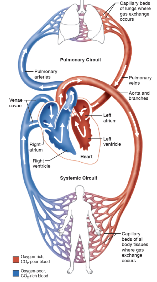
What is the organization of the cardiovascular system?
-There is an organization of the cardiovascular system so that the blood is able to circulate around the body, allowing for nutrient delivery and removal of waste and the travel of blood happens through the pulmonary circulation and the systematic circulation
What type of blood does the right side of the heart contain?
The right side of the heart contains deoxygenated blood; the heart pumps blood into the pulmonary circulation, where the circuit takes blood to the lungs, where it gains oxygen and loses carbon dioxide, from where the oxygenated blood is returned from the pulmonary circulation to the left side of the heart
What type of blood does the left side of the heart contain?
The left side of the heart contains oxygenated blood; the heart then pumps the oxygenated blood to the systematic circulation. this circuit takes blood to other tissues of the body, where it delivers nutrients and oxygen, but also picks up waste and CO₂, and the blood leaving the systemic circulation is then returned to the right side of the heart
What are the upper chambers of the heart?
-Right atrium: Receives blood returning from the systematic circulation
Left atrium: Receives blood returning from the pulmonary circulation

What are the lower chambers of the ventricles?
-Right ventricle: Pumps blood into the pulmonary circulation
-Left ventricle: Pumps blood into the systematic circulation
What is the difference between the arteries and the veins?
-The arteries carry blood away from the heart, whereas the veins carry blood towards to the heart
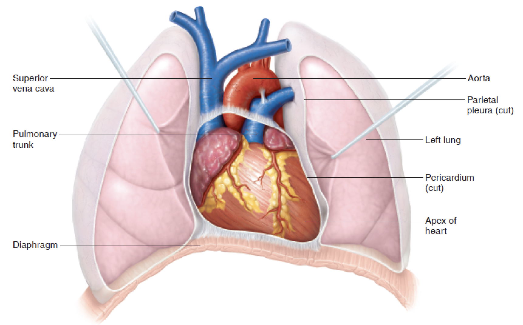
Where is the heart located?
the heart is located within the thorax, between the lungs and is anterior (front) to the vertebral column and posterior (back) to the sternum
What are the three layers of the heart wall?
Epicardium: External layer and forms the pericardium
Myocardium: Middle layer and makes up the majority of the heart. it is primary cardiac muscle and is responsible for cardiac contraction and pumping action of the heart
Endocardium: Innermost layer of the heart and provides a smooth layer lining inside the heart
Pericardium: Double walled sac, which helps protect the heart. the pericardial fluid between the 2 layers of the pericardium acts as lubricant, which reduced friction as the heart contracts the pericardium acts
What are the chambers of the heart?
Atria: Small chambers with thin walls, as they dont generate much force to push blood to adjacent ventricle
Right atrium: Receives blood from systematic circulation (via the inferior and superior vena cava), the blood then passes through the tricuspid valve to the right ventricle.
Left atrium: Receives blood from pulmonary circulation from pulmonary veins and this blood then passes through bicuspid valve to the left ventricle
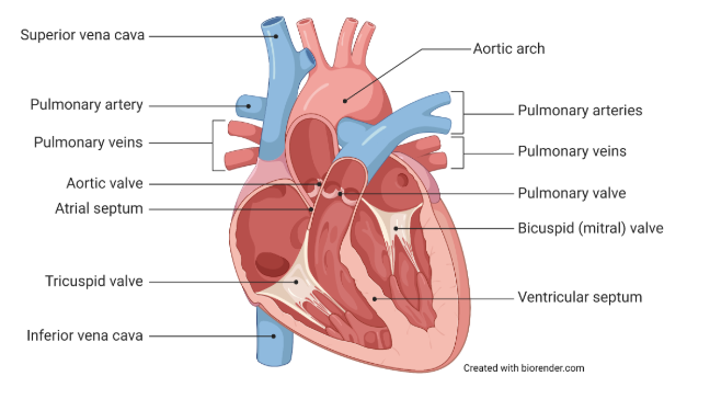
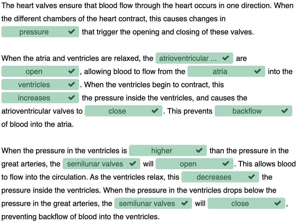
What are the atrioventricular valves? (AV)
-Located between the atria and ventricles, and are made up of flexible cusps,
-The right AV has 3 cusps called a tricuspid valve.
-The left AV has 2 cusps called a bicuspid/mitral valve
-When the heart is relaxing, the cusps hang into the ventricles and the valve is open, which allows blood to flow from the atria to the ventricles. when they contract the increased blood pressure in the ventricles pushes the cusps upward closing the valve
-The purpose of the AV valves is to prevent the backflow of blood from the ventricle to the atrium
What are the semilunar valves?
-Located between the ventricles and the large arteries, specifically the aortic valves that are located between the left ventricle and the aorta and the pulmonary valves, which are located between the right ventricle and the pulmonary trunk and pulmonary arteries
-The SL valves open and close based on changes in pressure. When the ventricles are contracting and the ventricle pressure is higher than the artery pressure, the SL valves open.
-As ventricles relax and pressure inside the heart decreases, the pressure in arteries forces valves closed, preventing backflow
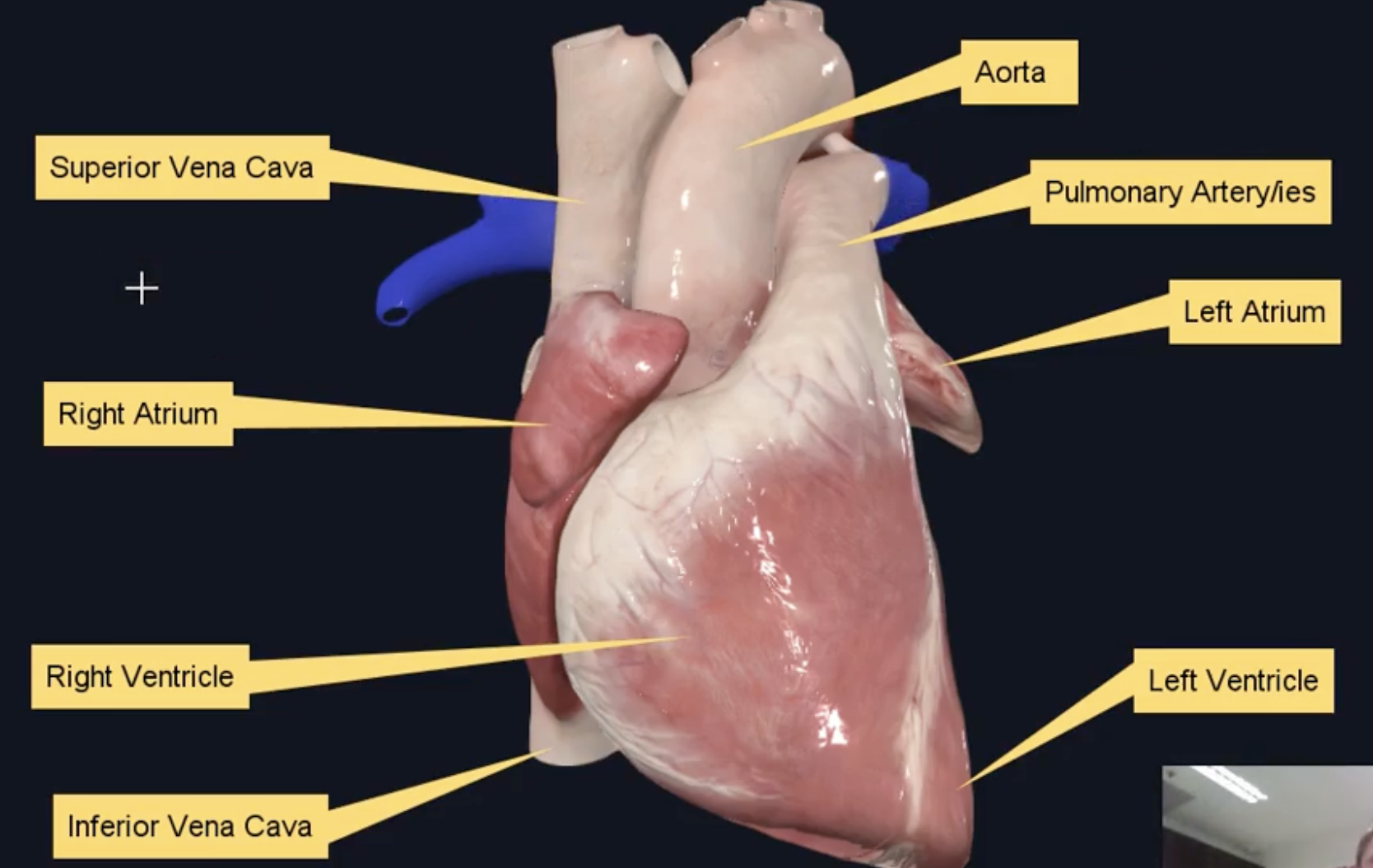
Anterior view of heart
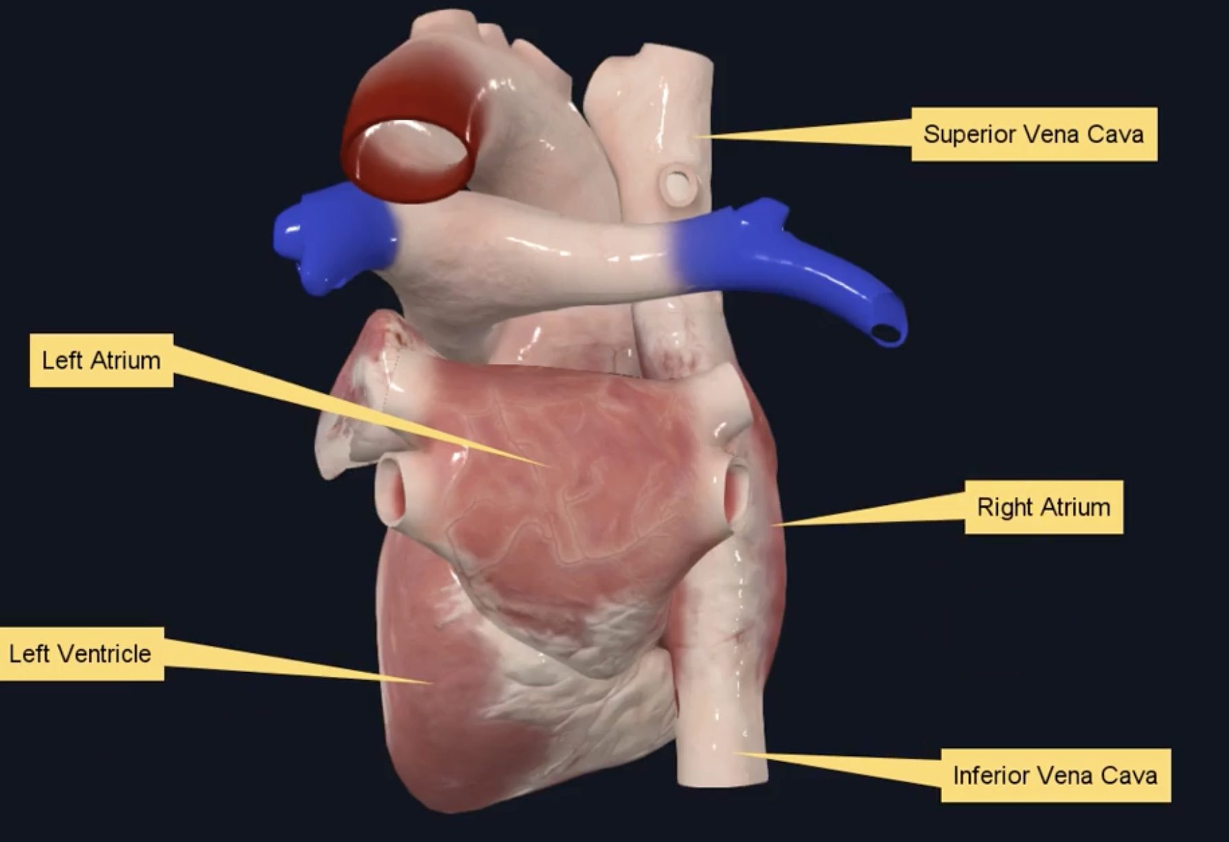
Posterior view of heart
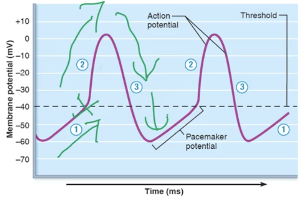
What are the attributes of action potentials in pacemaker cells?
1.Pacemaker potential: Slow spontaneous depolarization due to the slow entry of Na+
2.Depolarization: When the membrane potential reaches threshold, an action potential is fired, Ca2+ channels open, and the rapid entry of Ca2+ causes depolarization and rising phase of the action potential
3.Repolarization: Ca²⁺ + channels inactivate, and repolarization (falling phase of action potential) is due to the opening of K⁺ channels and exit of K⁺ from cell
What is the cardiac conduction system?
-In healthy heart, pacemaker cells are located in the sinoatrial node, which generates electrical impulses that travel through the heart.
-If pacemaker cells depolarize and fire action potentials more frequently, heart rate will increase
-If pacemaker cells depolarise and fire action potentials less frequently, heart rate will decrease
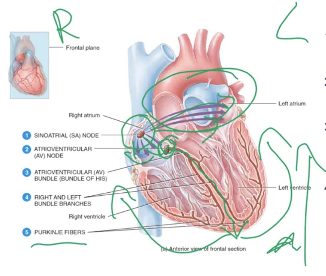
What are cardiac conduction pathways in the heart?
-Pathways that allow for the coordinated spread of electrical activity through the heart
1.Depolarization begins at the SA node and spreads through the atria
2. Depolarization spreads to the AV node via the internodal pathway
3.Depolarization moved through the septum (separates left and right side of the heart) to the heart apex via the AV bundle (Bundle of His) and bundle branches
4.Depolarization spreads from the apex through the ventricles via the purkinje fibres
What is an electrocardiogram(ECG) and its purpose?
-Uses recording electrodes placed on the body surface to measure the electrical activity of the heart.