PSLA CVS100 ex:3
 Call Kai
 Learn
Learn Practice Test
 Spaced Repetition
 Match
Match Flashcards
 Knowt Play
1/52

There's no tags or description
Looks like no tags are added yet.
Name | Mastery | Learn | Test | Matching | Spaced | Call with Kai | Chat |
|---|
No analytics yet
Send a link to your students to track their progress
53 Terms
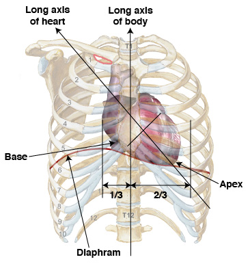
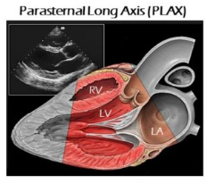
What is the Parasternal Long Axis View (PSLA)?
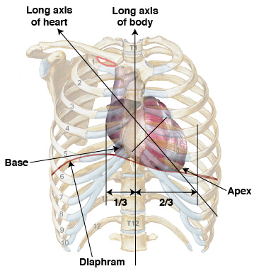
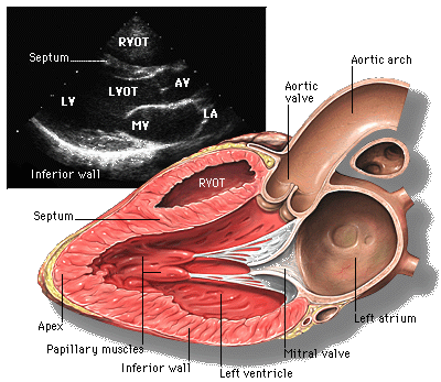
The PSLA view is generally the first view obtained in an echocardiography examination, providing real-time imaging of the heart by transecting it from the base to the apex (sliced in half), with the apex toward the left and the base toward the right.
What patient position is preferred for an echocardiography examination, and why?
The Left lateral decubitus position is preferred. The patient lays on their left side with their left arm extended over their head and their right arm at their side, as gravity displaces the cardiac apex closer to the chest wall.
How is a patient prepared for an echocardiography exam regarding monitoring?
A basic 3-lead EKG is applied to monitor the patient's heart rate and rhythm during the exam.
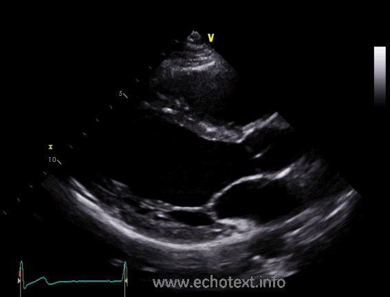
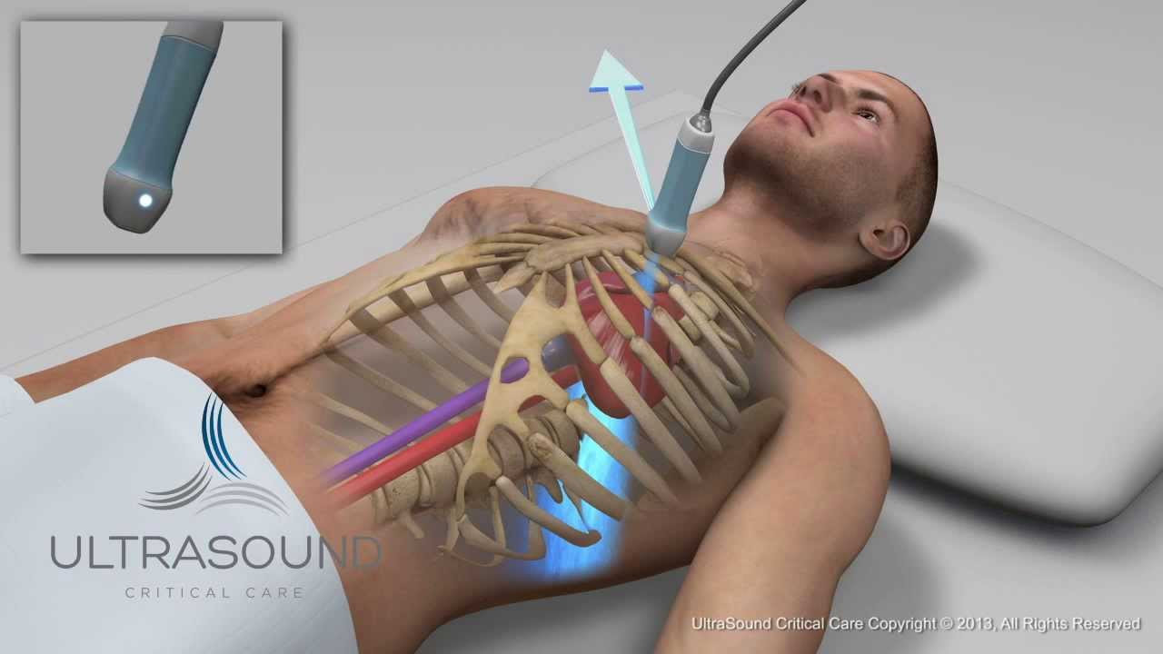
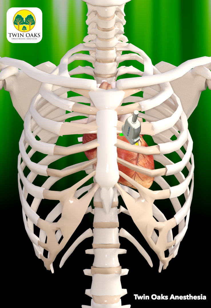
Where is the transducer initially placed for obtaining the PSLA view?
The transducer is placed to the left side of the sternum in the 2nd to 3rd intercostal space, with the index directed toward the patient's right shoulder.
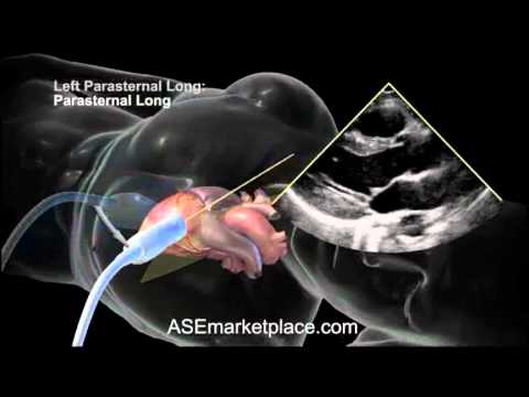
What is the typical index orientation for the PSLA view?
The index of the transducer points somewhat towards the patient's right shoulder, though the exact orientation depends on the axis of the patient's heart.
What is important to consider regarding the apex of the left ventricle in the PSLA view?
You usually will not be able to see the apex in this view. If you see it, rotate your index clockwise. You should try to display the LV as lengthened or long as possible.
What is the typical depth setting for echocardiography, and what influences it?
Depth is usually around 12-16 cm, but this is always dependent on the patient's anatomy.
Describe breathing techniques used during an echocardiography exam.
Patients should breathe normally. When lungs interfere, have the patient hold or expel all air and hold their breath. Experiment to obtain the best picture.
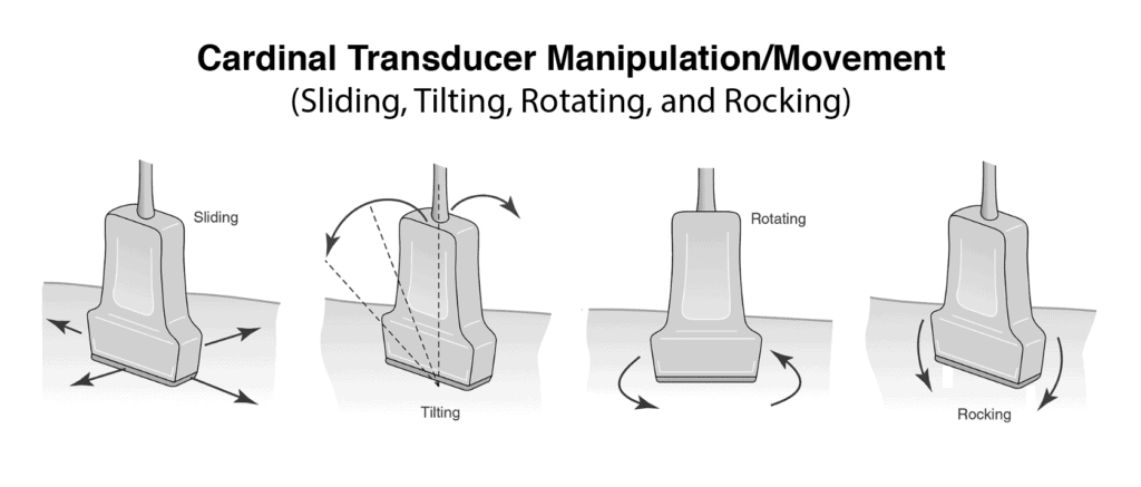
What is 'Tilt/Angle' movement of the transducer?
The transducer stays in the same imaging axis, but the face of the transducer is manipulated to demonstrate different imaging planes.
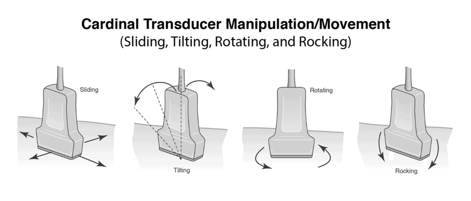
What is 'Rotate' movement of the transducer?
The transducer stays in the same position, but the index orientation is rotated to a different position.
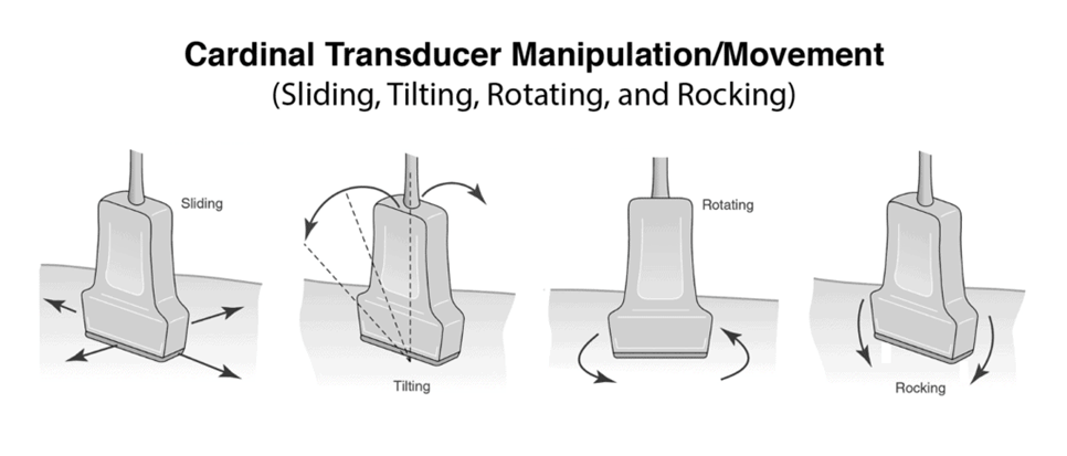
What is 'Rocking' movement of the transducer?
The transducer stays in the same imaging plane, but is moved towards or away from the index marker.
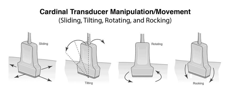
What is 'Slide' movement of the transducer and a clinical example?
Slide movement involves physically changing the location of the transducer. A clinical example is sliding the transducer from one rib space to another rib space.
What type of transducer is typically used for cardiac echocardiography, and what is its frequency range?
A phased array transducer is typically used for cardiac imaging, with frequencies ranging between 2MHz to 5MHz.
How does transducer frequency affect depth penetration and resolution?
A lower frequency has better depth penetration (ideal for adults) but poorer resolution, while higher frequencies offer better resolution but less depth penetration.
What MHz range is most frequently used for adult echocardiography, and when might 5 MHz be used?
2-3.5 MHz is most frequently used for adults. 5 MHz may be used for small adults, children, or when better resolution of the cardiac apex is needed to rule out a clot.
Define 'Homogeneous' in sonographic appearance.
Uniform gray scale.
Define 'Echogenic' in sonographic appearance.
Reflective.
Define 'Anechoic' in sonographic appearance.
Without echoes; appears black.
Define 'Hypoechoic' in sonographic appearance.
Low level echoes; appears darker gray.
Define 'Hyperechoic' in sonographic appearance.
Brighter echoes; appears almost white.
What is the most reflective structure in the heart according to sonographic appearance?
The pericardium, which appears almost white (hyperechoic).
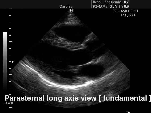
How do papillary muscles, myocardium, and valves appear sonographically?
Papillary muscles and myocardium are medium gray and homogeneous in echo texture. Valves are slightly more echogenic than the walls.
How do fluid-filled spaces like heart chambers and great vessels appear sonographically?
The area within the chambers and great vessels, as well as any other fluid space, is anechoic.
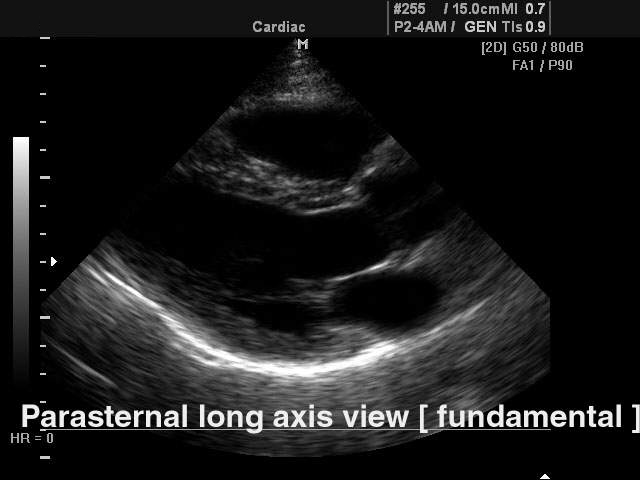
What are the criteria of quality for a good PSLA view?
The septum must be as horizontal as possible. You should not visualize the apex of the left ventricle (if seen, rotate index clockwise). You should see the aortic and mitral valves but not the tricuspid valve.
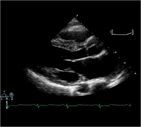
What is 'Overall Gain' optimization in 2D echo?
Adjusting the overall brightness so chamber cavities appear anechoic, while ventricular walls appear gray.
What are 'TGCs' (Time Gain Compensations), and how are they used?
TGCs compensate for attenuation loss. They are adjusted individually to optimize specific areas (nearfield, midfield, farfield) of the image.
What are 'LGCs'?
LGCs refer to Lateral Gain Compensations, used for the left and right sides of the image.
What is 'Attenuation' in ultrasound?
Attenuation refers to the inevitable loss of ultrasound wave amplitude or beam intensity.
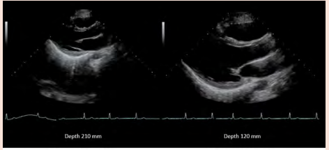
What is the optimal 'Depth of view' for a 2D echo image?
The image should be approximately 2/3rds of the screen (distance from the surface of the skin into the body).
Where should the 'Focal zone' (focus) be set during 2D optimization?
The focus should be set at the valve level, as it is the narrowest region of interest of the ultrasound beam that exhibits the best spatial resolution.
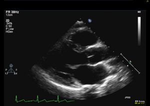
What can be evaluated concerning the Right Ventricle (RV) in the PSLA view?
RV dilatation and RV contractility.
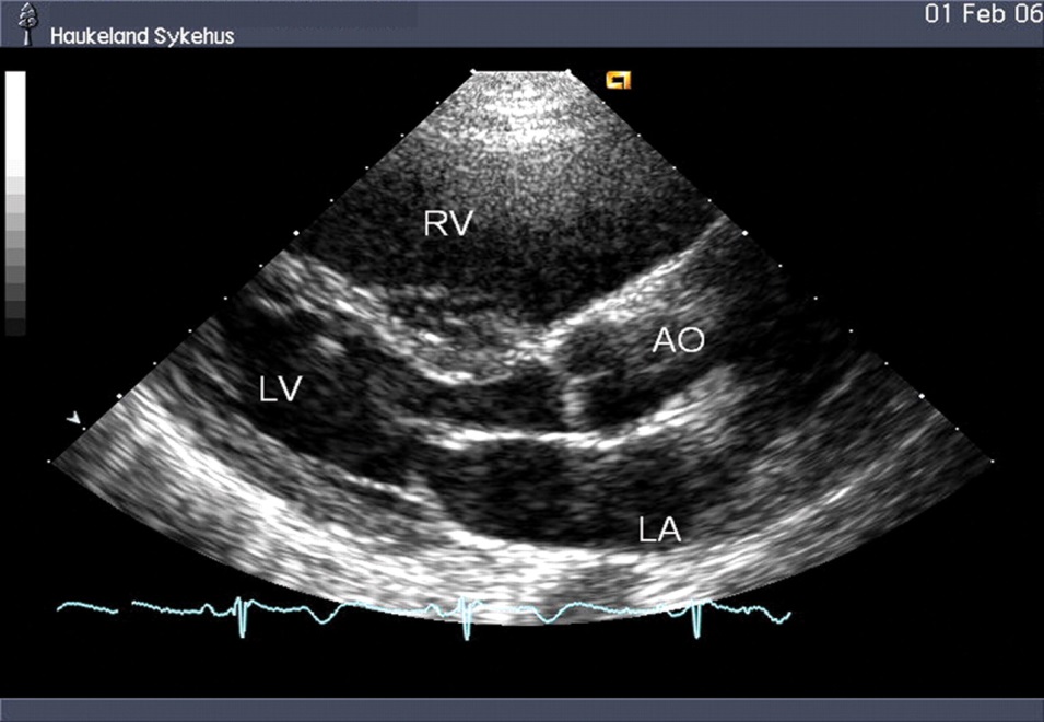
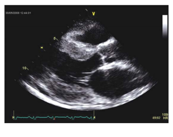
What is the PSLA view best for measuring regarding the Left Ventricle (LV)?
It is best for measuring the size of the LV and wall thickness (Hypertrophy) of the LV.
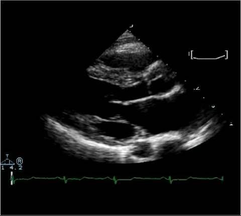
What specific walls of the LV can be assessed for contractility in PSLA?
The Interventricular Septum (IVS) and Left Ventricular Posterior Wall (LVPW).
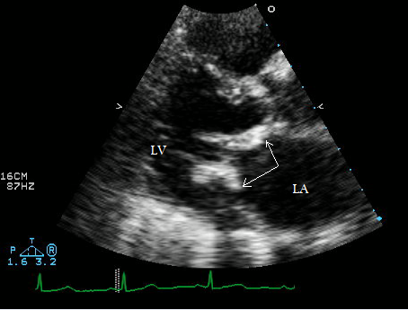
Which leaflets of the Mitral Valve (MV) are seen in the PSLA view?
The Anterior Mitral Leaflet (AML) on the top and the Posterior Mitral Leaflet (PML) on the bottom.
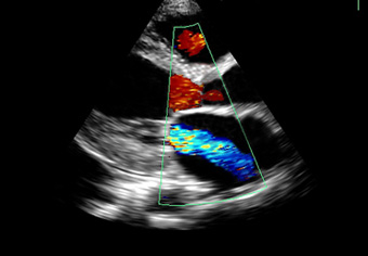
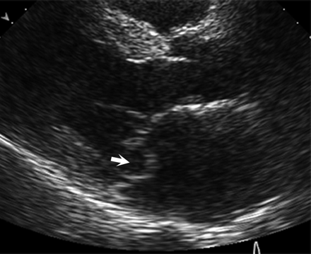
What can be evaluated regarding the Mitral Valve in PSLA?
Motion and thickness of the leaflets for calcifications or mitral valve prolapse, and with color, any MV regurgitation (backward flow into the atria).
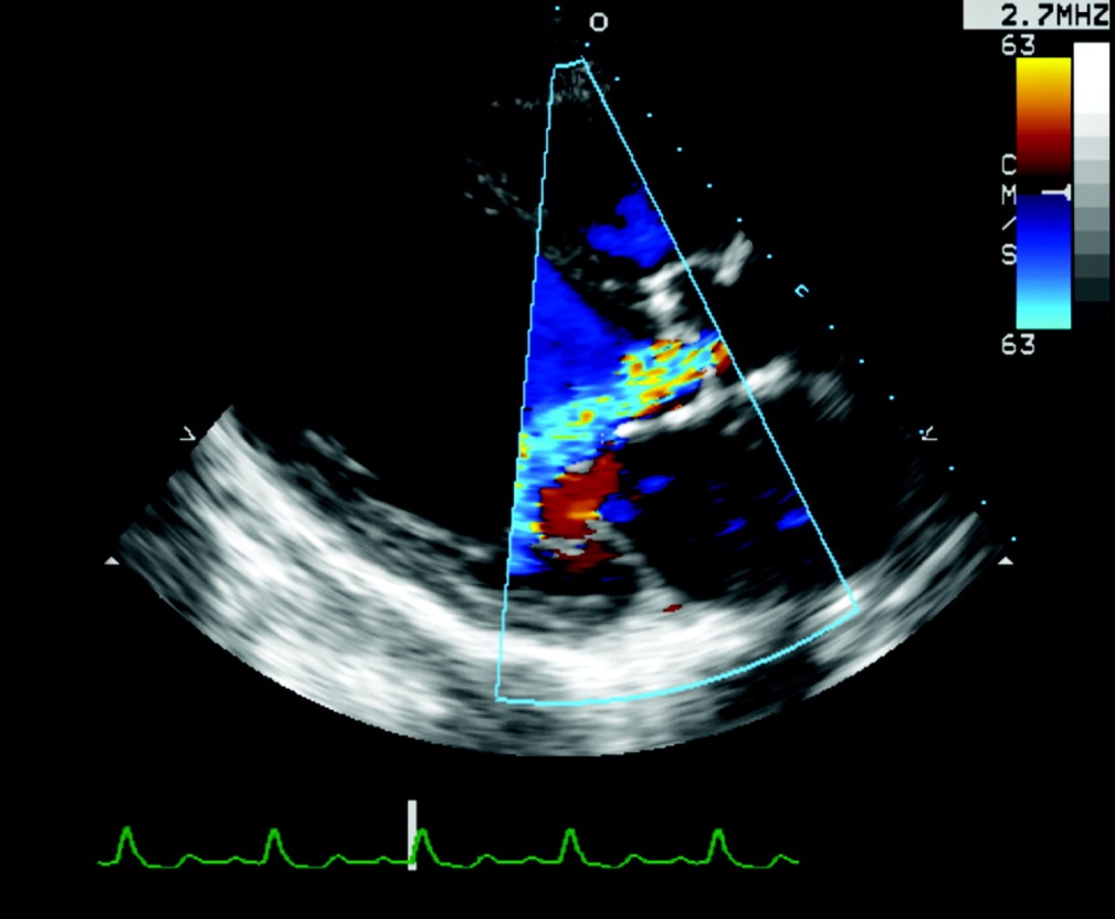
Which cusps of the Aortic Valve (AOV) are seen in the PSLA view?
Two of the three aortic cusps: the Right Coronary Cusp (RCC) on top and the Non Coronary Cusp (NCC) on the bottom.
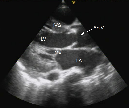
What can be evaluated regarding the Aortic Valve in PSLA?
The opening of the aortic cusps for suspected aortic stenosis (narrowing), calcifications, or restriction of valve opening. With color, aortic regurgitation (backward flow into the ventricle) can be assessed.
What can be measured and evaluated regarding the Aortic Root (AOR) in PSLA?
The aortic annulus, Left Ventricular Outflow Tract (LVOT), and the dimensions of the aortic root. The aortic walls should appear parallel.
What can be assessed regarding the Interventricular Septum (IVS) and Left Ventricular Posterior Wall (LVPW)?
Their thickness and contractility.
What is important to note about the size of the Left Atrium (LA) in PSLA?
It should be approximately the same size as the aortic root.
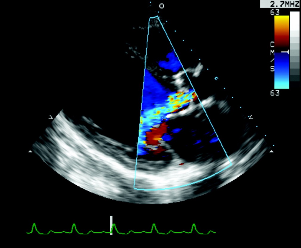
What is the Descending Aorta (DA) used for in PSLA?
The DA is seen below the LA and helps distinguish between a pericardial effusion and a pleural effusion.
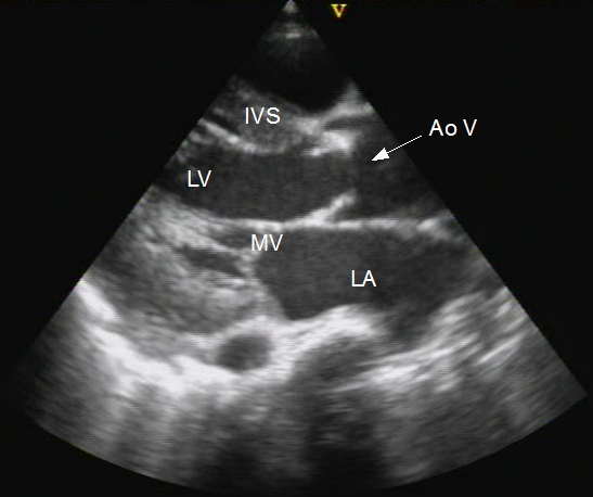
What are the anatomical structures typically seen in a PSLA view?
Anterior Right Heart Wall (ARHW), Right Ventricle (RV), Interventricular Septum (IVS), Left Ventricle (LV), Left Ventricular Posterior Wall (LVPW), Anterior Aortic Wall (AAW), Aortic Valve (AOV) (RCC/NCC), Posterior Aortic Wall (PAW), Mitral Valve (MV) (AML/PML), Left Atrium (LA), Descending Aorta (DA), Pericardium (PERI).
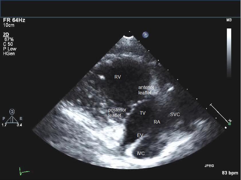
How is the Right Ventricular Inflow Tract (RVIT) view obtained from the PSLA view?
From the PSLA view, angle the beam medial and inferior towards the right hip.
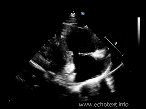
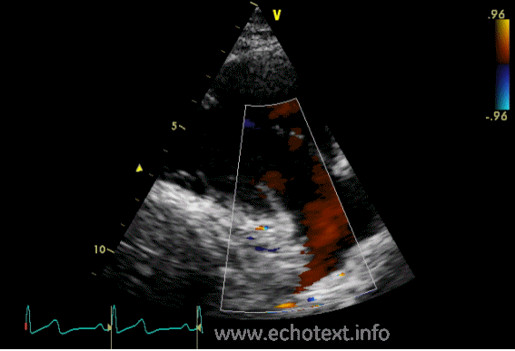
What anatomy is seen in the RVIT view, and what can it evaluate?
Anatomy seen: Right Atrium (RA), Right Ventricle (RV), Tricuspid Valve (TV) (anterior and posterior leaflets), and Eustachian Valve (EV). It is helpful in evaluating diseases of the TV and other diseases of the right heart, and with color, tricuspid regurgitation.
How is the Right Ventricular Outflow Tract (RVOT) view obtained from the PSLA view?
From the PSLA view, angle the beam superior and lateral towards the patient's left shoulder.
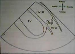
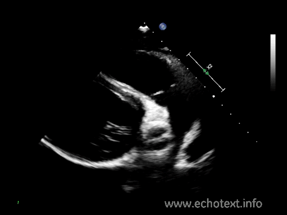
What anatomy is seen in the RVOT view?
Pulmonic Valve (PV), Right Ventricle (RV), and Main Pulmonary Artery (MPA).
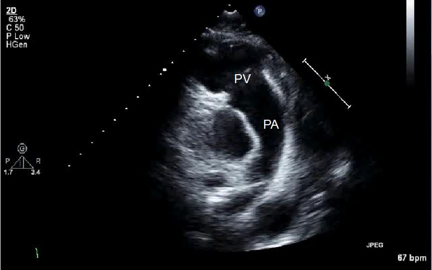
By what other names is the RVOT view known?
Conus Arteriosus or Infundibulum.
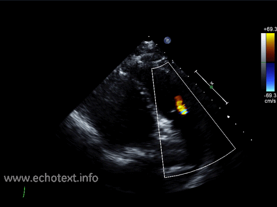
What can be evaluated using color Doppler in the RVOT view?
Pulmonic regurgitation (backward flow of blood into the right ventricle).
Name the three layers of the heart wall.
- Epicardium
- Myocardium
- Endocardium
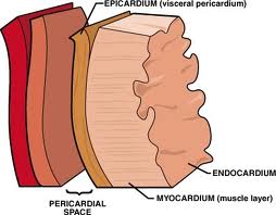
Describe the Epicardium.
The Epicardium is the thin, smooth outside layer that covers the surface of the heart and extends to the great vessels; it is the visceral layer of the serous pericardium.
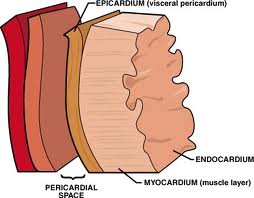
Describe the Myocardium.
The Myocardium is the thicker, muscular middle layer, which is the functional layer composed of striated muscle fibrils with contractile elements called myofibrils.
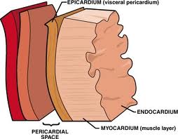
Describe the Endocardium.
The Endocardium is a thin layer of endothelium and underlying connective tissue. It lines the inner chambers of the heart, valves, chordae tendinae, and papillary muscles.
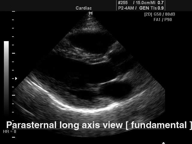
PSLA VIEW
Parasternal long access view