Benign Solid Breast Masses
1/61
There's no tags or description
Looks like no tags are added yet.
Name | Mastery | Learn | Test | Matching | Spaced |
|---|
No study sessions yet.
62 Terms
What is fibrocystic condition?
magnification of normal physiologic processes of breast tissue
The tissue in the fibrocystic condition is:
-very fibrous
-dense
-has many cysts
Fibrocystic Condition fluctuates with?
hormonal cycles
Fibrocystic Condition may _______ or _______ breast tumor
mimic or obscure
Fibrocystic Condition is typically a
bilateral process
What is the most common diagnosis at breast biospy?
fibrocystic condition
What are the clinical signs of fibrocystic condition?
-pain
-lumps or lumpy
What is the appearance of fibrocystic condition on mammogram?
-diffuse benign microcalcifications
-adenosis (breast glands are enlarged)
-multiple round masses
-very bright
What is the appearance of fibrocystic condition on Ultrasound?
-various appearances depending on stage
-multiple round masses/cysts
What is this image showing?
fibrocystic condition
What does fibrocystic condition look like microscopicly?
-apocrine metaplasia
-dilated acini
-fibrosis
-epithelial ductal hyperplasia (overgrowth of cells lining the breast)
-sclerosing adenosis (enlarged lobules)
Fibrocystic Condition and Breast Cancer Risk:
non-proliferative lesions
proliferative lesions without atypical cells
proliferative lesions with atypical cellular changes
What is the most common premenopausal benign tumor?
fibroadenoma (30%)
Fibroadenoma is
hormone dependent
What is the composition of fibroadenoma?
-connective
-epithelial
What is the appearance of a fibroadenoma on mammogram?
-radiopaque: brighter on xray/mammo
-oval or round mass
-circumscribed
-smooth margins
-may have lobulated contour or notching
-thin radiolucent halo
-eggshell calcification indicates older lesion
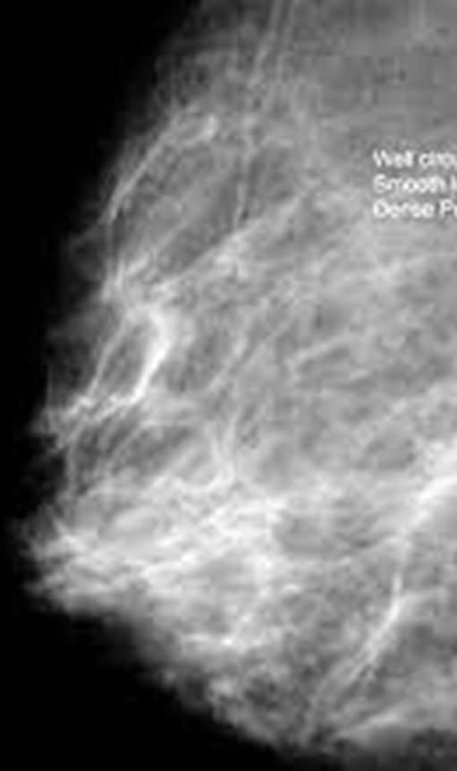
What is this image showing?
fibroadenoma on mammo
What is the sonographic appearance of an fibroadenoma?
-smooth walls
-oval to round
-lobulations
-homogeneous
-hypoechoic
-may enhance
-bilateral edge shadows
-horizontal
-slightly compressible
-mobile
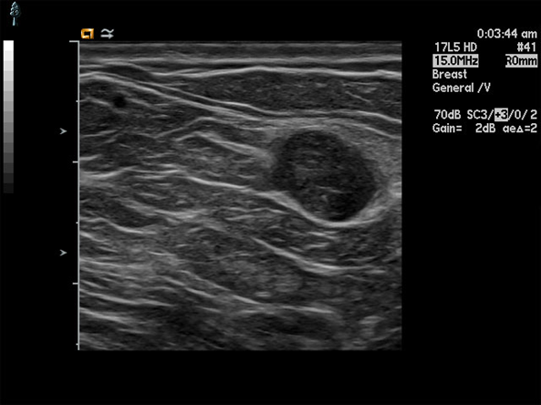
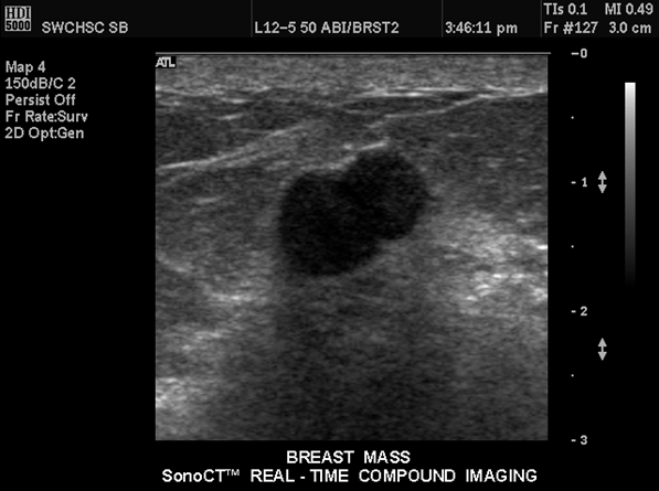
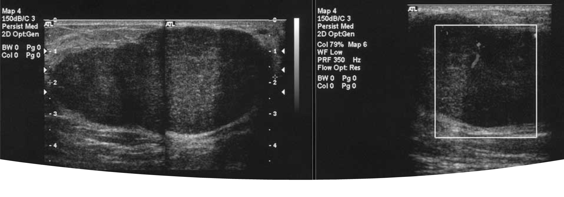
What are theses images showing?
fibroadenoma on ultrasound
What is this image showing?
a giant fibroadenoma/juvenile fibroadenoma
What is a juvenile fibroadenoma?
-10-20 years old
-extremely large: 6-8 cm
-excision (remove them because cause discomfort
What is a lipoma?
fatty tumor
-encapsulated fat cells
What are the clinical signs of a lipoma?
mobile and palpable
What are the mammo features of a lipoma?
-radiolucent
-smooth margins
they blend in very well
What are the sonographic features of a lipoma?
-isoechoic
-smooth borders
difficult to see on ultrasound
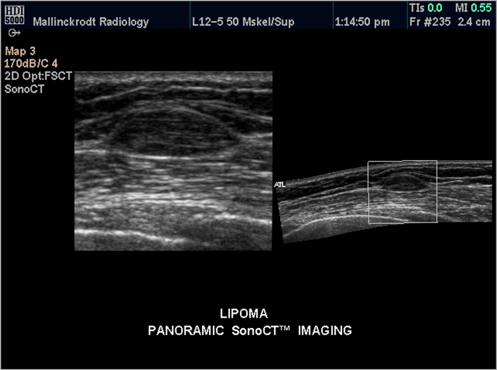
What is this image showing?
lipoma
What is a hot, red breast mean?
inflammation or an infection
What is inflammation of the breast?
mastitis -inflammatory breast cancer
What are the treatments for mastitis?
antibiotics and imaging
What is the sonographic appearance of mastitis?
-dominant atypical cystic lesion
-possible ductal dilation
What is acute mastitis caused from?
-infection
-trauma
-ductal obstruction
What are the clinical signs of mastitis?
-enlarged
-reddened
-tender
-confined to 1 area
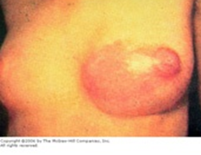
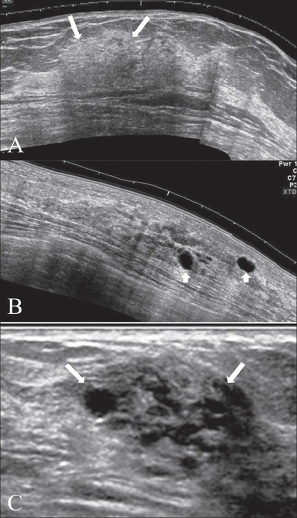
What are these images showing?
acute mastitis
What does chronic mastitis look like?
glandular tissue involvement
-mixed and diffuse echo pattern
-nipple discharge
-retracted/inverted nipple
Chronic Mastitis has what appearance on ultrasound?
heterogeneous

What are these images showing?
chronic mastitis
Acute Abscess is
poorly defined
Mature abscess is
well encapsulated
What are the clinical signs and symptoms of an abscess?
-pain
-swelling
-reddening
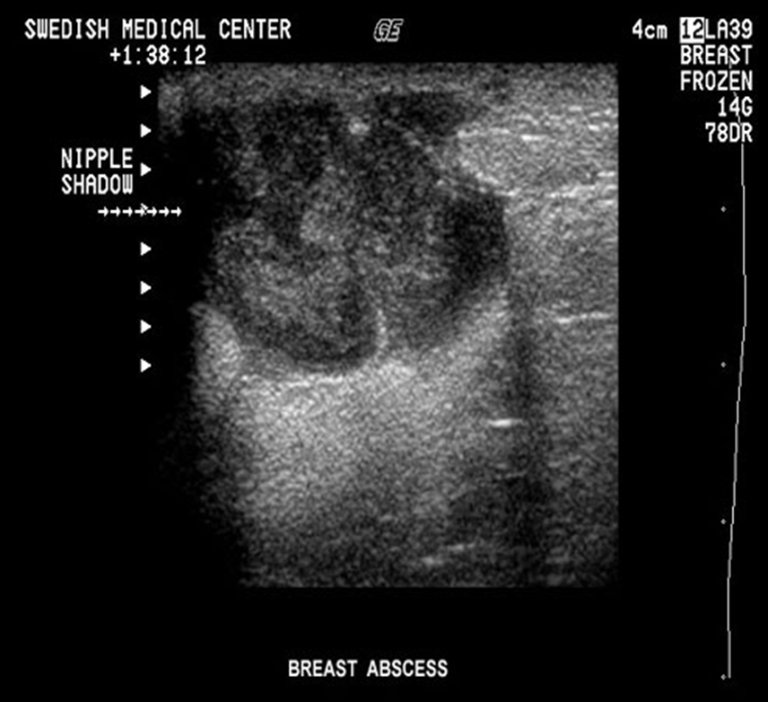
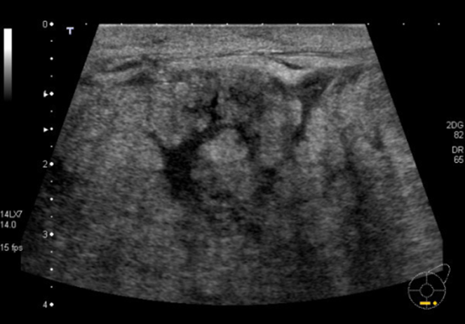
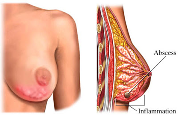
What is this image showing?
a breast abscess
What is the sonographic appearance of an abscess?
-diffuse mottled appearance
-irregular margins
-posterior enhancement
-low level internal echoes
-skin thickening
-hyperemia
What are phyllodes?
rare <1% of breast neoplasms
-transitional
Phyllodes are transitional meaning?
they can turn malignant 27% malignant
What age group does phyllodes fall under?
50-60 years old
What are the clinical features of phyllodes?
rapid growth
What are the sonographic features of phyllodes
similar to fibroadenoma
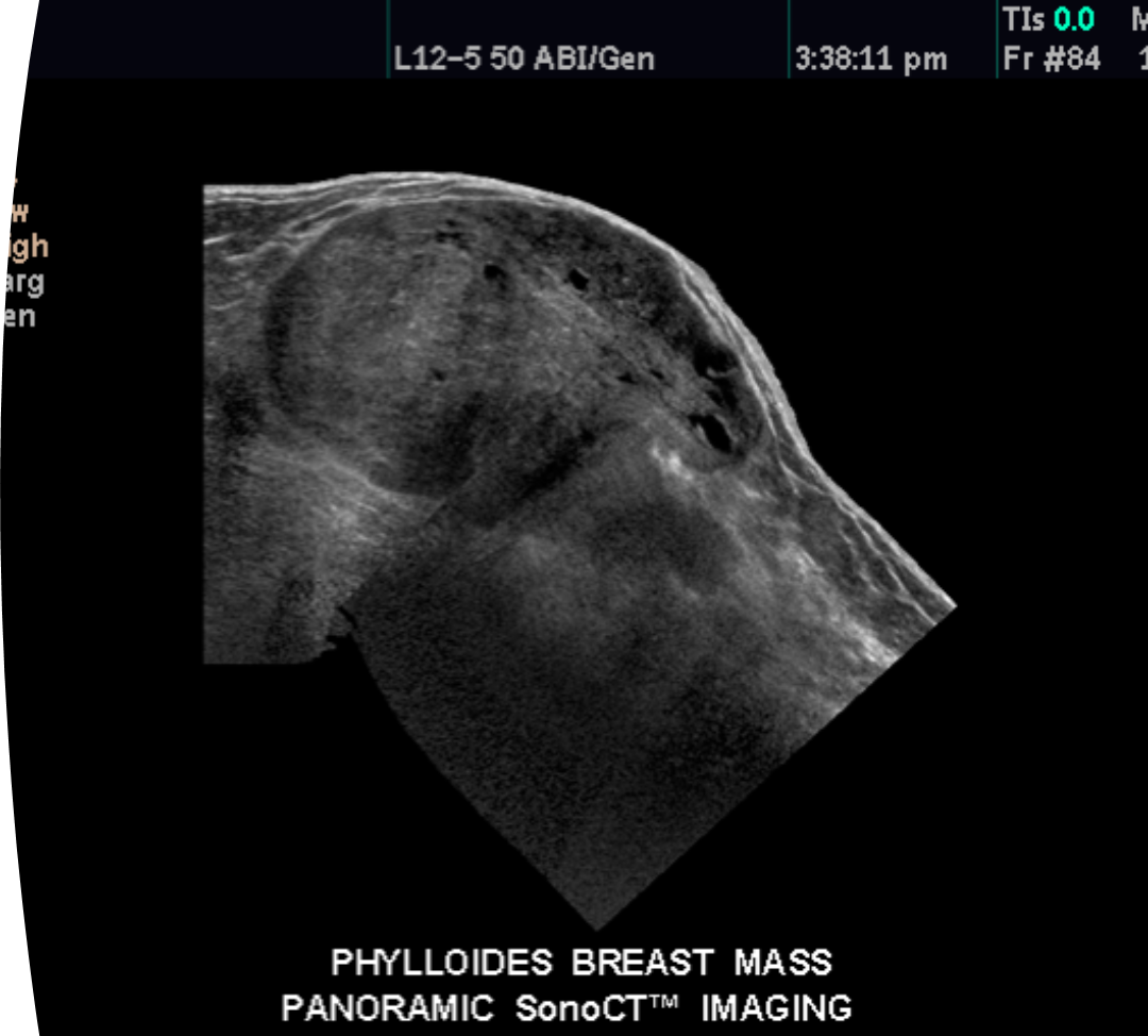
What is this image showing?
phyllodes
What is a small benign tumor within the acini (ducts) of the breast?
intraductal papilloma
What is the age group of intraductal papilloma?
35-55 years old
What are symptoms of intraductal papilloma?
nipple discharge
What are the sonographic features of intraductal papilloma?
-ductal dilation
-internal echoes
-blend in pretty well and are small
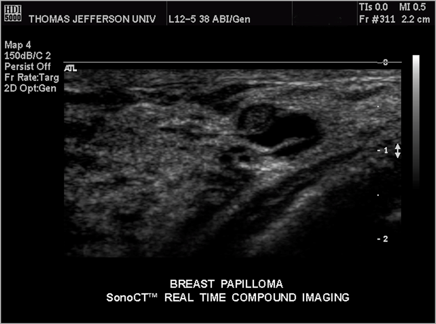
What is this image showing?
intraductal papilloma
What are traumatic changes of the breast?
-acute trauma
-post-surgical changes
-post-radiation changes
What are other benign findings of the breast?
lymph nodes and papilloma
What is common after surgeries and are caused by serum in tissues?
seroma
What are the sonographic features of a seroma?
fluid collection
septations
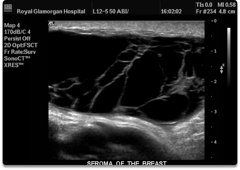
What is this image showing?
seroma
Post Surgical/Radiation Changes:
similar imaging appearance
different causes
-focal distortion
-irregular mass
should not have any blood flow within them
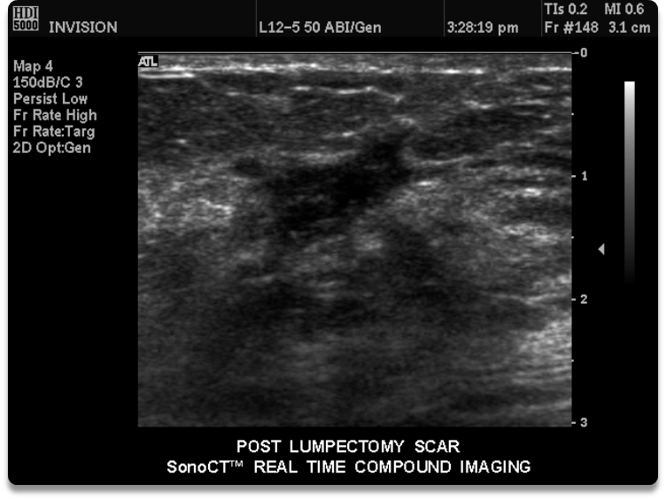
What is this image showing
a lumpectomy scar
What are benign breast tissue appear as?
displaced or compressed tissue
What does distorted breast tissue appear as
inflammation
post radiation
trauma
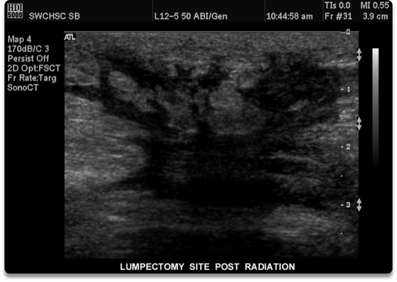
What is this image showing?
lumpectomy post radiation