Dr Priget Lectures
1/113
There's no tags or description
Looks like no tags are added yet.
Name | Mastery | Learn | Test | Matching | Spaced | Call with Kai | Chat |
|---|
No analytics yet
Send a link to your students to track their progress
114 Terms
name organelles in eukaryotic cells?
Nucleus
ER
Golgi apparatus
Lysosomes
Mitochondria
Plasma Membrane
Ribosomes
Vesicles
Cytoskeleton
Where do the proteins get made?
all proteins start in the cytosol made by ribosomes
some stay in cytosol
others go to the
-nucleus
-mitochondria
-ER → golgi → membrane or secreted out of cell
Types of ribosomes and how they sort proteins?
ER -bound ribosomes
deliver proteins for membranes or secretory pathways via co-translational insertion
Cytosolic Ribosomes
make proteins destined for the cytosol, mitochondria, nucleus or peroxisomes
What are the requirements for protein targeting?
signal address → encoded in amino acid sequence so it knows where its going
Receptor → recognises the signal (SRP)
Energy → ATP or GTP ( transfer the protein to new place)
Translocation Machinery → moves protein across or into membrane
Role of ATP in protein translocation
helps molecular chaperones that pull or push the proteins through the protein channel
Role of GTP in protein targeting
helps the timing and specificity of receptor binding and the release of the cargo
What are the different types of signal sequences?
ER signal Sequence
Mitochondrial signal
Nuclear localisation signal (NLS)
ER signal sequence
the signal is found in the N terminus (read while protein is being made and sent straight to the ER membrane)
Type: hydrophobic amino acids
cleaved off after import
Mitochondrial Signal Sequence
mitochondria have their own DNA but still import cytosol proteins
found in the N terminus
Type: Amphipathic alpha-helix (1 side = + charged, other side hydrophobic)
used by TCA cycle enzymes and matrix proteins
cleaved off after import by matric peptidase
Mitochondrial Matrix Protein
Nuclear Localisation Signal Sequence
proteins imported into the nucleus
signal sequence can be found anywhere on the protein
Type: short sequence with many Lysine (K) and Arginine (R)
not cleaved after import because needed for mitosis
How does a protein travel across the ER membrane?
Co-Translational Import
Co-Translational Import Process
ribosomes translate protein in cytosol
ribonucleoprotein SRP (Signal Recog Particle) pauses translation by binding to signal sequence
SRP guide ribosome to RER = bind to SRP receptor
The closed Sec61 channel is next to it and then receives the ribosome
channel opens and inserts signal peptide
SRP leaves protein using GTP energy
protein unpaused → ribosome translates → goes into the ER lumen
signal peptidase cleaves off signal peptide = protein free in ER.
ER Secretory Pathway
once its in the ER it has own transport network
stay in the vesicle and fuse with the golgi apparatus
golgi travel to final destination (Plasma membrane, Lysosome. Outside cell)
Pulse Chase Experiment
Prove Linear path of ER secretory pathway
Cells exposed to radiolabelled amino acids for 3 minutes → any proteins made in 3 mins had radioactivity especially in the RER
RA amino acids were washed and replaced with normal amino acids → no new RA protein made so RA could be observed → RA moved out of ER into golgi then secretory vesicles and out of cell
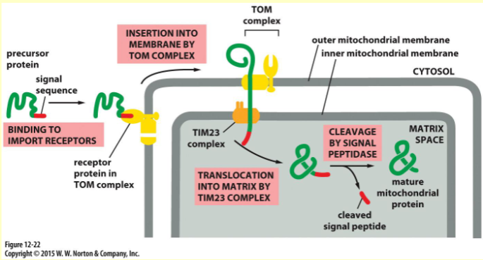
Mitochondrial Protein Import Process
Post - Translational = protein is done before sent to mitochondria
mitochondria has double membrane → to get to central matrix protein has to pass 2 translocase complexes (TOM & TIM)
chaperone proteins (Hsp70) help keep protein unfolded so it can fit TOM/TIM thin channels
moving unfolded protein needs ATP
ATP hydrolysis used by chaperones to pull proteins in
Membrane potential used in TIM complex
Nuclear Import Transport Process
nucleus wrapped in double membrane nuclear envelope but uses NPCs
NPCs acts like sieve:
proteins <40kDa can go through via passive diffusion w/o energy
proteins >40kDa are too big and need Active transport
to move
to move large proteins use importins and exportins
against conc gradient using GTP hydrolysis energy
what are importins?
specialised escort proteins that bind using to the nuclear localisation signal (NLS) of cytosolic protein and move it into protein
What are exportins?
specialised escort proteins that bind to a nuclear export signal (NES) to carry molecules (i.e. RNA) out of the nucleus
Compare the different types of Protein Import
Feature | ER Import | Mitochondrial Import | Nuclear Import |
Timing | Co-translational | Post-translational | Post-translational |
Protein Structure | Threaded while being made | Must be Unfolded | Can be Fully Folded |
Gateways Used | Sec61 Translocon | TOM & TIM complexes | Nuclear Pore Complex (NPC) |
Energy Source | Translation / GTP | ATP + Membrane Potential | GTP (via a molecular switch called Ran) |
examples of proteins using nuclear import?
Structural nucleus proteins (Histones, Nuclear lamins)
DNA/RNA Polymerase
ribosomal proteins
What is GTPase Ran
regulates nuclear import and export
acts as on/off switches
What is GTPase?
specialised enzyme that binds to GTP (Guanosine Triphosphate) = importins/exportins to function
can remove own phosphate group = GDP (Guanosine Diphosphate) = turn off importins/exportins function
What are the specific locations and functions of Ran-GAP?
Ran-GAP
located in the cytoplasm
hydrolyses GTP into GDP
What are the specific locations and functions of Ran-GEF?
located in the nucleus
makes Ran to release GDP do it can bind to fresh GTP
How do transport receptors know where they are?
Ran-GDP → cytoplasm
Ran-GTP → nucleus
Nuclear Export Process?
Exportin, Ran-GTP and the cargo with the NES signal all bind together in the nucleus
complex move through the NPC into cytoplasm
Ran-GTP stimulates Ran = GTP → GDP.
GDP removes self and complex disassembles = cargo released
Nuclear Import Process?
Importin, Ran-GTP and the cargo with the NLS signal all bind together to form complex
the complex moves through the NPC int the nucleus
In the Nucleus the Ran-GEF stimulates Ran to release GDP do it can bind to fresh GTP = cargo released.
How does Ran-GTP affect cargo binding differently for importin and exportin?
Importin → Ran-GTP displaces cargo in the nucleus
Exportin → stabilises cargo and binding in the nucleus
Are proteins functional when first made?
NO
must be modified, folded in the ER and the golgi
Function of ER in the secretory pathway?
Modification, Folding and Quality Control
handles co-translational transport
signal peptide cleaved
N-Linked glycosylation
disulfide bon formations
Function of golgi in the secretory pathway?
Modification and Sorting
Co-translational transport
modifies carbs and add targeting signals
Function of secretory vesicles in the secretory pathway?
final transport and secretion
What is glycosylation?
where carbs (glycans) are added to proteins or lipids = glycoproteins/ glycolipids
Why does glycosylation happen to proteins?
Prevents Aggregation
hydrophobic regions of unfolded proteins stick together
carbs = hydrophilic → make folding intermediate soluble
Helps Chaperon Binding
helps the chaperone proteins to bind and hold the sugar which help the protein fold
Help Protein stability
carbs block the extracellular proteases from reaching protein backbone = increases extracellular stability.
Mucus & Pathogen Protection
highly glycosylated proteins create thick protective layer = trap/ward off pathogens
Oligosaccharides on some cell surface glycoproteins have a role cell-cell adhesion
Types of glycosylation?
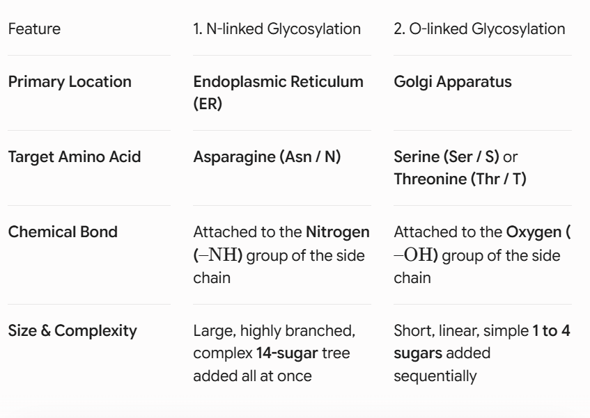
N - Linked
O - Linked
N - Linked glycolsylation process?
before protein translated cell builds large branched carb (14 sugars)
14 sugar precursor is made on the ER membrane called the Dolichol
Polypeptide chain goes through a translocon and membrane-bound enzyme called Oligosaccharyl Transferase scans chain
enzyme is looking for N-X-S/T (concensus motif)
N → Asparagine
X → any amino acid but NOT proline
S/T → Serine or Theorine
N-X-S/T passes into ER lumen and Oligosaccharyl Transferase cleaves off the 14 sugar and add it to the target protein on the -NH group
glycosidases start trimming sugars to monitor its folding correctly
moves via transport vesicles to the golgi apparatus
Why doesn’t Proline work in N-Linked Glycosylation?
Proline has a rigid structure and physically distorts the chain → blocks the enzyme
Oligosaccharyl Transferase is blocked form binding
protein = not glycosylated
Compare N and O - Linked glycosylation?
What does the drug Tunicamycin do?
drug inhibitor
blocks the synthesis of the dolichol-linked oligosaccharide precursor
proteins not glycosylated
BiP function?
chaperone protein
binds exposed hydrophobic regions
prevents misfolding
prevent exit of incorrect proteins
binding immunoglobin protein
Calnexin + Calreticulin
chaperone protein
binds glycosylated proteins
ensure proper folding
binds to oligosaccharides on incomplete folded proteins
What is a disulfide bond?
covalent bond between thiol -SH groups of two Cysteine amino acids
Oxidation reaction
stabilise tertiary and quaternary protein structures
where are disulfide bonds made?
oxidising environments like ER where enzymes like Protein disulfids isomerases (PDI) help correct bond formation.
Why are disulfide bonds important?
structurally stabilises the proteins final 3D shape = no unfold when outside the cell where drastic environmental changes
PDI function?
ER enzyme catalyses the chemical reaction and creates the disulfide bond
breaks incorrect covalent bonds formed
helps rearrange mismatched cysteine pairings
if folding protein mismatched/incorrect PDI cleaves incorrect bond = allow protein chain to rotate until the ideal native configuration is achieved.
Why do cytosolic proteins rarely contain disulfide bonds?
Cytosol highly reducing environment = prevents oxidation needed for disulfide bonds to form
oxidising environment is in the lumen of the ER
What is Protein Disulfide Isomerase?
PDI
enzyme that catalyses the oxidation reaction to make disulfide bonds
proline isomerisation?
Proline can exist in cis/trans forms
proline struggles to rotate and gets stuck switching between cis/trans
conversion is slow
a rate-limiting barrier to efficient protein folding
Peptidyl-prolyl isomerases?
PPIases
enzymes that speed up proline cis/trans conversion
this is needed for correct protein folding
acts as a chaperone protein
What if the protein is folded incorrectly?
Normal Process:
protein folded
checked by chaperone
exit the ER
Misfolded Protein:
Retained in ER
Refolded
OR destroyed
Name how the ER quality Control checks proteins?
Glycosylation-Based Quality control
Unfolded Protein Response (UPR)
ER - associated Degradation (ERAD)
Glycosylation-Based Quality control
glucosidase chop off two glucose molecules = 1 left
1 glucose is a tag for chaperone proteins Calnexin and Calreticulin → prevent clumping so it can fold
final glucose removed = chaperones removed
Sensor enzyme Glucosyltransferase evaluates released proteins
Folded correctly = exits ER
Folded Incorrectly
detects exposed hydrophobic patches
adds a UDP- glucose to the sugar backbone
protein goes through the Calnexin/Calreticulin stage for refold attempt
Unfolded Protein Response
emergency stress signal for when misfolded proteins build up in ER faster than cell can repair.
chaperone enzyme BiP binds to 3 transmembrane sensors (IRE1, PERK, ATF6) = turned off
misfolded proteins arrive → BiP leaves sensors and attaches to misfolded proteins = sensors turn on
Once activated IRE1 unmasks endoribonuclease domain → domain targets Xbp1 pre-mRNA in cytosol = cutting off intron
two remaining exons join together = frameshift mutation
unspliced Xbp1-u makes unstable protein
spliced Xbp1-S makes stable protein
Xbp1-s moves to nucleus → turn on genes that make more ER chaperones and folding catalysts
ER - Associated Degradation (ERAD)
If protein is still misfolded then destroyed
misfolded protein moved out of ER lumen back into cytosol via translocator complex
In cytosol, enzyme N-glycanase strips off sugar tags
E3 Ubiquitin Ligase tags the polypeptide with a polyubiquitin chain
Polyubiquitin chain is a marker for Proteasome which digests the protein = amino acids to reuse
Successfully folded protein go to…
transport via vesicles to golgi apparatus
Different compartments in the Golgi?
cis-Golgi (Entry)
medial - Golgi
Trans - Golgi (Exit)
each region has specific enzyme
Modification steps in the Golgi stacks after protein folding?
Golgi stacks remodel the sugar trees added to the protein in the ER
protein leaves ER as high-mannose oligosaccharide = not functional
Get modified:
Golgi Mannosidase removes extra mannose residue = makes space for new sugar to be added
N-acetyleglucosamine (GlcNAc) transferase I added a new GlcNAc sugar = make it complex glycoprotein
Golgi Mannosidase II remove more mannose residual
Golgi adds more sugars
GlcNAc
Galactose
Sialic Acid
results on complexed branched oligosaccharide
Lysosome function
digest proteins, DNA, lipids
can lead to cell damage/ death
How does the cell tell lysosomal enzymes and secreted enzymes apart?
GlcNAc - Phosphotransferase recognises signal patch and adds sugar = M6P
formed after protein folding
Where is the M6P tag found?
Cis Golgi Network
How does the M6P signal patch work?
M6P receptor binds to tagged enzyme
Clathrin coat forms - vesicle formed
Protein is packaged into vesicle
Vesicle goes to the endosome then lysosome
when in lysosome low pH = enzyme detach from receptor
receptor goes back to golgi
What happens if M6P is defective?
GlcNAc- phosphototransferase is defective
no Mp6 tag is added
lysosomal enzymes no recognised
get secreted outside the cell
lysosomes lack enzymes
waste builds up = cell damage
Inclusion Cell Disease
Inclusion Cell Disease
mutation in the GlcNac-phosphototransferase = cannot add M6P
Normal:
Lysosome enzyme made in ER → folded → signal patch forms → in Cis Golgi M6P tag added → in Trans Golgi M6P receptor binds → sent to lysosome
Disease:
no M6P
Enzyme is not recognised
enters default pathway → secreted out
Diagnostic Profile for Inclusion Cell Disease?
patients blood has high level of lysosomal enzymes
cellular lysosomes empty and non-functional
cells have build up debris
Diseases of disposal (Loss-of-Function)
ER quality control is too strict
Mechanism:
Protein fold slightly incorrect
chaperones detect defect
sent to ERAD
destroyed in cytosol
Example of Diseases of Disposal
Cystic Fibrosis
mutation in $\Delta\text{F508}$ = slight misfolding
ER detects → destroys via ERAD
no CTFR = no chloride channels
extra mucus made = lung disease
Toxic Gain of Function (Diseases of retention)
Misfolded proteins not removed efficiently = accumulate in cell
Mechanism:
Proteins misfold
Cannot exit ER
aggregates accumulates
ER become stressed = cell damage
Toxic
Toxic Gain of Function (Diseases of retention) Example
alpha 1 Antitrypsin Deficiency (Liver Failure)
misfolded protein accumulates in the ER
Forms aggregates that accumulates
kills Liver hepatocytes = widespread tissue destructions
Discuss how defects in protein processing and targeting in the secretory pathway can lead to disease?
Secretory Pathway important for protein structure and localisation
involves protein folding and modification in ER
further processing in Golgi
Explain what happens in ER
folding assisted with chaperone BiP, Calnexin, Calreticulin
additional modification = N-Linked Glycosylation and Disulfide bonds
Correctly folded leave ER
misfolded go ERAD pathway
ERAD may lead to diseases.
Liver Disease
Cystic Fibrosis
I- Cell diseases
Function of the Vesicle Coats?
bends the membrane = spherical bubble
Selectively capture and packages cargo proteins
Types of Vesicle Coats?
COPII
COPI
Clathrin
COPII
Anterogade
moves fresh cargo forward → ER to Golgi
Sar1 GTPase needed
COPI
Retrograde
Receives protein backwards → Golgi to ER
Arf1 GTPase needed
Clathrin
moves cargo between TGN, endosomes and plasma membrane
Describe the mechanism of COPII vesicle formation?
In ER membrane, Sec12 acts as Sar1-GEF protein and activates Sar1
Sar1-GDP to Sar1-GTP
Binding to GTP = exposes hydrophobic helix at th N-Terminus
Helix inserts into ER membrane
Sar1-GTP recruits
Sec23 → binds Sar1-GTP and slowly converts to Sar1-GDP via hydrolysis
Sec24 → binds sorting signals on cargo receptors
inner section pinned down → recruits outer structural framework
Sec13/Sec31 → quickens Sec23 GAP activity = Sar1GTP to Sar1GDP
Sar1-GDP loses structure, loses amphipathic helix and pulls off membrane
inner coat detaches
outer coat collapses
vesicle becomes uncoated
ready to fuse vesicle
What are RAB proteins?
small monomeric GTPases
Attached to membrane via lipid
Two types of RAB
What are the types of RAB proteins?
Rab-GTP → active
Rab-GDP → inactive
Examples of Rab
Rab1 - cisGolgi targeting
Rab5 - Early endosome
Rab7 - Late endosome
What do RAB proteins do?
acts as identifying markers for vesicles
ensure vesicles dock at correct membrane
What would happen without RAB proteins?
Vesicles would fuse randomly
cell organisation would collapse
How does RAB work as identifying markers?
Membrane Docking Sequence
Vesicle carries RAB-GTP
The target membrane has a RAB effector
large protein complex
Effector reaches across cytosol to the specific RAB-GTP on the incoming vesicle → pulls it towards membrane = tethering
RAB effector also binds to specific PIP
Vesicle can dock when correct RAB and PIP
SNARE proteins interact and fuse Vesicle with membrane.
What are PIPs?
lipids in membrane
helps RABs with vesicle localisation
How do PIPs helps RAB?
Mark the type of membrane that the Vesicle will dock with lipid identity code
The target membrane for the vesicle will have the correct RAB and PIP
Why can’t vesicles fuse without a SNARE protein?
Lipid Bilayer are surrounded with water and strongly resist fusion
What are SNARE proteins?
specialised molecular machine
Helps with vesicle fusion to mebrane
2 types
What are the types of SNARE Proteins
v - SNARE (vesicle SNAREs)
on vesicle membrane
e.g. Synaptobrevin
t- SNARE (target SNAREs)
on the target membrane
e.g. Syntaxin
What is the problem SNAREs must solve?
lipid bilayers naturally repel each other
fusion between vesicles and membranes need energy
How do the SNARE proteins work?
Zipping Mechanism
RAB proteins bring the vesicle close to the membrane → Tethering
v-SNARE on the vesicle meets matching t-SNARE on target membrane
v-SNARE helix interlocks with t-SNARE and wrap around each other
this interlocking = stable, 4 alpha helix bundle called trans-SNARE complex or SNAREpin
SNAREs continue interlocking = more energy released and membranes pulled closer and water molecules pushed out.
vesicle membrane and target membrane and outer lipid touch = Hemifusion
The membrane continue to open = channel appears between = fusion pore
fusion pore = cargo released from vesicle into target cell
After Fusion what happens to the the SNARE?
V and T SNAREs in the same membrane = cis-SNARE complex
Cell has to recycle SNAREs and cant dismantle coz its stable
protein called alpha SNAP wraps around cis-SNARE bundle
recruits big hexameric AAA+ ATPase enzyme called NSF
NSF binds to alpha SNAP
NSF uses energy from ATP hydrolysis to unravel the helixes
Individual SNARE proteins released
Why do SNARE proteins need ATP?
SNARE interlocking provides the energy needed for the membrane fusions
When recycling the SNARE, NSF uses this ATP to separate SNAREs after fusion
NSF full name?
N-ethylmaleimide Sensitive Factor
Vesicle Sequence Steps?
Vesicle uncoating
RAB targeting with PIPs
Tethering by RAB
v-SNARE bind to to t SNARE
Trans-SNARE complex forms
Interlocking helices = membranes together
Hemifusion
Fusion Pore opens
Cargo delivered
Cis SNARE complex remains in cell
alpha SNAP binds
NSF hydrolyses ATP
ATP break cis SNAP complex
SNARE recycled
How do vesicles release neurotransmitters?
RAB targeting
SNARE interlocking
Membrane fusion
cargo release in fusion pore
cargo is neurotransmitter
What is Botulinum Toxin?
bacterial protease that is a neurotoxin
AKA Botox
What does Botulinum Toxin target?
SNAP-25
Syntaxin
What does Botulinum Toxin do?
cleave SNARE complex
no trans-SNARE complex forms
no hemifusion of membranes
no fusion pore
No ACh released
muscles don’t receive stimulation
muscles become weak and relaxed
What does Tetanus Toxin do?
cleaves SNARE proteins
No fusion of membranes
no fusion pore = no cargo released
no inhibitory neurons released = motor neurons fire = muscle is rigid and contracted.
What are the mutations that affect cargo accumulation?
Sec61
SAR1
RAB1
v-SNARE
cargo accumulation: Sec61 mutation
Sec61 is in the ER channel
prevent translocation into ER
Secretory proteins stay in the cytosol
Cargo accumulation: Sar1 or Sec12
Sec12 = GEF for Sar1
Sar1= starts COPII coat assembly
mutation prevents COP11 coat assembling and vesicle budding
Secretory proteins accumulate in ER lumen