Calcaneus Anatomy Terms and Definitions for Medicine Study
 Call Kai
 Learn
Learn Practice Test
 Spaced Repetition
 Match
Match Flashcards
 Knowt Play
1/73

There's no tags or description
Looks like no tags are added yet.
Name | Mastery | Learn | Test | Matching | Spaced | Call with Kai | Chat |
|---|
No analytics yet
Send a link to your students to track their progress
74 Terms
Come. To. Colorado. Next. Christmas
Mnemonic for the Tarsals?
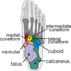
Tarsal bones
the seven bones of the ankle

Calcaneus (os calcis)
Largest and strongest bone of the foot
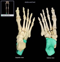
Superior or proximal surface of Calcaneus
Which view of the calcaneus is this?
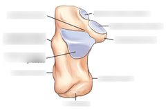
Posterior articular facet
Largest facet on calcaneus, articulates with talus

Anterior and middle articular facet
smaller facets that articulate with the talus to form the talocalcaneonavicular joint

Sustentaculum tali
Means "support for the talus", Located on the medial proximal aspect
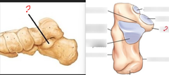
Calcaneus sulcus
deep depression between the posterior and middle articular facets
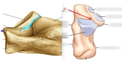
Tuberosity of calcaneus
Common site for bone spurs, Achilles tendon attaches
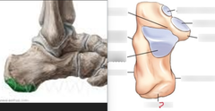
Peroneal trochlea (trochlear process)
Visualized laterally on an axial projection
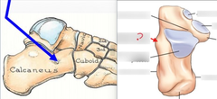
Sinus tarsi (tarsal sinus)
Visualized laterally, Ligaments pass through, opening in the subtalar joint, formed by a depression of the talus and calcaneal sulcus
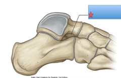
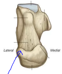
Lateral process of calcaneus
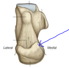
Medial process of calcaneus
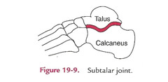
Talocalcaneal joint (subtalar joint)
articulation of the inferior surface of the talus and the superior surface of the calcaneus
-Cuboid-Anteriorly
-Talus-Superiorly
(Forms the subtalar (talocalcaneal) joint)
Which bones articulate with the calcaneus?
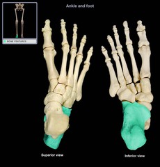
Calcaneus (os calcis)
5?

Sustentaculum tali
1?

Talocalcaneal joint (subtalar joint)
8?
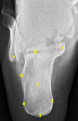
Tuberosity 5th metatarsal
7?
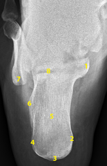
Peroneal trochlea (trochlear process)
6?
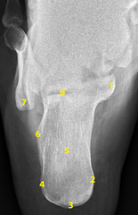
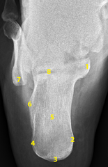
Lateral process of calcaneus
4?
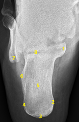
Medial process of calcaneus
2?
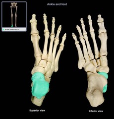
Talus (astragalus bone)
2nd largest tarsal bone, Located between lower leg and calcaneus, Weight of the body is transmitted by this bone through the ankle and talocalcaneal joints
Tibia-Superiorly
Fibula-Superiorly
Calcaneus-Inferior
Navicular-Anterior
Which bones articulate with the Talus?

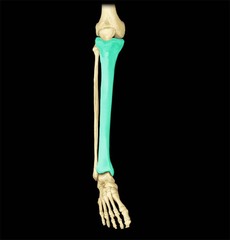
Tibia
the medial and larger bone of the lower leg
lateral condyle of tibia
articulates with lateral condyle of femur

proximal tibiofibular joint
joint between fibula (head) and tibia (lateral condyle)

intercondyle eminence of tibia (tibial spine) (medial and lateral intercondylar tubercle)
Between the articular facets on the superior articular facet of the tibia

Tibial plateau (articular facets of tibia)
opposed ends of the bones, covered with hyaline cartilage

medial condyle of the tibia
articulates with medial condyle of femur

Tibial Tuberosity
point where the patellar ligament attaches

Anterior crest
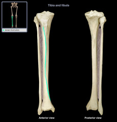
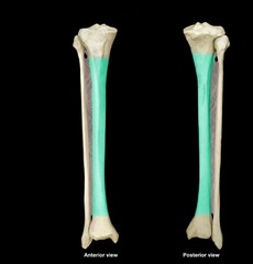
Body or shaft of tibia
fibular notch
lateral notch on the distal end tibia where tibia and fibula articulate
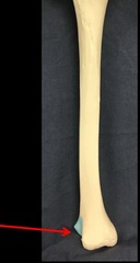
Distal tibiofibular joint
articulation between the distal fibula and the fibular notch of the tibia

Medial malleolus
Easily palpated on medial side of ankle
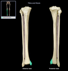
Anterior Tubercle
Expanded process at the distal anterior & lateral tibia, Articulates with the superolateral talus, Partially overlaps the fibula anteriorly, (distal portion of tibia)
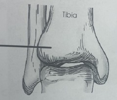
Tibial Plafond
Distal tibial joint surface, Forms the roof of the ankle mortise joint
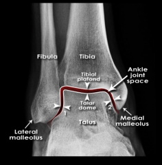
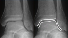
Mortise space
"socket" or three-sided opening The superior Talus fits in here • Not seen on a true AP in full
fibula

apex of fibula (styloid process)

head of fibula
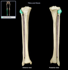
neck of fibula

body or shaft of fibula

lateral malleolus
bony prominence on the lateral side of the fibula (forms ankle)
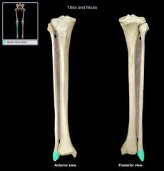
distal end of fibula

Tibia: Proximally & Distally
Talus: Distally
Which bone articulates with the fibula?

1. tibia
2. fibula
3. talus
What 3 bones form the ankle joint?

Tibia
1?
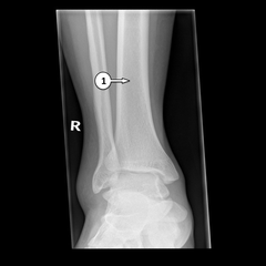
Fibula
2?
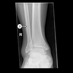
Anterior Tubercle
3?
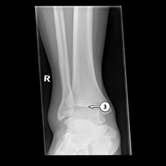
Medial malleolus
4?

lateral malleolus
5?

Mortise space
6? (joint space)
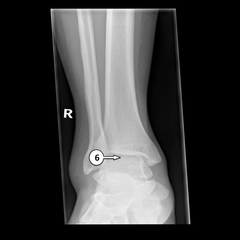
Sustentaculum tali
11?

Calcaneus (os calcis)
14?
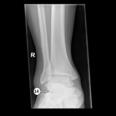
Inferior surface of the tibia (tibial plafond / tibial facets)
When performing an axial projection of the ankle, what should you see?
Fiberglass: 3-4
Small to medium: 5-7
Large plaster cast: 8-10
How much do you change the kVp for casts?
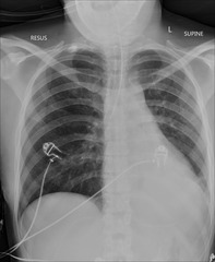
radiopaque objects, clothing removed placed in gown , long hair, oxygen tubing, and pacemaker leads to avoid reduced detail/scatter
During patient preparation, what must be removed?
1. Limit the number of exposures
2. Collimation
3. shielding (led apron)
4. Position correctly
What 4 things help protect you and the patient against radiation?
BEFORE the examination
When must the anatomical marker be placed?
kVp: 65-75 (increase for other foot projections)
mAs: depends
Grid: No
SID: 40in
What technical factors are needed for a good ankle/calcaneus (kVp, grid, mA, SID)?
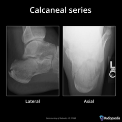
Plantodorsal (axial)
Dorsoplantar (axial)
Lateral
Routine projections of the calcaneus?
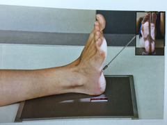
What is the evaluation criteria and procedure for an Plantodorsal axial Calcaneus X-ray?
kVp: 65-75
Grid: No
CR: base of 3rd metatarsal, 40° caudad from long axis of foot
Patient position: supine or seated on table with leg fully extended, Dorsiflex foot, align ankle joint to CR and IR, plantar surface near perpendicular
Evaluation criteria: entire calcaneus (tuberosity posteriorly to subtalar joint anteriorly)
What is the evaluation criteria and procedure for an Dorsoplantar axial Calcaneus X-ray?
kVp: 65-75
Grid: No
CR:
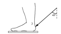
STANDING: direct CR to level of base of 5th metatarsal, 45° anteriorly and directed through posterior surface of flexed ankle
PRONE: CR directed to midpoint of the IR (will emerge at the level of the base of the 5th MT, Caudal angle 40˚ to long axis of foot
Patient position:
STANDING: standing upright with weight placed on affected foot, place opposite foot one step forward and lean forward as far as possible
PRONE: lying down foot elevated with plantar surface on IR
Evaluation criteria: entire calcaneus and tuberosity posteriorly
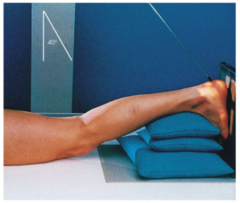
What is the evaluation criteria and procedure for an Lateral (mediolateral) Calcaneus X-ray?
kVp: 65-75
Grid: No
CR: 1 in inferior to medial malleolus
Patient position: lateral recumbent position, recumbent, Flex knee on affected limb 45˚
Evaluation criteria: calcaneus demonstrated in profile with talus and distal tib-fib.

AP
AP Mortise
Lateral (mediolateral)
Ankle Routine projections?
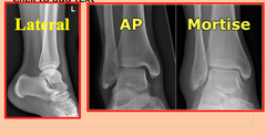
What is the evaluation criteria and procedure for an AP Ankle X-ray?
kVp: 60-75
Grid: No
CR: directed midway between malleoli
Patient position: Patient supine, leg fully extended, Do not force dorsiflexion, Foot & ankle in true AP
Evaluation criteria: Distal ⅓ of tibia and fibula demonstrated, Proximal ½ of metatarsals included, Medial and superior aspect of ankle joint open

What is the evaluation criteria and procedure for an AP Mortise X-ray?
kVp: 60-75
Grid: No
CR: directed midway between malleoli
Patient position: Patient supine, leg fully extended, Do not dorsiflex foot allow it to remain in natural position, Internally rotate leg & foot 15-20°
Evaluation criteria: Distal ⅓ of tibia and fibula demonstrated, Proximal ½ of metatarsals included, Medial and superior aspect of ankle joint open
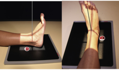
What is the evaluation criteria and procedure for a Lateral (mediolateral)
Ankle X-ray?
kVp: 60-75
Grid: No
CR: directed at Medial Malleolus
Patient position: Patient in a lateral recumbent position, Flex affected limb 45˚, Dorsiflex foot so that plantar surface is at a right angle to leg or as far as patient can tolerate (DO NOT FORCE)
Evaluation criteria: Entire talus and calcaneus visualized, Lateral malleolus superimposed over posterior half of tibia
Oblique 45°
AP Stress (Inversion & Eversion)
AP & Lateral Weight Bearing
Ankle special projections?
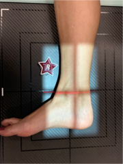
What is the evaluation criteria and procedure for an AP Oblique ankle
X-ray?
kVp: 60-75
Grid: No
CR: directed midway between malleoli
Patient position: Supine, legs fully extended, dorsiflex foot to 80-85° and rotate leg and foot medially 45°
Evaluation criteria: Distal tibiofibular joint open, Distal ⅓ of tibia and fibula demonstrated, Proximal ½ of metatarsals included
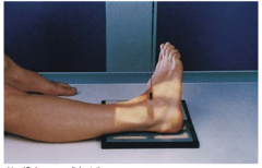
What is the evaluation criteria and procedure for an AP stress ankle
X-ray?
kVp: 60-75
Grid: No
CR: directed midway between malleoli
Patient position: supine, legs fully extended, dorsiflex foot near right ankle lower leg as far as possible, then plantarflex and laterally bend as far as possible
Evaluation criteria: Distal aspect of tibia and fibula demonstrated and Ankle joint to center of collimation field
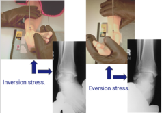
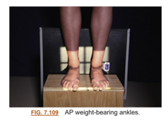
What is the evaluation criteria and procedure for an AP bilateral and lateral Weight Bearing X-ray?
kVp: 60-75
Grid: No
CR: directed midway between malleoli for AP and Medial malleolus for lateral
Patient position: both ankles in view, for the lateral do one ankle at a time
Evaluation criteria: Distal aspect of tibia and fibula demonstrated and Ankle joint to center of collimation field