Binocular sensory adaptation
1/21
There's no tags or description
Looks like no tags are added yet.
Name | Mastery | Learn | Test | Matching | Spaced | Call with Kai |
|---|
No analytics yet
Send a link to your students to track their progress
22 Terms
Changes in strabismic eye to avoid diplopia and confusion
Binocular sensory adaptation: Harmonious anomalous retinal correspondence, Unharmonious anomalous retinal correspondence, Suppression
Monocular sensory adaptation: Eccentric fixation, amblyopia
Differentiate diplopia and confusion
Diplopia: eye sees double images of 1 object, object imaged on non-corresponding retinal points
Confusion: 2 images superimposed, object seen by strabismic eye’s fovea and fellow eye’s fovea are different, both foveas are corresponding points, causing images of OU to be superimposed
Early onset strabismus adaptations to avoid diplopia and confusion
Binocular vision not fully developed, sensory adaptation to avoid symptoms: strabismus, ARC, amblyopia, EF
Recent onset strabismus in adults
Binocular vision system past its plasticity period, cannot adopt sensory adaptation to avoid diplopia and confusion
they will have normal retinal correspondence: both fovea share the same visual direction
will adopt abnormal head posture and cover one eye
Suppression
active inhibition resulting in loss of awareness of visual impression for 1 eye in binocular vision
young enough patient with sensory plasticity will develop HARC
Physiological suppression
present in normal BSV to prevent awareness of physiological diplopia and retinal rivalry
Pathological suppression
developed to overcome binocular diplopia, confusion, incompatible images (retinal images of different size and shapes, preventing central fusion)
Anomalous retinal correspondence purpose
For a point of the retina of a good eye to correspond with a new point of the retina of a strabismic eye
Alternative to suppression, only present under binocular viewing conditions
Key requirements of HARC
Unilateral strabismus
Small angle strabismus
Constant strabismus
Early onset strabismus (plastic visual system)
Objective angle and subjective angle of ARC
Objective angle: manifest deviation of strabismus, measured objectively (cover test)
Subjective angle: angle of strabismus perceived by patient (Maddox rod, von graefe)
Angle of anomaly
Angle which retinal correspondence has shifted from normal
Objective angle-subjective angle
Angle of anomaly: 0 (NRC)
Not equal to 0 (UARC)
Equals of objective angle with allowance of 4 (HARC)
Unharmonious anomalous retinal correspondence
rare
objective angle: angle of new strabismus
subjective angle: difference between angle of old and new strabismus
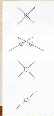
Presence of suppression tests
Bagolini striated lens
Worth 4-dot test
Mallett modified OXO test
Worth 4-dot tests
If px has strabismus but sees 4 dots=ARC
4 dots without strabismus=Normal
5 dots=crossed/uncrossed diplopia
Interpret this:
Bifoveal or anomalous BSV
Heterotropia with NRC and diplopia
Central or paracentral suppression scotoma in one eye
Central and peripheral suppression of one line image
Draw out all possible results of near Mallett modified OXO test
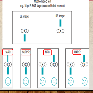
Mallett unit suppression test
Wear polaroid glasses
test calibrated at 35cm
RE see red letters
LE see yellow letters
OU see green letters
Mallett unit suppression test procedure
Get patient to wear polaroid glasses
Read down the chart— if patient does not read all letters at the bottom row
Occlude better eye and ask them to read the smallest letter they see
Compare the acuity in binocular and monocular conditions
Difference in readings= foveal suppression in minutes of arc
Depth of suppression tests
ND filter
Sbisa bar: if suppression still present at number 7-9, deep suppression indicated
Bagolini lens with ND filter
ND filter placed on unsuppressed eye
ND filter increased by 0.3 log units until suppressed eye sees an image
Suppression depth—>log unit before reversal
Step 1: If left eye suppressed and strabismic, image only seen by right eye
Step 2: Increase density of ND filter on right eye, until streak seen by left eye appears
Step 3: Further increase ND filter will cause retinal illuminance of suppressed eye to be better, resulting in suppression of right eye. Left eye now assumes primary position and RE becomes strabismic
Sbisa bar procedure
Place bar on unsuppressed eye, only unsuppressed eye can see a red spot of light
Slowly increase density of the sbisa bar until suppressed eye can see a white spot of light (diplopia occurs)
Continue to increase the density of the filter until image of the unsuppressed eye cannot be seen
Good eye is now suppressed and shows strabismus, Suppressed eye shifts to primary position
Log unit recorded before reversal
Management of suppression
Aim: encourage patient to become aware of suppressed image and integrate it correctly with image from other eye
Method of treatment should: Ensure simultaneous stimulation of foveal areas/corresponding points of both eyes, angle of squint must be relieved
Treatment for anti-suppression: relieve deviation so that eye can fuse both images (red-green TV trainer)