Muscarinic Acetylcholine receptor
 Call Kai
 Learn
Learn Practice Test
 Spaced Repetition
 Match
Match Flashcards
 Knowt Play
1/11

There's no tags or description
Looks like no tags are added yet.
Name | Mastery | Learn | Test | Matching | Spaced | Call with Kai | Chat |
|---|
No analytics yet
Send a link to your students to track their progress
12 Terms
Muscarinic Ach Receptor
GPCR (7 transmembrane pass receptor)
Class A - member of the rhodopsin like rceeptors
4+3 arrangement of the alpha helices
Cavity in the centre of the helical bundle where the molecule will bind (apart from rhodopsin where the ligand doesn’t have to diffuse in and out)
Activated by muscarine and inhibited by atropine
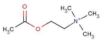
Similar binding site geometry – quaternary ammonium group, acetyl group
Functions by binding to the G protein – in the case of muscarinic receptors are linked to Gi or Gq
Cloning the mAchR
Range of different subtypes – M1 and M2 which are present in different locations
M1 in the cerebral cortex primarily and is the most abundant in the CNS
M2 in the cardiac tissue primarily and is most abundant in the PNS
Also identified 3 different subtypes M3, M4 and M5 which also have quite specific distribution 🡪 important to distinguish between these at the molecular level to target them
Understanding the structure
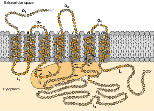
N terminus outside the cell
7 TM domains linked by short linkages outside the cell which allow easy access to the binding site from ligands and are frequently glycosylated or involved in disulphide bonds
3 loop regions inside the cell – 3rd one is quite large to allow interaction surface to the binding site and the G protein. Also can be phosphorylated for inactivation
Intracellular C terminal
G protein coupling
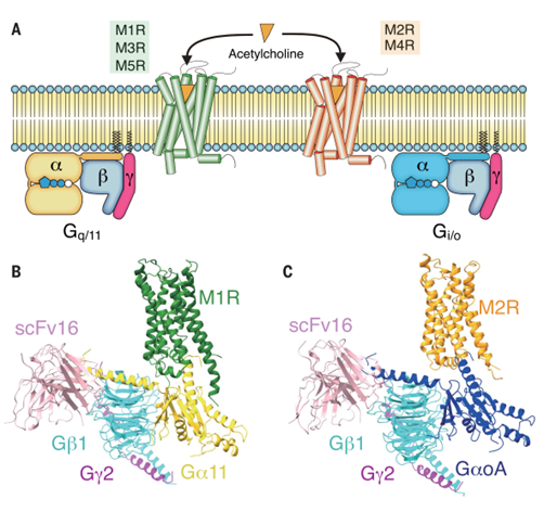
Heterotrimeric G protein which upon activation there’s an exchange of GTP for GDP and dissociation of alpha and gamma subunit which influence the secondary signalling pathway
M1, M3 and M5 go through the Gq/11 pathway which causes:
Increased PLC beta
Increase in Ca2+
Increase in MAPK
Decreased M current
M2 and M4 go through the Gi/o pathway which causes:
Decrease in adenylyl cyclase
Increases MAPK
Increases G protein activated inward rectifier potassium channels (GIRK)
Decreases voltage gated potassium channels
cell hyperpolarisation
Structure activity relationship studies
Take a receptor and bind it to different drugs and understand how the pharmacophores are involved in binding
Carbachol a useful molecule because its not broken down readily – can then change the length of chains to see how this effects binding
2 order magnitude drop in affinity when you remove the carbonyl group suggests that it may be involved in binding
Decrease in affinity when hydrophobic chain is replaced with a methyl group suggesting that the hydrophobic surface drives binding
However, this carbonyl group alone is not responsible for sufficient binding (even with a sulphur molecule to rescue the geometry) showing that this hydrogen bonding is important for binding
If we knock out potential hydrogen bonding partners, we reduce the affinity of Acetylcholine for its receptor
Ach binding site
Aspartate at position 147 on helix 3 which forms an ionic interaction with quaternary ammonium group (not the cation pi interactions like the nAchR) and loss of this prevents binding
Lots of hydroxyl groups which are well positioned to form H bonds to the carbonyl group
Hydrophobic surface also interacts with the receptor and a key length of ligand which allows the hydrophobic group to bind favourably to the receptor
Hydrophobic interaction supports the binding – binding the drug and replacing the water molecules is entropically favourable
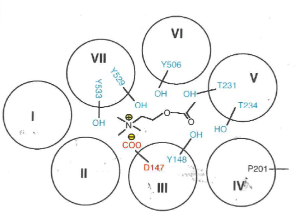
Subtype selectivity from X ray structures
We know that these are spatially localised and activate different pathways 🡪 want to understand how to selectively target these
Crystal structures of rat M2 and M3 subtypes in the presence of their agonist QNB or tiotropium
Similarities between M2 and M3 subtypes
Long C terminal tail which is an 8th amphipathic helix - this is important for interacting with the G protein
7 transmembrane domains (3+4 bundle)
Large intracellular loops are less well defined because they’re very mobile and hard to tie down in crystal structure which is a challenge to understand G protein coupling
Differences in drug binding site
M3 binding site is larger – if we know how these binding sites vary, we can use this to design drugs to target this
Spatial arrangements of compounds within the binding site and location of pharmacophores are similar
Also a similar distribution of the residues forming the binding pocket
Aspartic acid close to the N atom which supports the structure-activity relation studies
Bind very similar classes of molecules and have very similar binding sites
Molecular dynamics
Receptors have a ligand binding site, but also a range of other binding sites/ conformational changes that it must undergo
Once you have a crystal structure, you can do molecular dynamics (essentially solving F=ma at the molecular level)
We know what the atomic weight of atoms are and we know what the forces which are acting on them (when you pull atoms are pulled apart, forces will pull them back together)
E = Ebond + E charge + EHbond + E torsion to fork out F which can feed into F=ma
Equations allow us to work out the forces that are acting on these systems and we can evolve this with respect to time (working out acceleration)
Instead of focusing on the agonist binding site we can look for other binding sites within the protein which may help to differentiate them
Found a vestibule region which could bind a whole host of other molecules ad showed differences between M2 and M3, suggesting diversity in chemical functionality
Cryo-EM studies of the mAchR – How do the ligand recognise different G proteins?
G proteins bind through the intracellular loops
One of the common internal loop motifs are the dry motifs which is an ionic lock made up of an Aspartate, an Arginine and a tyrosine which locks the receptor into the inactive state
Between M2 and M3 the interaction with this ionic lock is the same
This lock is the first thing that you have to break before you can get the G protein to bind to its receptor
Activation mechanism is also the same: the binding site located in the helical bundle which causes a movement of the TM domains which is an outward movement of TM domains 5 and 6
G protein selectivity
Interface region between the receptor and G protein – interaction between the C terminal helix of alpha subunit of G protein and a cavity which has opened up as a result of the outward movement of TMD5 and TMD6
Conserved residues in the two G proteins
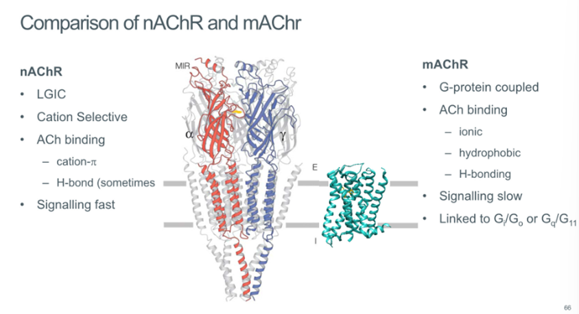
Comparison of nAchR and mAchR
nAchR | mAchR |
LGIC | GPCR |
Ach Binding:
| Ach binding:
|
Fast signaling – transmembrane domains “locked” and springs open as soon as these loop residues removed | Slow signalling – triggered by movement of helix, binding to G protein, large domain conformational changes |
Fluxes cations | Linked to Gi/Go or GqG11 |