6- Nerve impulses, Synaptic transmission, Skeletal muscles
1/32
There's no tags or description
Looks like no tags are added yet.
Name | Mastery | Learn | Test | Matching | Spaced |
|---|
No study sessions yet.
33 Terms
Describe the structure of a myelinated motor neurone
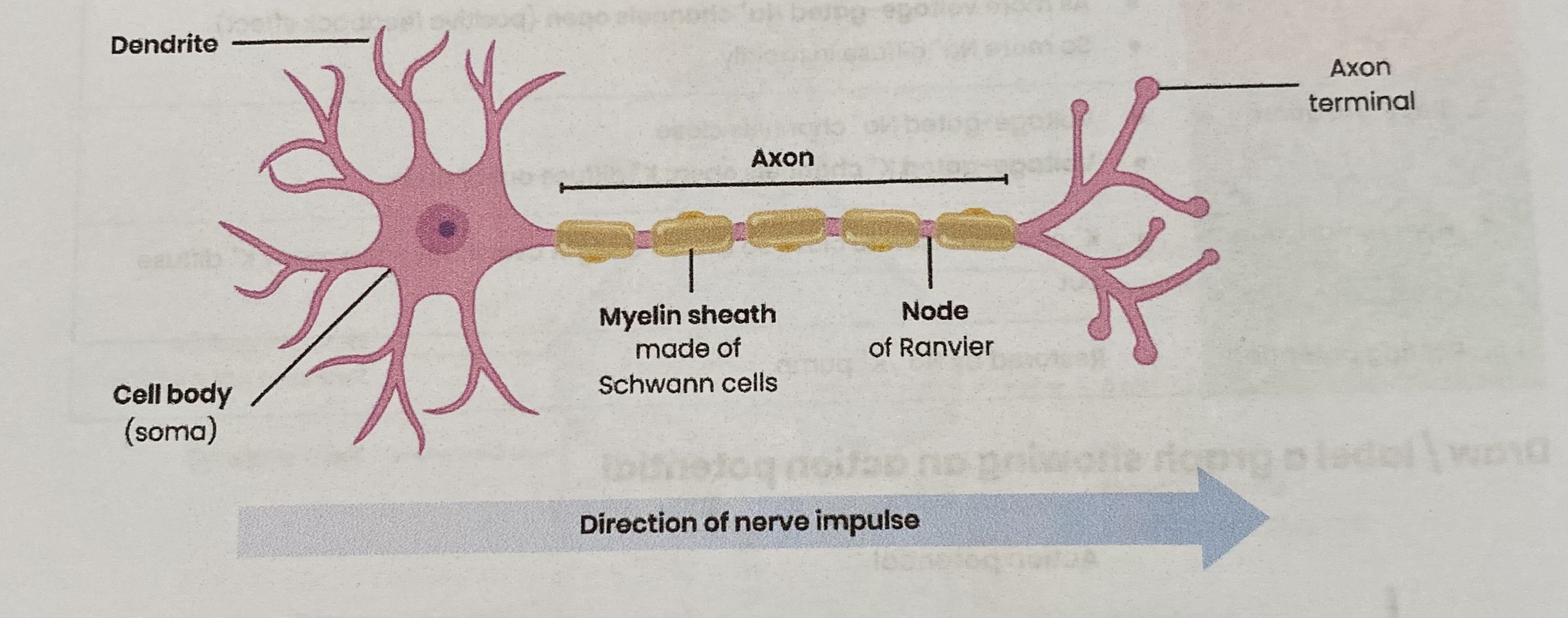
Describe resting potential
Inside of axon has a negative charge relative to outside (as more positive ions outside compared to inside)
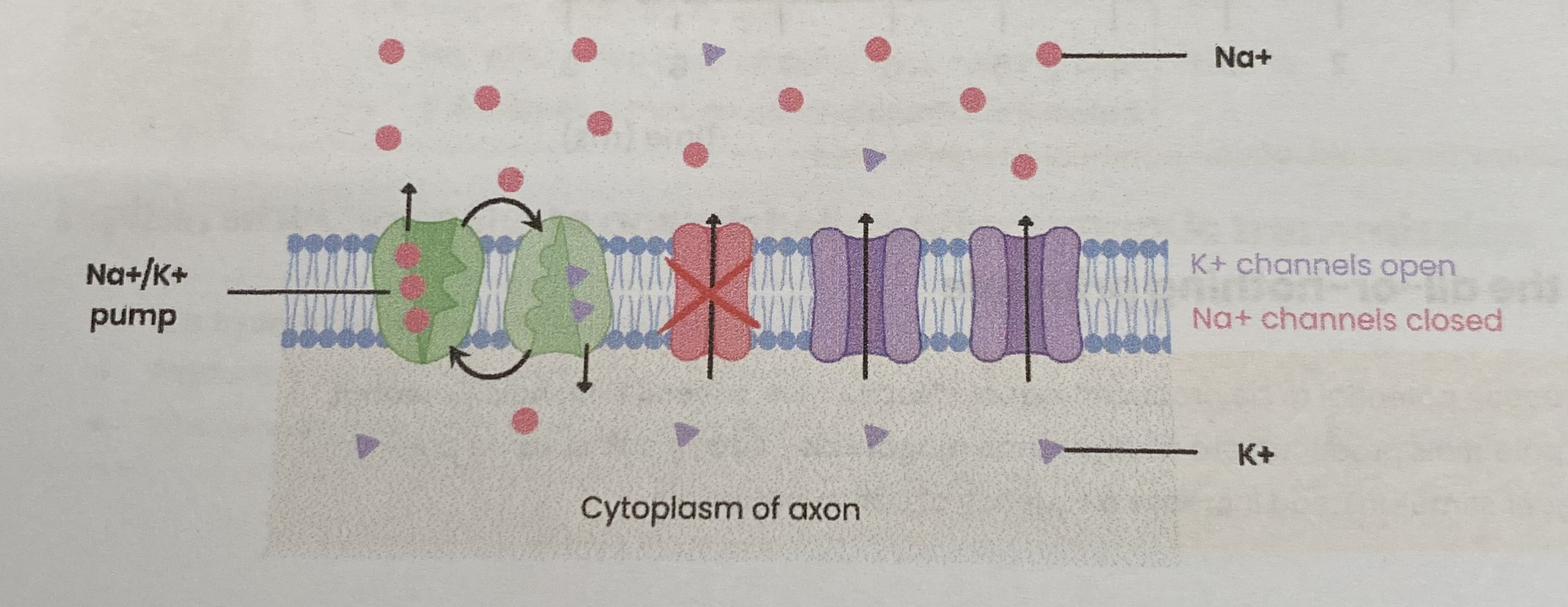
Explain how a resting potential is established across the axon membrane in a neurone
Na+/ K+ pump actively transports:
(3) Na+ out of axon AND (2) K+ into axon
Creating an electrochemical gradient:
higher K+ conc inside AND higher Na+ conc outside
Differential membrane permeability:
more permeable to K+= move out by facilitated diffusion
less permeable to Na+ (closed channels)
What are the stages of depolarisation and generation of an action potential?
Stimulus
Depolarisation
Repolarisation
Hyperpolarisation
Resting potential
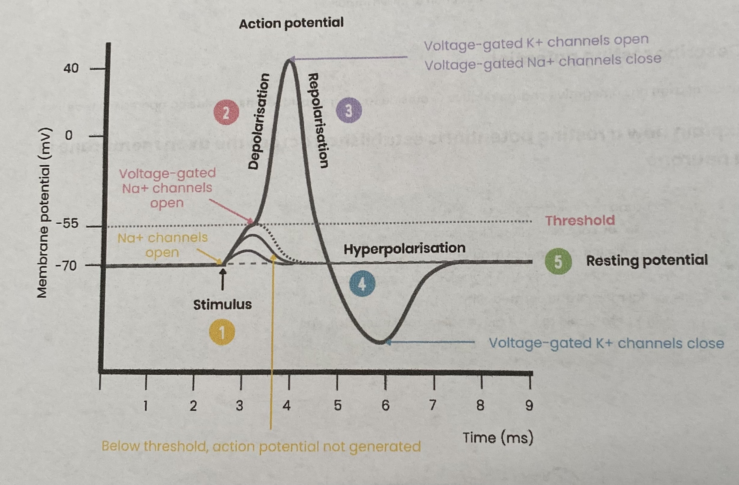
Explain how changes in membrane permeability lead to depolarisation and the generation of an action potential
Stimulus
Na+ channels open; membrane permeability to Na+ increases
Na+ diffuse into axon down electrochemical gradient (causing depolarisation)
Depolarisation
If threshold potential reached, an action potential is generated
As more voltage-gated Na+ channels open (positive feedback effect)
So more Na+ diffuse in rapidly
Repolarisation
Voltage-gated Na+ channels close
Voltage-gated K+ channels open; K+ diffuse out of axon
Hyperpolarisation
K+ channels slow to close so there’s a slight overshoot- too many K+ diffuse out
Resting potential
Restored by Na+/ K+ pump
Describe the all-or- nothing principle
For an action potential to be produced, depolarisation must exceed threshold potential
Action potentials produced are always same magnitude/ size/ peak at same potential
bigger stimuli instead increase frequency of action potential
Explain how the passage of an action potential along non- myelinated axons results in nerve impulses
Action potential passes as a wave of depolarisation
Influx of Na+ in one region increases permeability of adjoining region to Na+ by causing voltage-gated Na+ channels to open so adjoining region depolarises
Explain how the passage of an action potential along myelinated axons results in nerve impulses
Myelination provides electrical insulation
Depolarisation of axon at nodes of Ranvier only
Resulting in saltatory conduction (local currents circuits)
So there is no need for depolarisation along whole length of axon
Suggest how damage to the myelin sheath can lead to slow responses and/ or jerky movement
Less/ no saltatory conduction; depolarisation occurs along whole length of axon
so nerve impulses take longer to reach neuromuscular junction; delay in muscle contraction
Ions/ depolarisation may pass/ leak to other neurones
causing wrong muscle fibres to contract
Describe the nature of the refractory period
Time taken to restore axon to resting potential when no further action potential can be generated
As Na+ channels are closed/ inactive/ will not open
Explain the importance of the refractory period
Ensures discrete impulses are produced (action potentials don’t overlap)
Limits frequency of impulse transmission at a certain intensity (prevents over reaction to stimulus)
higher intensity stimulus causes higher frequency of action potentials
but only up to certain intensity
Also ensures action potentials travel in one direction- can’t be propagated in a refractory region
In the second half of the refractory period an action potential can be produced but requires greater stimulation to reach threshold
Describe the factors that affect speed of conductance
Myelination:
depolarisation at NOdes of Ranvier only= saltatory conduction
impulse doesn’t travel/ depolarise whole length of axon
Axon diameter:
bigger diameter means less resistance to flow of ions in cytoplasm
Temperature:
increases rate of diffusion of Na+ and K+ as more kinetic energy
but proteins/ enzymes could denature at a certain temperature
Describe the structure of the synapse

What are cholinergic synapses?
synapses that use the neurotransmitter acetylcholine (ACh)
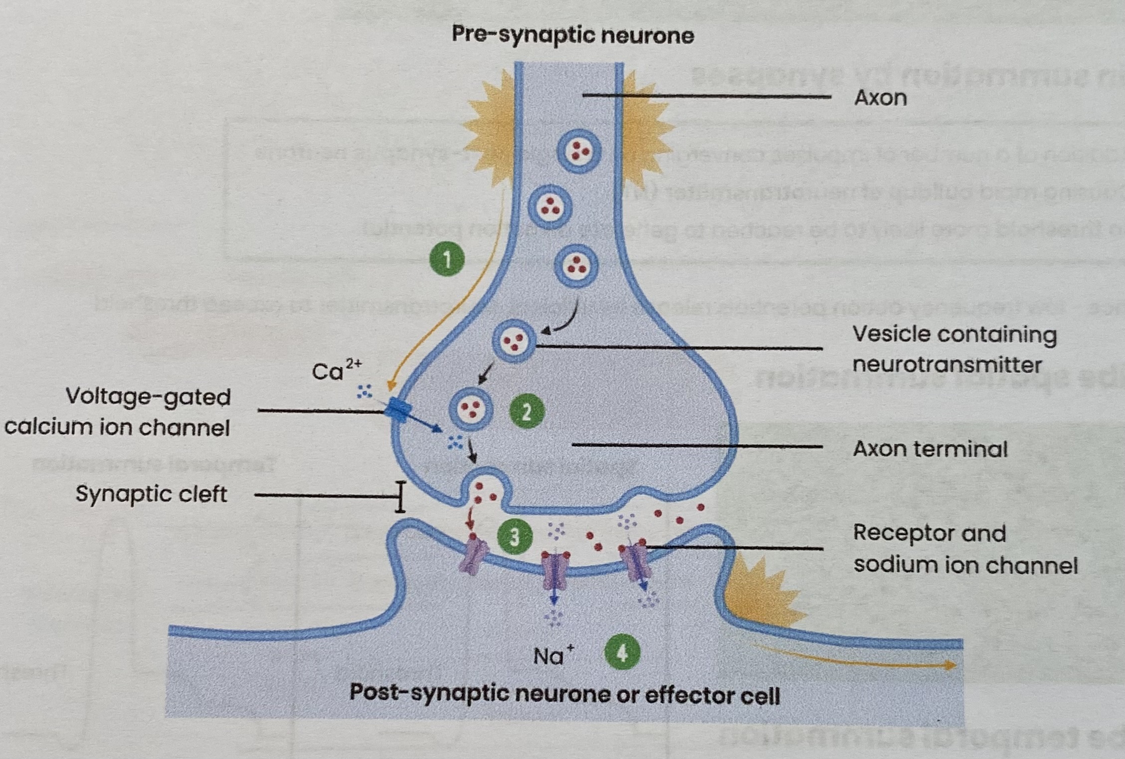
Describe transmission across a cholinergic synapse
AT PRE-SYNAPTIC NEURONE:
Depolarisation of pre-synaptic membrane causes opening of voltage-gated Ca2+ channels
Ca2+ diffuse into pre-synaptic neurone/ knob
Causing vesicles containing ACh to move and fuse with pre-synaptic membrane
releasing ACh into the synaptic cleft (by exocytosis)
AT POST-SYNAPTIC NEURONE:
ACh diffuses across synaptic cleft to bind to specific receptors on post- synaptic membrane
Causing Na+ channels to open
Na+ diffuse into post- synaptic knob causing depolarisation
if threshold is met, an action potential is initiated
Explain what happens to acetylcholine after synaptic transmission
It is hydrolysed by acetylcholinesterase
Products are reabsorbedby the presynaptic neurone
To stop overstimulation- if not removed it would keep binding to receptors, causing depolarisation
Explain how synapses result in unidirectional nerve impulses
neurotransmitter only made in/ released from pre-synaptic neurone
receptors only on post-synaptic membrane
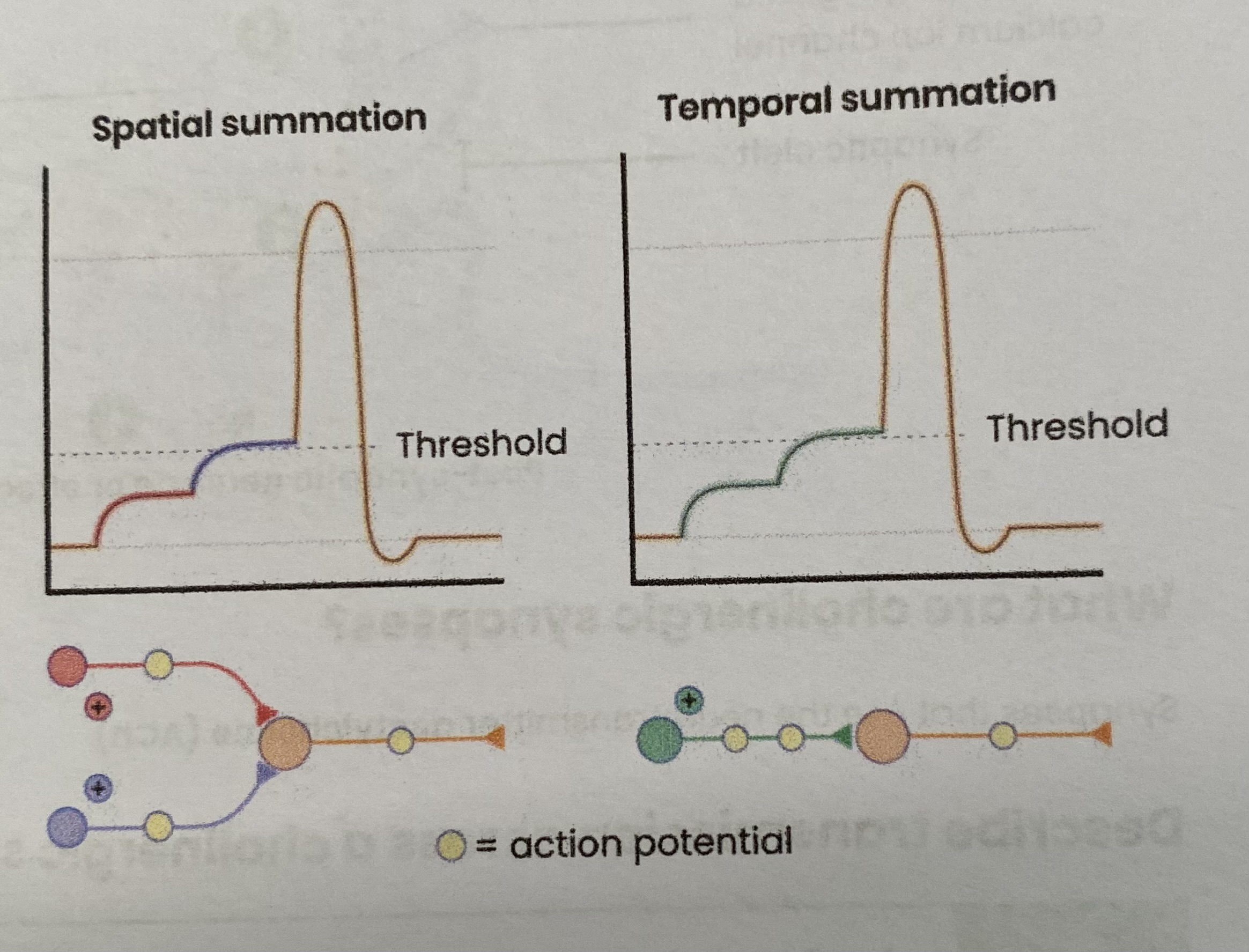
Explain summation by synapses
Addition of a number of impulses converging on a single post-synaptic neurone
causing rapid buildup of neurotransmitter (NT)
so threshold more likely to be reached to generate an action potential
Importance- low frequency action potentials release insufficient neurotransmitter to exceed threshold
Describe spatial summation
Many pre-synaptic neurones share one synaptic cleft/ post- synaptic neurone
Collectively release sufficient neurotransmitter to reach threshold to trigger an action potential

Describe temporal summation
one pre-synaptic neurone releases neurotransmitter many times over a short time
sufficient neurotransmitter to reach threshold to trigger an action potential
Describe inhibition by inhibitory synapses
Inhibitory neurotransmitters hyperpolarise postsynaptic membrane as:
Cl- channels open= Cl- diffuse in
K+ channels open= K+ diffuse out
This means inside of axon has a more negative charge relative to outside/ below resting potential
So more Na+ required to enter for depolarisation
Reduces likelihood of threshold being met/ action potential formation at post- synaptic membranes
Importance- both excitatory and inhibitory neurones forming synapses with the same post- synaptic membrane gives control of whether it ‘fires’ an action potential
Describe the structure of a neuromuscular junction
Very similar to a synapse except:
receptors are on muscle fibre sarcolemma instead of postsynaptic membrane and there are more
muscle fibre forms clefts to store enzyme e.g. acetylcholinesterase to break down neurotransmitter
Compare transmission across cholinergic synapses and neuromuscular junctions
In BOTH: transmission is unidirectional
CHOLINERGIC SYNAPSE:
neurone to neurone (or effectors, glands
neurotransmitters can be excitatory or inhibitory
action potential may be initiated in postsynaptic neurone
NEUROMUSCULAR JUNCTION:
(motor) neurone to muscle
always excitatory
action potential propagates along sarcolemma dow T tubules
Use examples to explain the effect of drugs on a synapse
Some drugs stimulate the nervous system, leading to more action potentials, e.g.;
similar shape to neurotransmitter
stimulate release of more neurotransmitter
inhibit enzyme that breaks down neurotransmitter= Na+ continues to enter
Some drugs inhibit the nervous system, leading to fewer action potentials, e.g.;
inhibit release of neurotransmitter e.g. prevent opening of calcium ion channels
block receptors by mimicking shape of neurotransmitter
Describe how muscles work
work in antagonistic pairs= pull in opposite directions e.g. biceps/ triceps
one muscle contracts (agonist), pulling on bone/ producing force
one muscle relaxes (antagonist)
skeleton is incompressible so muscle can transmit force to bone
ADVANTAGE- the second muscle required to reverse movement caused by the first (muscles can only pull) and contraction of both muscles helps maintain posture
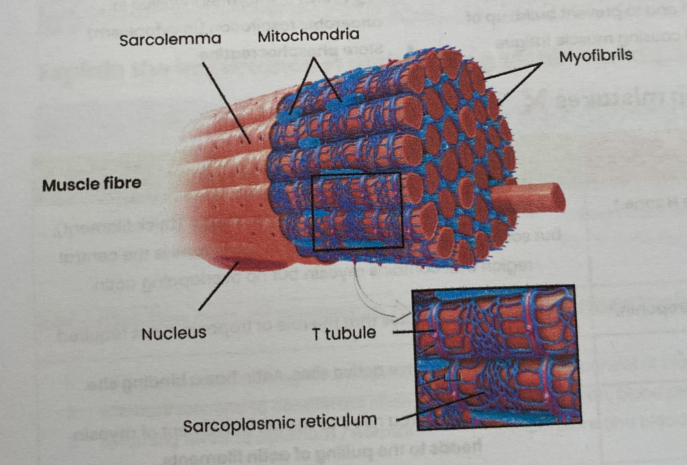
Describe the gross and microscopic structure of skeletal muscle
Made of many bundles of muscle fibres (cells) packaged together
Attached to bones by tendons
Muscle fibres contain:
sarcolemma (cell membrane) which folds inwards (invagination) to form transverse (T) tubules
sarcoplasm (cytoplasm)
multiple nuclei
many myofibrils
sarcoplasmic reticulum (endoplasmic reticulum)
many mitochondria
Describe the ultrastructure of a myofibril
Made of 2 types of long protein filaments, arranged in parallel
myosin- thick filament
actin- thin filament
Arranged in functional units called sacromeres
ends- Z-line/ disc
middle- M-line
H zone- contains only myosin
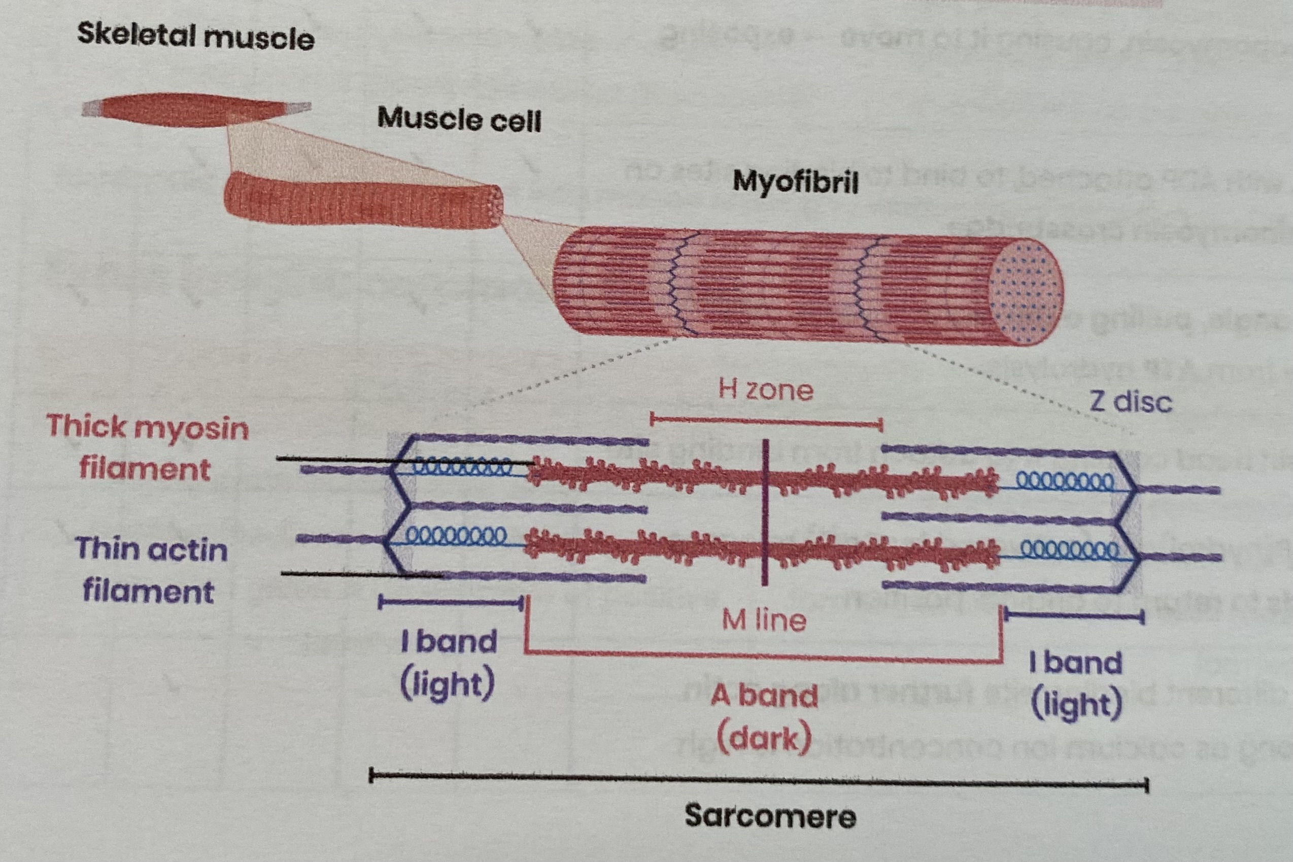
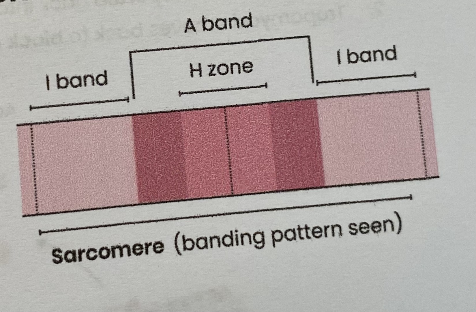
Explain the banding pattern to be seen in myofibrils
I-bands= light bands containing only thin actin filaments
A-bands= dark bands containing thick myosin filaments (and some actin filaments)
H zone contains only myosin
darkest region contains overlapping actin and myosin
Give an overview of muscle contraction
Myosin heads slide actin along myosin causing the sacromere to contract
Simulanteous contraction of many sacromeres causes myofibrils and muscle fibres to contract
When sacromeres contract (shorten)…
H zones get shorter
I band get shorter
A band stays the same
Z lines get closer
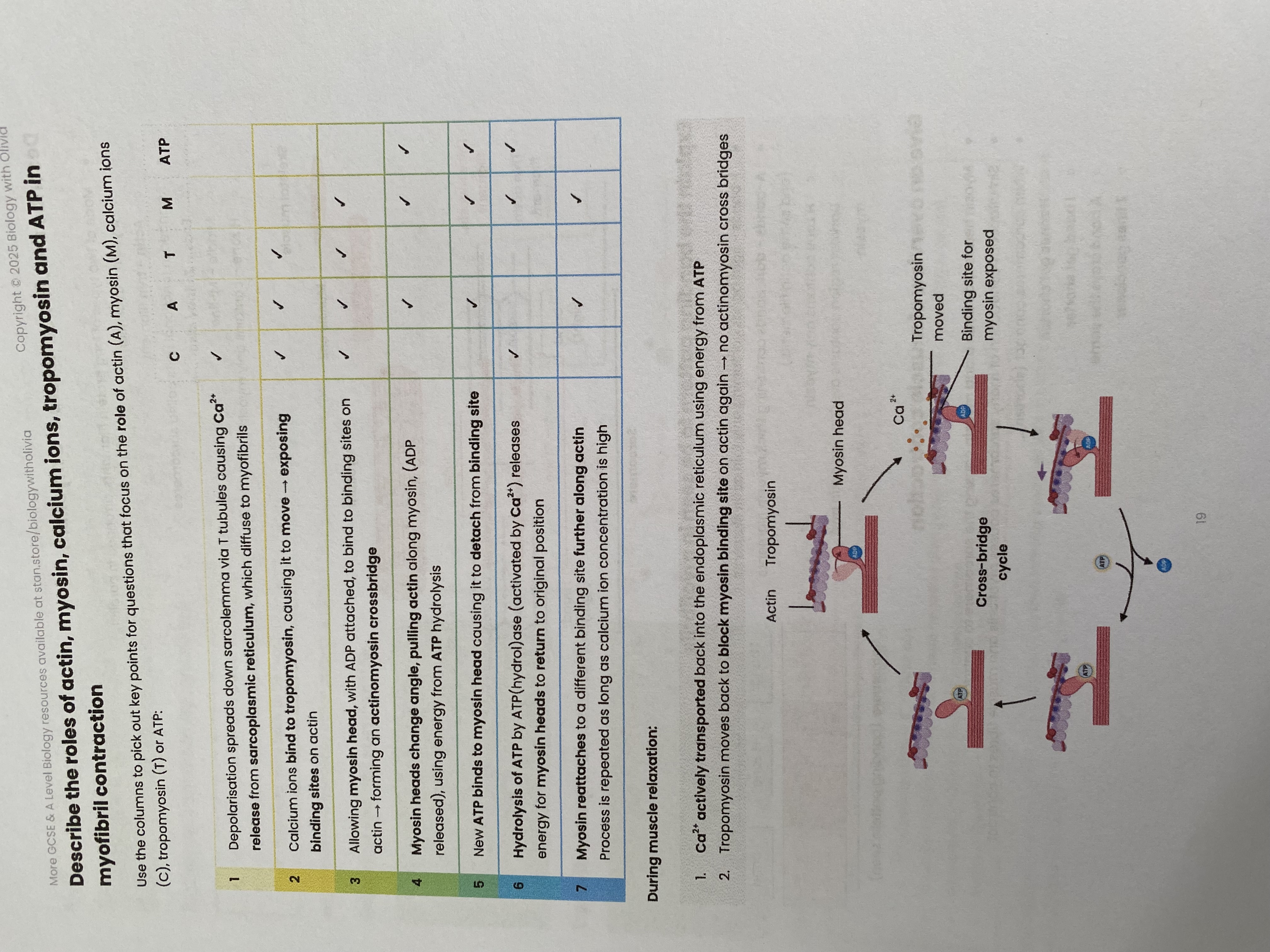
Describe the roles of actin, myosin, calcium ions, tropomyosin and ATP in myofibril contraction
Describe the role of phosphocreatine in muscle contraction
A source of inorganic phosphate (Pi) = rapidly phosphorylates ADP to regenerate ATP
ADP+ phosphocreatine = ATP + creatine
Runs out after a few seconds= used in short bursts of vigorous exercise
Anaerobic and alactic
Compare the structure, location and general properties of slow and fast skeletal muscle fibres (slow twitch)
GENERAL PROPERTIES:
specialised for slow, sustained contractions (e.g. posture, long distance running)
produce more ATP slowly (mostly) from aerobic respiration
fatigues slowly
LOCATION:
high proportion in muscles used for posture e.g. back, calves
legs of long distance runners
STRUCTURE:
high conc. of myoglobin= stores oxygen for aerobic respiration
many mitochondria= high rate of aerobic respiration
many capillaries= supply high conc. of oxygen/ glucose for aerobic respiration and to prevent build-up of lactic acid causing muscle fatigue
Compare the structure, location and general properties of slow and fast skeletal muscle fibres (fast twitch)
GENERAL PROPERTIES:
specialised for brief, intensive contractions (e.g. sprinting)
produce less ATP rapidly (mostly) from anaerobic respiration
fatigues quickly due to high lactate concentration
LOCATION:
high proportion in muscles used for fast movement e.g. biceps, eyelids
legs of sprinters
STRUCTURE:
low levels of myoglobin
lots of glycogen= hydrolysed to provide glucose for glycolysis/ anaerobic respiration which is inefficient so large quantities of glucose required
high conc. of enzymes involved in anaerobic respiration (in cytoplasm)
store phosphocreatine